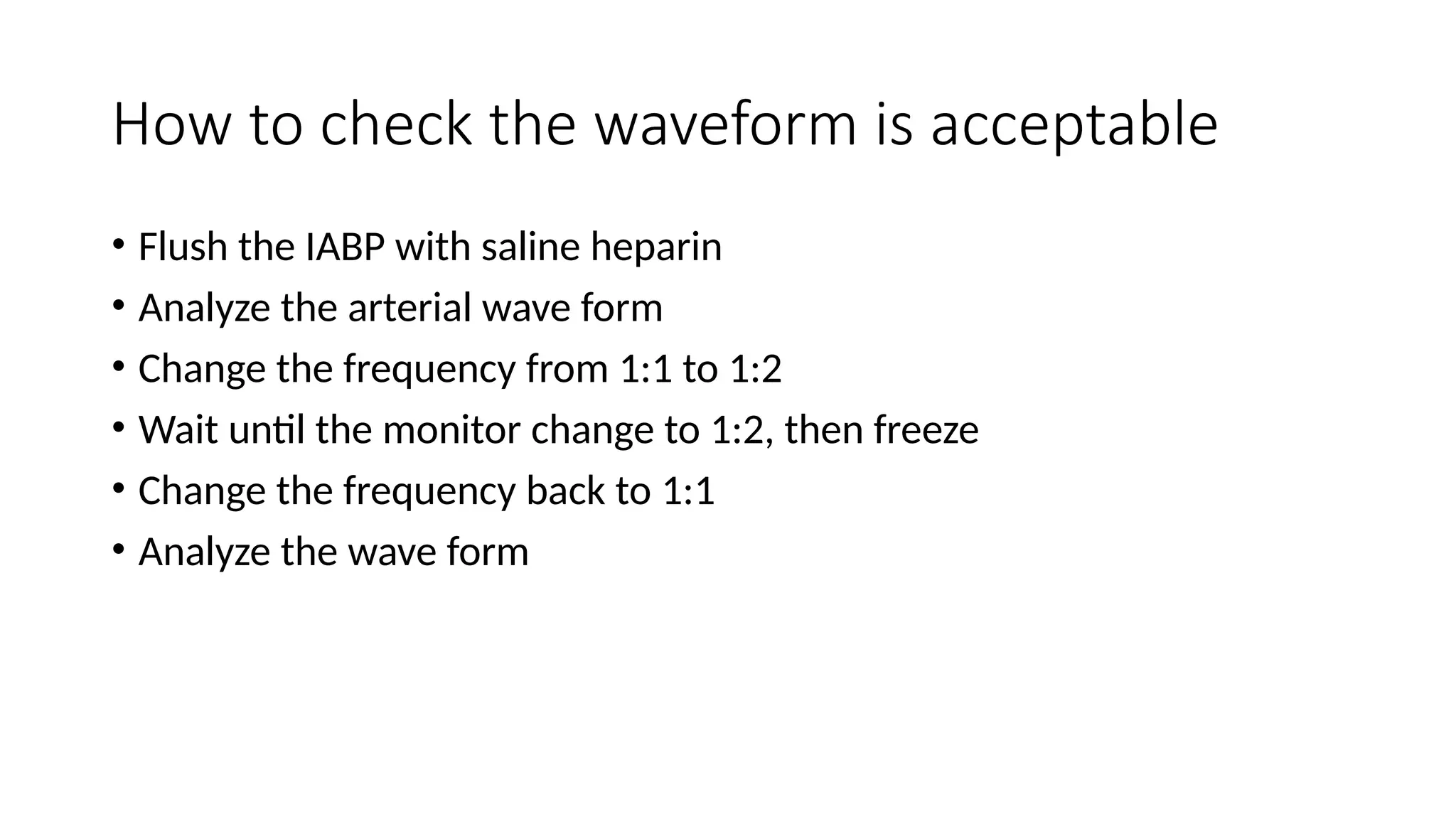
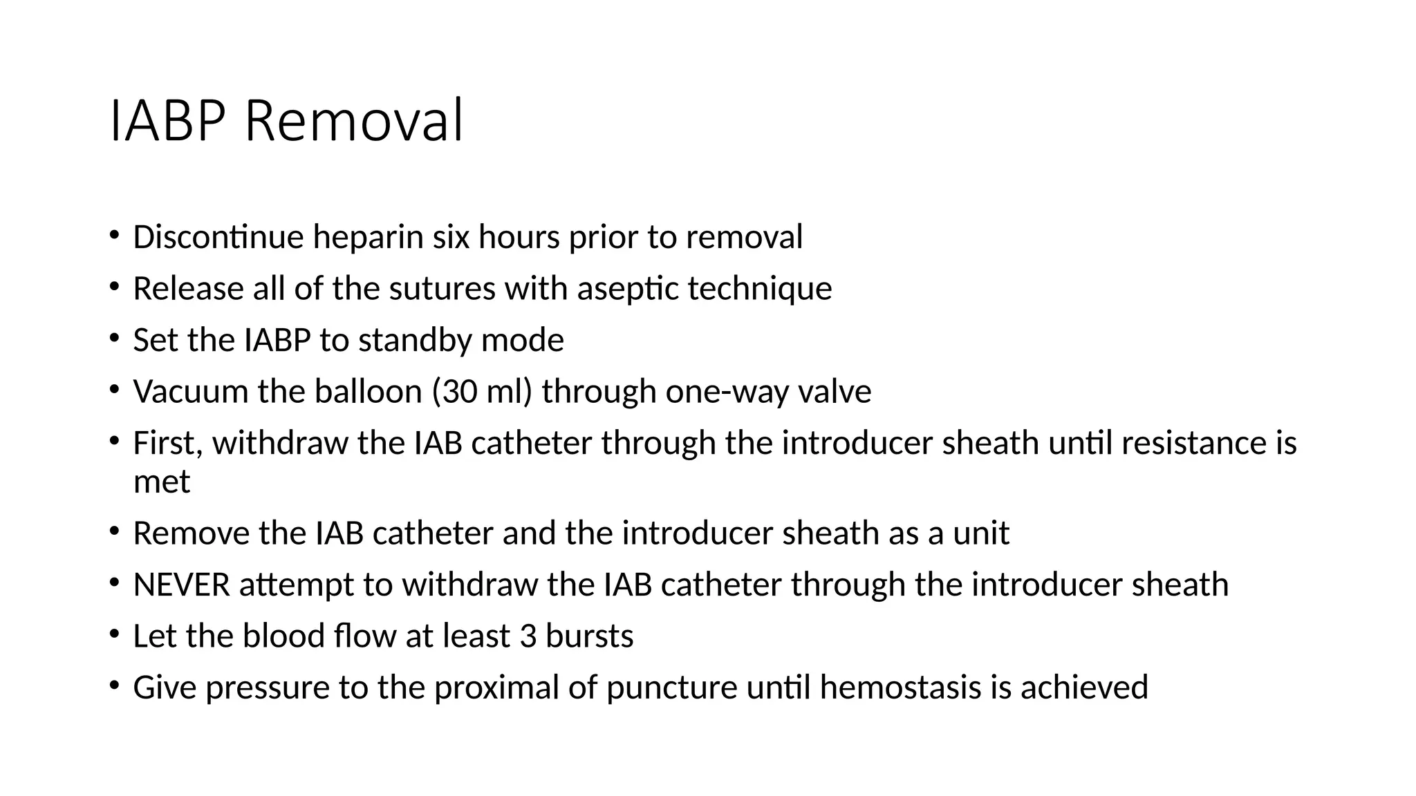
Tugas Stase CVCU
Intraaortic Ballon Pump
Yordan W. Ashari
Pembimbing
dr. Setyasih Anjarwani, Sp.JP(K)
Outline
• History
• What is IABP
• Indication and Contraindication
• Placement
• Weaning
• Removal
• Complication
History
Adrian Kantrowitz, 1918-2008
A thoracic surgeon, who
performed the first human
heart transplant in the
United States in 1967
Back in 1951, he
discovered that it was
possible to manipulate
blood flow in the aorta so
as to pulse it into the
coronary arteries during
diastole and increased the
coronary and general
circulation, while
decreasing demand on the
heart.
This principle of "diastolic
augmentation" or
"counterpulsation" would
be the basis for
Kantrowitz's left ventricular
assist devices (LVAD) and
his intraaortic balloon
pump.
Basic Principle of
Counterpulsation
• Counterpulsation is a term that describes
balloon inflation in diastole and deflation in
early systole.
• Balloon inflation causes ‘volume
displacement’ of blood within the aorta,
both proximally and distally.
• This leads to a potential increase in coronary
blood flow and potential improvements in
systemic perfusion by augmentation of the
intrinsic ‘Windkessel effect’, whereby
potential energy stored in the aortic root
during systole is converted to kinetic energy
with the elastic recoil of the aortic root
Windkessel
effect
How it works
How it works
Expected Changes with IABP Support in
Hemodynamic profile
• Decrease SBP by 20%
• Increase in aortic diastolic pressure by 30% (increase coronary blood
flow)
• Increase in MAP
• Reduction of the HR by 20%
• Decrease the mean PCWP by 20%
• Elevation in CO by 20%
Indication
• Medical Indication
• Cardiogenic Shock
• Pre-Shock Syndrome
• Unstable (refractory) Angina
• Coronary Angiography
• Coronary Angioplasty
• Thrombolytic Therapy
• High risk interventional procedures
• Bridging device for
• Other Mechanical Assist Devices
Contraindication
• Hemodynamically significant
aortic regurgitation
• Dissecting Aortic /Aneurysm
• Severe calcific atherosclerotic
peripheral vascular disease
• End Stage Terminal Disease
• Severe Clotting Disorders
• Severe Obesity
• Skin to femoral artery distance
exceeds 5 cm
the IAB…. To Transducer/arterial line
Extension
to Helium
To Helium
with One
way Valve
Syringe for
vacuuming
the
balloon
The
Balloon
ECG
IAB sizing chart
Preparing the IAB Catheter
IAB Insertion
IAB Insertion (1)
Positioning
• The tip of the balloon should be
just distal (1-2cm) to the takeoff
of the left subclavian artery
• Position should be confirmed by
fluoroscopy (at ICS 2-3) or chest
X-ray (2 cm above the carina)
Initial Set-Up
• Once connected properly the console would show ECG and pressure
waveforms
• Make sure the setting is at “AUTO”
• Usually IABP started at 1:1 or 1:2 augmentation
• Keep the augmentation at maximum
Trigger Modes
• Trigger:
• Event the pump uses to identify the onset of cardiac cycle
• Pump must have consistent trigger in order to provide patient assist
• If selected trigger not detected, counterpulsation will be interrupted
• ECG  uses the slope of QR segment to detect trigger point
• Pressure systolic upstroke of the arterial pressure wave form
How to check the waveform is acceptable
• Flush the IABP with saline heparin
• Analyze the arterial wave form
• Change the frequency from 1:1 to 1:2
• Wait until the monitor change to 1:2, then freeze
• Change the frequency back to 1:1
• Analyze the wave form
1. Check the dicrotic
notch. See if
augmentation start at
that point, this should
produce a sharp “V” at
inflation
2. Check if diastolic
augmented wave is >>
systolic wave
3. Confirmed if end
diastolic wave following
the augmented wave is
less than an non
augmented wave
Factors Affecting Diastolic Augmentation
• Patient
• Heart Rate
• Mean Arterial Pressure
• Stroke Volume
• Systemic Vascular Resistance
• IAB Catheter
• Position of IAB
• IAB not unfolded
• Kinking IAB catheter
• IAB leak
• IABP
• Timing
• Position of IAB augmentation
control
Timing – Early Inflation
• Inflation of the IAB prior to aortic valve
closure
• Waveform Characteristic
• Inflation of IAB prior to dicrotic notch
• Diastolic augmentation encoaches onto
systole
• Physiologic effect
• Potential premature closure to aortic valve
• Potential increase in LVEDP/PCWP
• Increase left ventricular wall stress or
afterload
• Reducing cardiac output
• Aortic regurgitation
Timing – Late Inflation
• Inflation of the IAB markedly
after closure of the aortic valve
• Waveform Characteristic
• Inflation of IAB after the dicrotic
notch
• Absence of sharp V
• Physiologic Effects
• Suboptimal diastolic augmentation
• Suboptimal coronary artery
perfusion
Timing – Early Deflation
• Premature deflation of the IAB during
the diastolic phase
• Waveform characteristic:
• Deflation of the IAB is seen as a sharp
drop following diastolic augmentation
• Sub-optimal diastolic augmentation
• Assisted systolic pressure may rise
• Physiologic effect:
• Sub-optimal coronary perfusion
• Angina may occur as a result of
retrograde coronary blood flow
• Sub-optimal afterload reduction
Timing – Late Deflation
• Late deflation of the IAB during the
diastolic phase
• Waveform characteristic:
• Assisted aortic end diastolic pressure
may be equal to the unassisted aortic
end diastolic pressure
• Diastolic augmentation may appear
widened
• Physiologic effect:
• Afterload reduction is essentially absent
• IAB may impede left ventricular ejection
and increase afterload
Complication
• Limb ischemia
• Bleeding and insertion site
• Groin hematomas
• Aortic perforation / dissection
• Renal failure and bowel ischemia (due to migration of IAB)
• HIT
• infection
Patient management during IABP support
• Anticoagulation ??
• Kogan et al  heparin should not be used in patient requiring IABP after
cardiac surgery
• Jiang et al  the used of heparin asscociated with higher bleeding incidence,
but no difference in incidence of limb ischemia
• CXR evaluation
• Check lower limb pulse every 2 hours
Patient management during IABP support (1)
• Hip flexion is restricted, and head of the bed should not be elevated
beyond 30°
• Never leave the patient with standby or 1:2 or 1:3 mode for more
than 20 minutes
• Echo  evaluate VTI variation daily
• Wean the IABP as early as possible as longer duration ias associated
with higher incidence of limb complication
Weaning of IABP
• Timing:
• Patient should be stable for 12-24 hours
• Decrease inotropic support
• Decrease pump ratio
• Decrease augmentation
• Monitor patient closely
• If patient become unstable, weaning should be immediately
discontinued
IABP Removal
• Discontinue heparin six hours prior to removal
• Release all of the sutures with aseptic technique
• Set the IABP to standby mode
• Vacuum the balloon (30 ml) through one-way valve
• First, withdraw the IAB catheter through the introducer sheath until resistance is
met
• Remove the IAB catheter and the introducer sheath as a unit
• NEVER attempt to withdraw the IAB catheter through the introducer sheath
• Let the blood flow at least 3 bursts
• Give pressure to the proximal of puncture until hemostasis is achieved
Conclussion
• The intra-aortic balloon pump (IABP) is the oldest and most widely
used pVAD currently in use for left ventricular support
• it works on the principle of counterpulsation to pressure unload
the heart and, to a lesser extent, increase coronary perfusion
• It is important for cardiologist, to understand the indication,
contraindication, complication of IABP
"When we first did it, people
thought it was silly. You really don't
have a good idea unless people
think it's silly.“
Adrian Kantrowitz
Indication
• Mechanical Defects
• Valve Stenosis
aortic / mitral
• Mitral Valvular Insufficiency
• Ventricular Septal Defect
• Ruptured Papillary Muscle
• Mechanical complications
post MI
Indication
• Surgical Indication
• Post Surgical Myocardial
Dysfunction
• Weaning from CPB
• Cardiac Support following
correction of anatomical defects
• Maintenance of graft patency post
CABG
• Pulsatile flow during CPB
Contraindication
• Hemodynamically significant
aortic regurgitation
• Dissecting Aortic /Aneurysm
• Severe calcific atherosclerotic
peripheral vascular disease
• End Stage Terminal Disease
• Severe Clotting Disorders
• Severe Obesity
• Skin to femoral artery distance
exceeds 5 cm
Indications, Contraindications, and
Complications

Materi intra Aortic Balloon pulsation.pptx

  • 1.
    Tugas Stase CVCU IntraaorticBallon Pump Yordan W. Ashari Pembimbing dr. Setyasih Anjarwani, Sp.JP(K)
  • 2.
    Outline • History • Whatis IABP • Indication and Contraindication • Placement • Weaning • Removal • Complication
  • 3.
  • 4.
    Adrian Kantrowitz, 1918-2008 Athoracic surgeon, who performed the first human heart transplant in the United States in 1967 Back in 1951, he discovered that it was possible to manipulate blood flow in the aorta so as to pulse it into the coronary arteries during diastole and increased the coronary and general circulation, while decreasing demand on the heart. This principle of "diastolic augmentation" or "counterpulsation" would be the basis for Kantrowitz's left ventricular assist devices (LVAD) and his intraaortic balloon pump.
  • 5.
    Basic Principle of Counterpulsation •Counterpulsation is a term that describes balloon inflation in diastole and deflation in early systole. • Balloon inflation causes ‘volume displacement’ of blood within the aorta, both proximally and distally. • This leads to a potential increase in coronary blood flow and potential improvements in systemic perfusion by augmentation of the intrinsic ‘Windkessel effect’, whereby potential energy stored in the aortic root during systole is converted to kinetic energy with the elastic recoil of the aortic root
  • 6.
  • 7.
  • 8.
  • 10.
    Expected Changes withIABP Support in Hemodynamic profile • Decrease SBP by 20% • Increase in aortic diastolic pressure by 30% (increase coronary blood flow) • Increase in MAP • Reduction of the HR by 20% • Decrease the mean PCWP by 20% • Elevation in CO by 20%
  • 11.
    Indication • Medical Indication •Cardiogenic Shock • Pre-Shock Syndrome • Unstable (refractory) Angina • Coronary Angiography • Coronary Angioplasty • Thrombolytic Therapy • High risk interventional procedures • Bridging device for • Other Mechanical Assist Devices
  • 12.
    Contraindication • Hemodynamically significant aorticregurgitation • Dissecting Aortic /Aneurysm • Severe calcific atherosclerotic peripheral vascular disease • End Stage Terminal Disease • Severe Clotting Disorders • Severe Obesity • Skin to femoral artery distance exceeds 5 cm
  • 13.
    the IAB…. ToTransducer/arterial line Extension to Helium To Helium with One way Valve Syringe for vacuuming the balloon The Balloon ECG
  • 14.
  • 15.
  • 16.
  • 17.
  • 18.
    Positioning • The tipof the balloon should be just distal (1-2cm) to the takeoff of the left subclavian artery • Position should be confirmed by fluoroscopy (at ICS 2-3) or chest X-ray (2 cm above the carina)
  • 19.
    Initial Set-Up • Onceconnected properly the console would show ECG and pressure waveforms • Make sure the setting is at “AUTO” • Usually IABP started at 1:1 or 1:2 augmentation • Keep the augmentation at maximum
  • 21.
    Trigger Modes • Trigger: •Event the pump uses to identify the onset of cardiac cycle • Pump must have consistent trigger in order to provide patient assist • If selected trigger not detected, counterpulsation will be interrupted • ECG  uses the slope of QR segment to detect trigger point • Pressure systolic upstroke of the arterial pressure wave form
  • 26.
    How to checkthe waveform is acceptable • Flush the IABP with saline heparin • Analyze the arterial wave form • Change the frequency from 1:1 to 1:2 • Wait until the monitor change to 1:2, then freeze • Change the frequency back to 1:1 • Analyze the wave form
  • 27.
    1. Check thedicrotic notch. See if augmentation start at that point, this should produce a sharp “V” at inflation 2. Check if diastolic augmented wave is >> systolic wave 3. Confirmed if end diastolic wave following the augmented wave is less than an non augmented wave
  • 29.
    Factors Affecting DiastolicAugmentation • Patient • Heart Rate • Mean Arterial Pressure • Stroke Volume • Systemic Vascular Resistance • IAB Catheter • Position of IAB • IAB not unfolded • Kinking IAB catheter • IAB leak • IABP • Timing • Position of IAB augmentation control
  • 30.
    Timing – EarlyInflation • Inflation of the IAB prior to aortic valve closure • Waveform Characteristic • Inflation of IAB prior to dicrotic notch • Diastolic augmentation encoaches onto systole • Physiologic effect • Potential premature closure to aortic valve • Potential increase in LVEDP/PCWP • Increase left ventricular wall stress or afterload • Reducing cardiac output • Aortic regurgitation
  • 31.
    Timing – LateInflation • Inflation of the IAB markedly after closure of the aortic valve • Waveform Characteristic • Inflation of IAB after the dicrotic notch • Absence of sharp V • Physiologic Effects • Suboptimal diastolic augmentation • Suboptimal coronary artery perfusion
  • 32.
    Timing – EarlyDeflation • Premature deflation of the IAB during the diastolic phase • Waveform characteristic: • Deflation of the IAB is seen as a sharp drop following diastolic augmentation • Sub-optimal diastolic augmentation • Assisted systolic pressure may rise • Physiologic effect: • Sub-optimal coronary perfusion • Angina may occur as a result of retrograde coronary blood flow • Sub-optimal afterload reduction
  • 33.
    Timing – LateDeflation • Late deflation of the IAB during the diastolic phase • Waveform characteristic: • Assisted aortic end diastolic pressure may be equal to the unassisted aortic end diastolic pressure • Diastolic augmentation may appear widened • Physiologic effect: • Afterload reduction is essentially absent • IAB may impede left ventricular ejection and increase afterload
  • 34.
    Complication • Limb ischemia •Bleeding and insertion site • Groin hematomas • Aortic perforation / dissection • Renal failure and bowel ischemia (due to migration of IAB) • HIT • infection
  • 35.
    Patient management duringIABP support • Anticoagulation ?? • Kogan et al  heparin should not be used in patient requiring IABP after cardiac surgery • Jiang et al  the used of heparin asscociated with higher bleeding incidence, but no difference in incidence of limb ischemia • CXR evaluation • Check lower limb pulse every 2 hours
  • 36.
    Patient management duringIABP support (1) • Hip flexion is restricted, and head of the bed should not be elevated beyond 30° • Never leave the patient with standby or 1:2 or 1:3 mode for more than 20 minutes • Echo  evaluate VTI variation daily • Wean the IABP as early as possible as longer duration ias associated with higher incidence of limb complication
  • 37.
    Weaning of IABP •Timing: • Patient should be stable for 12-24 hours • Decrease inotropic support • Decrease pump ratio • Decrease augmentation • Monitor patient closely • If patient become unstable, weaning should be immediately discontinued
  • 38.
    IABP Removal • Discontinueheparin six hours prior to removal • Release all of the sutures with aseptic technique • Set the IABP to standby mode • Vacuum the balloon (30 ml) through one-way valve • First, withdraw the IAB catheter through the introducer sheath until resistance is met • Remove the IAB catheter and the introducer sheath as a unit • NEVER attempt to withdraw the IAB catheter through the introducer sheath • Let the blood flow at least 3 bursts • Give pressure to the proximal of puncture until hemostasis is achieved
  • 39.
    Conclussion • The intra-aorticballoon pump (IABP) is the oldest and most widely used pVAD currently in use for left ventricular support • it works on the principle of counterpulsation to pressure unload the heart and, to a lesser extent, increase coronary perfusion • It is important for cardiologist, to understand the indication, contraindication, complication of IABP
  • 40.
    "When we firstdid it, people thought it was silly. You really don't have a good idea unless people think it's silly.“ Adrian Kantrowitz
  • 42.
    Indication • Mechanical Defects •Valve Stenosis aortic / mitral • Mitral Valvular Insufficiency • Ventricular Septal Defect • Ruptured Papillary Muscle • Mechanical complications post MI
  • 43.
    Indication • Surgical Indication •Post Surgical Myocardial Dysfunction • Weaning from CPB • Cardiac Support following correction of anatomical defects • Maintenance of graft patency post CABG • Pulsatile flow during CPB
  • 44.
    Contraindication • Hemodynamically significant aorticregurgitation • Dissecting Aortic /Aneurysm • Severe calcific atherosclerotic peripheral vascular disease • End Stage Terminal Disease • Severe Clotting Disorders • Severe Obesity • Skin to femoral artery distance exceeds 5 cm
  • 48.