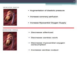
The major physiological effects of counterpulsation include:
A) increased coronary artery perfusion, increased preload, decreased after load, decreased myocardial oxygen consumption
2
Intra aortic ballooncounter pulsation( IABP):
Most common and widely available methods of mechanical
circulatory support
Temporary support for the left ventricle by mechanically
displacing blood within the aorta
Concepts:
- Systolic unloading
- Diastolic augmentation
Traditionally used in surgical and non surgical patients
with cardiogenic shock
3.
3
Indications for IABP
1.Cardiogenic shock:
2. In association with CABG :
Preoperative insertion
- Patients with severe LV dysfunction
- Patients with intractable ischemic arrhythmias
Postoperative insertion
- Postcardiotomy cardiogenic shock
- Associated with acute MI
- Mechanical complications of MI - MR , VSD
3. In association with nonsurgical revascularization:
-Hemodynamically unstable infarct patients
-High risk coronary interventions
- severe LV dysfunction, LMCA, complex coronary artery disease
4. Stabilization of cardiac transplant recipient before insertion of VAD
Post infarction angina
Ventricular arrhythmias relathed to ischemia
10
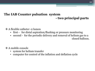
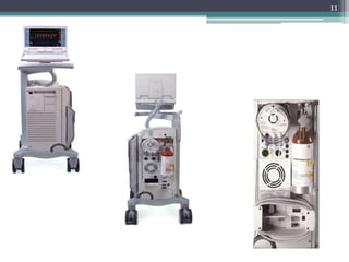
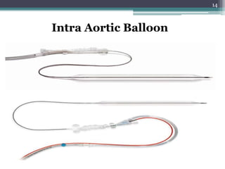
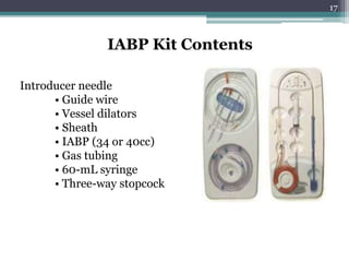
The IAB Counterpulsation system
- two principal parts
A flexible catheter -2 lumen
• first - for distal aspiration/flushing or pressure monitoring
• second - for the periodic delivery and removal of helium gas to a
closed balloon.
A mobile console
• system for helium transfer
• computer for control of the inflation and deflation cycle
12
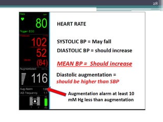
Expected changes withIABP support in hemodynamic profile in
patients with Cardiogenic shock
- Decrease in SBP by 20 %
- Increase in aortic Diastolic Press. by 30 % ( raise coronary blood flow)
- Increase in MAP
- Reduction of the HR by 20%
-Decrease in the mean PCWP by 20 %
- Elevation in the COP by 20%
13.
13
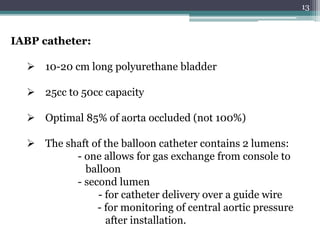
IABP catheter:
10-20cm long polyurethane bladder
25cc to 50cc capacity
Optimal 85% of aorta occluded (not 100%)
The shaft of the balloon catheter contains 2 lumens:
- one allows for gas exchange from console to
balloon
- second lumen
- for catheter delivery over a guide wire
- for monitoring of central aortic pressure
after installation.
15
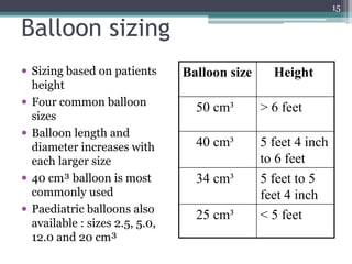
Balloon sizing
Sizingbased on patients
height
Four common balloon
sizes
Balloon length and
diameter increases with
each larger size
40 cm³ balloon is most
commonly used
Paediatric balloons also
available : sizes 2.5, 5.0,
12.0 and 20 cm³
Balloon size Height
50 cm³ > 6 feet
40 cm³ 5 feet 4 inch
to 6 feet
34 cm³ 5 feet to 5
feet 4 inch
25 cm³ < 5 feet
16.
16
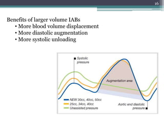
Benefits of largervolume IABs
• More blood volume displacement
• More diastolic augmentation
• More systolic unloading
18
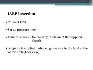
Connect ECG
Set uppressure lines
Femoral access – followed by insertion of the supplied
sheath
0.030 inch supplied J-shaped guide wire to the level of the
aortic arch (LAO view)
- IABP insertion
19.
19
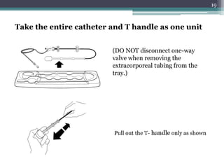
Take the entirecatheter and T handle as one unit
(DO NOT disconnect one-way
valve when removing the
extracorporeal tubing from the
tray.)
Pull out the T- handle only as shown
20.
20
• Remove stylet/aspirate/Flush
•Insert the balloon only over the guide wire
• Hold the catheter close to skin insertion point
• Advance in small steps of 1 to 2 cm at a time and
stop if any resistance.
• The IABP should advance freely
Inserting the Balloon catheter
- Many vascular complications occur during insertion itself
- Resistance during insertion either indicates PVOD, or dissection
- Kinking of IABP » improper inflation/deflation
21.
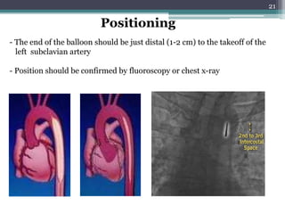
21
- The endof the balloon should be just distal (1-2 cm) to the takeoff of the
left subclavian artery
- Position should be confirmed by fluoroscopy or chest x-ray
Positioning
22.
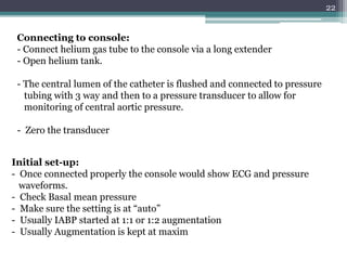
22
Connecting to console:
-Connect helium gas tube to the console via a long extender
- Open helium tank.
- The central lumen of the catheter is flushed and connected to pressure
tubing with 3 way and then to a pressure transducer to allow for
monitoring of central aortic pressure.
- Zero the transducer
Initial set-up:
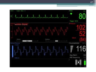
- Once connected properly the console would show ECG and pressure
waveforms.
- Check Basal mean pressure
- Make sure the setting is at “auto”
- Usually IABP started at 1:1 or 1:2 augmentation
- Usually Augmentation is kept at maxim
25
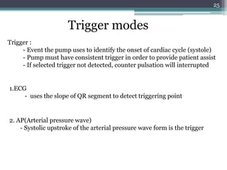
Trigger modes
Trigger :
-Event the pump uses to identify the onset of cardiac cycle (systole)
- Pump must have consistent trigger in order to provide patient assist
- If selected trigger not detected, counter pulsation will interrupted
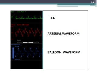
1.ECG
- uses the slope of QR segment to detect triggering point
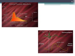
2. AP(Arterial pressure wave)
- Systolic upstroke of the arterial pressure wave form is the trigger
26.
26
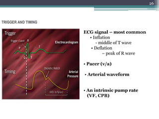
ECG signal –most common
• Inflation
- middle of T wave
• Deflation
– peak of R wave
• Pacer (v/a)
• Arterial waveform
• An intrinsic pump rate
(VF, CPB)
31
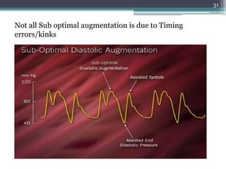
Not all Suboptimal augmentation is due to Timing
errors/kinks
32.
32
Factors affecting diastolicaugmentation
Patient
- Heart rate
- Mean arterial pressure
- Stroke volume
- Systemic vascular resistance
Intra aortic balloon catheter
- IAB in sheath
- IAB not unfolded
- IAB position
- Kink in the IAB catheter
- IAB leak
- Low helium concentration
Intra aortic balloon pump
- Timing
- Position of IAB augmentation control
33.
33
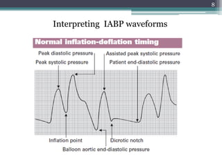
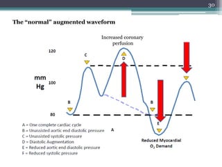
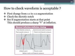
How to checkwaveform is acceptable ?
First change from 1:1 to 1:2 augmentation
Check the dicrotic notch
See if augmentation starts at that point
This should produce a sharp “V” at inflation.
34.
34
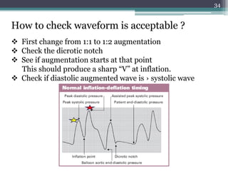
How to checkwaveform is acceptable ?
First change from 1:1 to 1:2 augmentation
Check the dicrotic notch
See if augmentation starts at that point
This should produce a sharp “V” at inflation.
Check if diastolic augmented wave is › systolic wave
35.
35
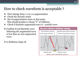
How to checkwaveform is acceptable ?
First change from 1:1 to 1:2 augmentation
Check the dicrotic notch
See if augmentation starts at that point
This should produce a sharp “V” at inflation.
Check if diastolic augmented wave is › systolic wave
Confirm if end diastolic wave
following the augmented wave
is less than an non augmented
wave.
Is Deflation slope ok
37
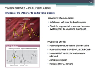
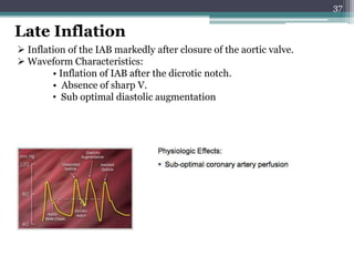
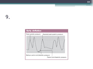
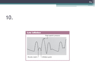
Late Inflation
Inflationof the IAB markedly after closure of the aortic valve.
Waveform Characteristics:
• Inflation of IAB after the dicrotic notch.
• Absence of sharp V.
• Sub optimal diastolic augmentation
39
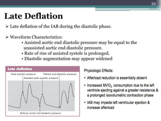
Late Deflation
Latedeflation of the IAB during the diastolic phase.
Waveform Characteristics:
• Assisted aortic end diastolic pressure may be equal to the
unassisted aortic end diastolic pressure.
• Rate of rise of assisted systole is prolonged.
• Diastolic augmentation may appear widened
41
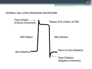
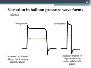
Variation in balloonpressure wave forms
Increased duration of
plateau due to longer
diastolic phase
Decreased duration
of plateau due to
shortened diastolic
phase
42.
42
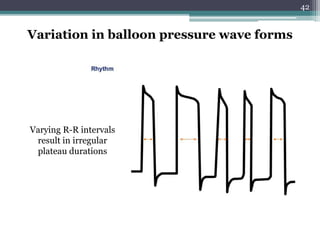
Variation in balloonpressure wave forms
Varying R-R intervals
result in irregular
plateau durations
43.
43
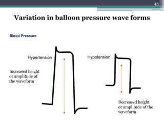
Variation in balloonpressure wave forms
Increased height
or amplitude of
the waveform
Decreased height
or amplitude of the
waveform
44.
44
Variation in balloonpressure wave forms
Gas leak
Leak in the closed system causing the
balloon pressure waveform to fall below
zero baseline..
- due to a loose connection
- a leak in the IAB catheter
- H2O condensation in the external tubing
- a patient who is tachycardiac and febrile which causes increased gas
diffusion through the IAB membrane
45.
45
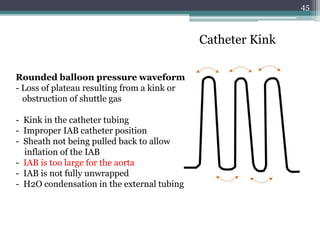
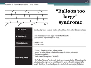
Catheter Kink
Rounded balloonpressure waveform
- Loss of plateau resulting from a kink or
obstruction of shuttle gas
- Kink in the catheter tubing
- Improper IAB catheter position
- Sheath not being pulled back to allow
inflation of the IAB
- IAB is too large for the aorta
- IAB is not fully unwrapped
- H2O condensation in the external tubing
47
Patient Management DuringIABP support
Anticoagulation-- maintain apTT at 50 to 70 seconds
CXR daily – to R/O IAB migration
Check lower limb pulses - 2 hourly.
- If not palpable » ? - vascular obstruction
- thrombus, embolus, or dissection
(urgent surgical consultation)
Prophylactic antibiotics NOT INDICATED
Hip flexion is restricted, and the head of the bed should not be
elevated beyond 30°.
48.
48
Never leavein standby by mode for more than 20 minutes >
thrombus formation
Daily
– Haemoglobin (risk of bleeding or haemolysis)
– Platelet count (risk of thrombocytopenia)
– Renal function (risk of acute kidney injury secondary to distal
migration of IABP catheter)
Wean off the IABP as early as possible as longer duration is associated
with higher incidence of limb complications
Patient Management During IABP support
50
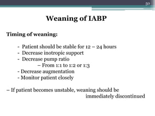
Weaning of IABP
Timingof weaning:
- Patient should be stable for 12 – 24 hours
- Decrease inotropic support
- Decrease pump ratio
– From 1:1 to 1:2 or 1:3
- Decrease augmentation
- Monitor patient closely
– If patient becomes unstable, weaning should be
immediately discontinued
51.
51
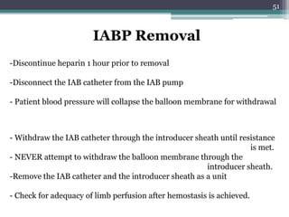
IABP Removal
-Discontinue heparin1 hour prior to removal
-Disconnect the IAB catheter from the IAB pump
- Patient blood pressure will collapse the balloon membrane for withdrawal
- Withdraw the IAB catheter through the introducer sheath until resistance
is met.
- NEVER attempt to withdraw the balloon membrane through the
introducer sheath.
-Remove the IAB catheter and the introducer sheath as a unit
- Check for adequacy of limb perfusion after hemostasis is achieved.
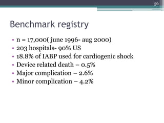
Benchmark registry
• n= 17,000( june 1996- aug 2000)
• 203 hospitals- 90% US
• 18.8% of IABP used for cardiogenic shock
• Device related death – 0.5%
• Major complication – 2.6%
• Minor complication – 4.2%
56
57.
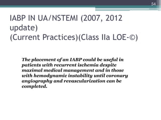
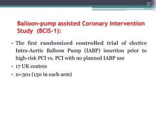
Balloon-pump assisted CoronaryIntervention
Study (BCIS-1):
• The first randomized controlled trial of elective
Intra-Aortic Balloon Pump (IABP) insertion prior to
high-risk PCI vs. PCI with no planned IABP use
• 17 UK centres
• n=301 (150 in each arm)
57
58.
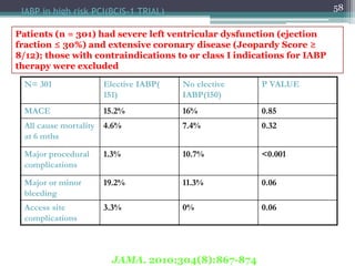
58IABP in highrisk PCI(BCIS-1 TRIAL)
N= 301 Elective IABP(
151)
No elective
IABP(150)
P VALUE
MACE 15.2% 16% 0.85
All cause mortality
at 6 mths
4.6% 7.4% 0.32
Major procedural
complications
1.3% 10.7% <0.001
Major or minor
bleeding
19.2% 11.3% 0.06
Access site
complications
3.3% 0% 0.06
Patients (n = 301) had severe left ventricular dysfunction (ejection
fraction ≤ 30%) and extensive coronary disease (Jeopardy Score ≥
8/12); those with contraindications to or class I indications for IABP
therapy were excluded
JAMA. 2010;304(8):867-874
59.
59
Conclusions of longterm results of
BCIS1 trial(2012-2013)
In patients with severe ischemic cardiomyopathy treated
with PCI, all cause-mortality was 33% at 51 months
(median)
Elective IABP use during PCI was associated with an
observed 34% reduction in long-term all-cause mortality
60.
60
Counterpulsation Reduces InfarctSize Acute Myocardial
Infarction (CRISP AMI) trial.
Intra-aortic balloon pump counterpulsation prior to PCI in
patients with ST segment elevation MI without shock does
not reduce infarct size as measured by MRI
63
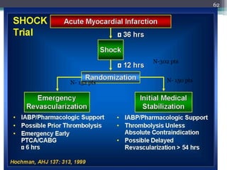
SHOCK Trial
Primary andSecondary Endpoints
0
20
40
60
80
30 Days 6 months
Immediate
Revascularization
Strategy
Medical Stabilization
as an Initial Strategy
Primary
Endpoint
Secondary
Endpoint
Mortality(%)
46.7
%
56.0
% 50.3
%
63.1
%
P=.11
P= .027
Hochman et al, NEJM 1999; 341:625.
64.
64
Impact of thrombolysis,intra-aortic balloon
pump counterpulsation, and their
combination in cardiogenic shock
complicating acute myocardial infarction: a
report from the SHOCK Trial Registry
65.
65
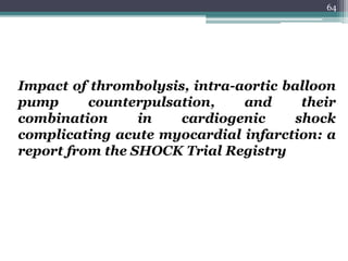
SHOCK Registry: Impactof Thrombolytics
and IABP
0
20
40
60
80
47%
52%
%
P<0.0001
63%
77%
Thrombolytics
+ IABP
No
Thrombolytics
+ IABP
Thrombolytics
+ No IABP
Neither
Hochman et al, NEJM 1999; 341:625
Conclusion
• The useof intraaortic balloon counterpulsation
did not significantly reduce 30-day mortality in
patients with cardiogenic shock complicating
acute myocardial infarction for whom an early
revascularization strategy was planned.
67
• 1. Majorphysiological effects of counter pulsation
include?
▫ A) increased coronary artery perfusion, increased preload,
decreased after load, decreased myocardial oxygen consumption
▫ B) increased coronary artery perfusion, increased preload,
increased after load, decreased myocardial oxygen consumption
▫ C) increased coronary artery perfusion, decreased preload,
decreased after load, increased myocardial oxygen consumption
▫ D) increased coronary artery perfusion, decreased preload,
decreased after load, decreased myocardial oxygen consumption
69
70.
2. The dicroticnotch on the arterial wave form
reflects
A) aortic valve opening
B) aortic valve closure
C) isovolumetric contraction
D)rapid ejection
70
71.
3. Expected changeswith IABP support in hemodynamic
profile in patients with Cardiogenic shock include all
except?
A) Decrease in SBP by 20 %
B) Increase in aortic DP by 30 %
C) Decrease in MAP by 10%
D) Reduction of the HR by 20%
E)Decrease in the mean PCWP by 20 %
71
72.
4. late inflationof the balloon can result in?
A) premature augmentation
B) increased augmentation
C) decreased augmentation
D) increased coronary perfusion
72
73.
5. A roundedballoon pressure wave form
indicate?
A) helium leak
B) power failure
C) hypovolemia
D) balloon occluding the aorta
73
74.
6. width ofballoon pressure wave form
corresponds to
A) length of systole
B) length of diastole
C) arterial pressure
D) helium level
74
75.
7. true statement
a)Dicrotic notch- land mark used to set deflation
b) Deflation is timed to occur during period of iso
volumetric contraction
c) Most common trigger used is arterial pressure
wave method
d) Internal trigger mode is acceptable to use in a
patient with normal sinus rhythm
75
76.
8. true statement
A)pacing spikes are automatically rejected in ECG
triggered modes
B) pacing trigger modes can be used in a patient
of 50% paced rhythm
C) Varying R-R interval result in regular plateau
durations in Balloon pressureWave form
76
• 1. Majorphysiological effects of counter pulsation
include?
▫ A) increased coronary artery perfusion, increased preload,
decreased after load, decreased myocardial oxygen consumption
▫ B) increased coronary artery perfusion, increased preload,
increased after load, decreased myocardial oxygen consumption
▫ C) increased coronary artery perfusion, decreased preload,
decreased after load, increased myocardial oxygen consumption
▫ D) increased coronary artery perfusion, decreased preload,
decreased after load, decreased myocardial oxygen consumption
80
81.
2. the dicroticnotch on the arterial wave form
reflects
A) aortic valve opening
B) aortic valve closure
C) isovolumetric contraction
D)rapid ejection
81
82.
3. Expected changeswith IABP support in hemodynamic
profile in patients with Cardiogenic shock include all
except?
•A) Decrease in SBP by 20 %
•B) Increase in aortic DP by 30 %
•C) Decrease in MAP by 10%
•D) Reduction of the HR by 20%
•E)Decrease in the mean PCWP by 20 %
82
83.
4. late inflationof the balloon can result in?
• A) premature augmentation
• B) increased augmentation
• C) decreased augmentation
• D) increased coronary perfusion
83
84.
5. A roundedballoon pressure wave form
indicate?
• A) helium leak
• B) power failure
• C) hypovolemia
• D) balloon occluding the aorta
84
85.
6. width ofballoon pressure wave form
corresponds to
• A) length of systole
• B) length of diastole
• C) arterial pressure
• D) helium level
85
86.
7. true statement
a)Dicrotic notch- land mark used to set deflation
b) Deflation is timed to occur during period of iso
volumetric contraction
c) Most common trigger used is arterial pressure
wave method
d) Internal trigger mode is acceptable to use in a
patient with normal sinus rhythm
86
87.
8. true statement
A)pacing spikes are automatically rejected in ECG
triggered modes
B) pacing trigger modes can be used in a patient
of 50% paced rhyth
C) Varying R-R interval result in regular plateau
durations in Balloon press. Wave form
87


![INTRA AORTIC BALLON PUMP [IABP].ppt news](https://cdn.slidesharecdn.com/ss_thumbnails/intraaorticballonpumpiabp-240723151642-9cbe1595-thumbnail.jpg?width=640&height=640&fit=bounds)


