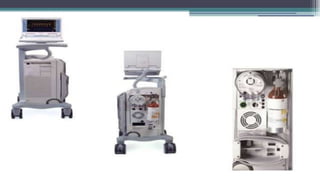
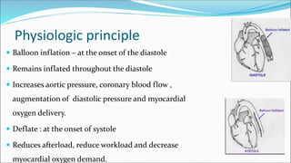
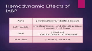
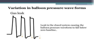
PRINCY FRANCIS M
II MSc (N)
JMCON
Dr. Adrian Kantrowitz – 1952
Augmentation of coronary blood flow by retardation of
the arterial pressure pulse in animal models.
1979 : Bergman - first percutaneous insertion of IABP
1985 : First IAB model
definition
IABP is a mechanical device that increases myocardial oxygen
perfusion and indirectly increases cardiac output through afterload
reduction.
IABP therapy is referred to as counter pulsation because the timing of
balloon inflation in opposite to ventricular contraction
PURPOSe
The primary goals of IABP treatment are:
• To increase myocardial oxygen supply and to decrease myocardial oxygen
demand.
• Improvement of cardiac output (CO), an increase of coronary perfusion
pressure.
INDICATION
• Cardiac failure.
• Refractory Unstable angina.
• Perioperative treatment of complications due to myocardial infarction.
(ventricular aneurysm with ventricular dysrhythmia, acute ventricular septal
defect, acute mitral valve function, cardiogenic shock)
• As a bridge to cardiac transplantation
• Preoperative, intraoperative and postoperative cardiac surgery
• High risk interventional cardiology procedures
contraindication
• Severe aortic insufficiency
• Aortic aneurysm
• Aortic dissection
• Aortic stents
• Bilateral femoral popliteal bypass grafts for severe PVD
• Irreversible brain damage
• Limb ischemia
• Thrombo embolism
 Biocompatible, non thrombogenic
material
 Catheter is 10- 20 cm long
polyurethane bladder with 25- 50cc
capacity.
IABP parts
1. A flexible catheter -2 lumen
 For distal aspiration/flushing or pressure monitoring
 For the periodic delivery and removal of helium gas to a closed balloon.
2. A mobile console
 System for helium transfer
 Computer for control of the inflation and deflation cycle.
• f
Physiologic principle
 Balloon inflation – at the onset of the diastole
 Remains inflated throughout the diastole
 Increases aortic pressure, coronary blood flow ,
augmentation of diastolic pressure and myocardial
oxygen delivery.
 Deflate : at the onset of systole
 Reduces afterload, reduce workload and decrease
myocardial oxygen demand.
• p
Hemodynamic effect
• Aorta : Systolic pressure, diastolic pressure
• Left ventricle : Systolic pressure, end diastolic volume, Volume,
Wall tension
• Heart : afterload, CO, oxygen demand
• Blood flow: coronary blood flow
IABP KIT CONTENTS
• Introducer needle
• Guide wire
• Vessel dilators
• Sheath
• IABP (34 or 40cc)
• Gas tubing
• 60-mL syringe
• Three-way stopcock
BALLOON SIZING
 Sizing based on patients height
 Four common balloon sizes
 Balloon length and diameter
increases with each larger size
 40 cm³ balloon is most commonly
used.
IABP INSERTION
• Connect to ECG
• Set up pressure line
• Select IABP balloon size
• IABP catheter is inserted
percutaneously into femoral artery
through an introducer sheath using
Modified seldinger technique.
• Subclavian, axillary, brachial or illiac
artery
 The catheter is secured in position by suturing
 Outer lumen for delivery of gas and inner lumen for arterial pressure
monitoring.
 The balloon inflates after the aortic valve closure and deflate
immediately before the opening of the aortic valve.
 IABP timing were in relation to cardiac cycle .
IABP WAVEFORM
• dfgd
Trigger modes
• Event used by pump to identify the onset of cardiac
cycle
• ECG PATTERN: The height, width and slope of a positively or negatively deflected QRS
complex are analysed by the IABP machine. This is the preset (default) trigger mode.
• ECG PEAK: The height and slope of a positively or negatively deflected QRS complex
are analysed by the IABP machine. This is the trigger mode of choice in wide complex
rhythms.
• A-FIB: The QRS complex is analysed in the same manner as in the peak mode. This is
the trigger mode of choice in varying R-R intervals as in atrial fibrillation.
• V PACE: Ventricular signal is used as the trigger signal. This is the trigger mode of
choice in 100% ventricular or AV paced rhythms.
• A PACE: Atrial spike is used as the trigger signal. This is the trigger mode of choice in
100% atrial paced rhythms.
• ARTERIAL PRESSURE: Systolic upstroke of the arterial pressure waveform as the trigger
signal. This is the trigger mode of choice where ECG signals are distorted or
unavailable.
• INTERNAL: The balloon inflates and deflates at a preset rate regardless of the patient's
cardiac activity. This mode is used in situations where there is no cardiac output or
ECG is unavailable.
• xd
Timingerrors
Early inflation
Late inflation
Early deflation
Late deflation
Early inflation
PHYSIOLOGIC EFFECT :
• Potential premature closure of aortic valve
• Increase afterload
• Aortic regurgitation
• dvd
PHYSIOLOGIC EFFECTS :
• Suboptimal coronary perfusion
• Suboptimal afterload reduction
• Vdvfs
Physiologic effect
• Absent afterload reduction and may increase afterload
• cxvv
• dsf
• fds
• rgs
• fds
Rounded balloon pressure waveform
• Loss of plateau resulting from a kink or
obstruction of gas
• Improper catheter position
• IABP is too large for aorta
• zds
Patient management during IABP support
 Check chest X ray daily
 Provide anticoagulation and maintain aPTT at 50 – 70 sec.
 Check lower limb pulses 2 hourly.
 Hip flexion is restricted and head of the bed should not be elevated
beyond 30 degree
 Prophylactic antibiotics
 Use pressure relieving mattress
 Never leave in standby mode for > 20 mts
 Daily check heamoglobin, platelet count and renal function.
 Wean off the IABP as early as possible
 Change occlusive dressing daily
WEANING OF IABP
• FZ
Wave forms
• ref
- Discontinue heparin 1 hour prior to removal
- Disconnect the IAB catheter from the IAB pump
- Patient blood pressure will collapse the balloon membrane for
withdrawal
- Withdraw the IAB catheter through the introducer sheath until
resistance is met.
- NEVER attempt to withdraw the balloon membrane through the
introducer sheath.
- Remove the IAB catheter and the introducer sheath as a unit
- Check for adequacy of limb perfusion after hemostasis is achieved.
- Apply constant pressure to the insertion site for a minimum of 30 mts
- Check distal pulses frequently
COMPLICATIONS
• Limb Ischaemia
• Bleeding from site & internal
• Thrombosis
• Aortic perforation and / dissection
• Renal failure and bowel ischemia
• Neurologic complication include paraplegia
• Heparin induced thrombocytopenia
• Aortic valve rupture
• Infection/Sepsis
• Balloon leak or rupture
Nursing management
• Conduct detailed cardiovascular assessment
• Monitor ECG frequently
• Assess for adequate tissue perfusion
• Adopt prophylactic measures to prevent atelectasis,
respiratory tract infection and aspiration.
• Avoid prolonged hypotension and shock, which may affect
renal function.
• Provide psychosocial support to patient and family-
minimize stress, disorientation and sleep deprivation.
• https://www.youtube.com/watch?v=nGfo1JZuFps
• https://www.youtube.com/watch?v=o11fhdVOYWA

Iabp

  • 2.
    PRINCY FRANCIS M IIMSc (N) JMCON
  • 3.
    Dr. Adrian Kantrowitz– 1952 Augmentation of coronary blood flow by retardation of the arterial pressure pulse in animal models. 1979 : Bergman - first percutaneous insertion of IABP 1985 : First IAB model
  • 4.
    definition IABP is amechanical device that increases myocardial oxygen perfusion and indirectly increases cardiac output through afterload reduction. IABP therapy is referred to as counter pulsation because the timing of balloon inflation in opposite to ventricular contraction
  • 5.
    PURPOSe The primary goalsof IABP treatment are: • To increase myocardial oxygen supply and to decrease myocardial oxygen demand. • Improvement of cardiac output (CO), an increase of coronary perfusion pressure.
  • 6.
    INDICATION • Cardiac failure. •Refractory Unstable angina. • Perioperative treatment of complications due to myocardial infarction. (ventricular aneurysm with ventricular dysrhythmia, acute ventricular septal defect, acute mitral valve function, cardiogenic shock) • As a bridge to cardiac transplantation • Preoperative, intraoperative and postoperative cardiac surgery • High risk interventional cardiology procedures
  • 7.
    contraindication • Severe aorticinsufficiency • Aortic aneurysm • Aortic dissection • Aortic stents • Bilateral femoral popliteal bypass grafts for severe PVD • Irreversible brain damage • Limb ischemia • Thrombo embolism
  • 8.
     Biocompatible, nonthrombogenic material  Catheter is 10- 20 cm long polyurethane bladder with 25- 50cc capacity.
  • 9.
    IABP parts 1. Aflexible catheter -2 lumen  For distal aspiration/flushing or pressure monitoring  For the periodic delivery and removal of helium gas to a closed balloon. 2. A mobile console  System for helium transfer  Computer for control of the inflation and deflation cycle.
  • 10.
  • 11.
    Physiologic principle  Ballooninflation – at the onset of the diastole  Remains inflated throughout the diastole  Increases aortic pressure, coronary blood flow , augmentation of diastolic pressure and myocardial oxygen delivery.  Deflate : at the onset of systole  Reduces afterload, reduce workload and decrease myocardial oxygen demand.
  • 12.
  • 13.
    Hemodynamic effect • Aorta: Systolic pressure, diastolic pressure • Left ventricle : Systolic pressure, end diastolic volume, Volume, Wall tension • Heart : afterload, CO, oxygen demand • Blood flow: coronary blood flow
  • 14.
    IABP KIT CONTENTS •Introducer needle • Guide wire • Vessel dilators • Sheath • IABP (34 or 40cc) • Gas tubing • 60-mL syringe • Three-way stopcock
  • 15.
    BALLOON SIZING  Sizingbased on patients height  Four common balloon sizes  Balloon length and diameter increases with each larger size  40 cm³ balloon is most commonly used.
  • 16.
    IABP INSERTION • Connectto ECG • Set up pressure line • Select IABP balloon size • IABP catheter is inserted percutaneously into femoral artery through an introducer sheath using Modified seldinger technique. • Subclavian, axillary, brachial or illiac artery
  • 18.
     The catheteris secured in position by suturing  Outer lumen for delivery of gas and inner lumen for arterial pressure monitoring.  The balloon inflates after the aortic valve closure and deflate immediately before the opening of the aortic valve.  IABP timing were in relation to cardiac cycle .
  • 19.
  • 20.
    Trigger modes • Eventused by pump to identify the onset of cardiac cycle
  • 21.
    • ECG PATTERN:The height, width and slope of a positively or negatively deflected QRS complex are analysed by the IABP machine. This is the preset (default) trigger mode. • ECG PEAK: The height and slope of a positively or negatively deflected QRS complex are analysed by the IABP machine. This is the trigger mode of choice in wide complex rhythms. • A-FIB: The QRS complex is analysed in the same manner as in the peak mode. This is the trigger mode of choice in varying R-R intervals as in atrial fibrillation. • V PACE: Ventricular signal is used as the trigger signal. This is the trigger mode of choice in 100% ventricular or AV paced rhythms. • A PACE: Atrial spike is used as the trigger signal. This is the trigger mode of choice in 100% atrial paced rhythms. • ARTERIAL PRESSURE: Systolic upstroke of the arterial pressure waveform as the trigger signal. This is the trigger mode of choice where ECG signals are distorted or unavailable. • INTERNAL: The balloon inflates and deflates at a preset rate regardless of the patient's cardiac activity. This mode is used in situations where there is no cardiac output or ECG is unavailable.
  • 22.
  • 23.
  • 24.
    Early inflation PHYSIOLOGIC EFFECT: • Potential premature closure of aortic valve • Increase afterload • Aortic regurgitation
  • 25.
  • 26.
    PHYSIOLOGIC EFFECTS : •Suboptimal coronary perfusion • Suboptimal afterload reduction
  • 27.
    • Vdvfs Physiologic effect •Absent afterload reduction and may increase afterload
  • 28.
  • 29.
  • 30.
  • 31.
  • 32.
  • 33.
    Rounded balloon pressurewaveform • Loss of plateau resulting from a kink or obstruction of gas • Improper catheter position • IABP is too large for aorta
  • 34.
  • 35.
    Patient management duringIABP support  Check chest X ray daily  Provide anticoagulation and maintain aPTT at 50 – 70 sec.  Check lower limb pulses 2 hourly.  Hip flexion is restricted and head of the bed should not be elevated beyond 30 degree
  • 36.
     Prophylactic antibiotics Use pressure relieving mattress  Never leave in standby mode for > 20 mts  Daily check heamoglobin, platelet count and renal function.  Wean off the IABP as early as possible  Change occlusive dressing daily
  • 37.
  • 38.
  • 39.
    - Discontinue heparin1 hour prior to removal - Disconnect the IAB catheter from the IAB pump - Patient blood pressure will collapse the balloon membrane for withdrawal - Withdraw the IAB catheter through the introducer sheath until resistance is met. - NEVER attempt to withdraw the balloon membrane through the introducer sheath. - Remove the IAB catheter and the introducer sheath as a unit - Check for adequacy of limb perfusion after hemostasis is achieved. - Apply constant pressure to the insertion site for a minimum of 30 mts - Check distal pulses frequently
  • 40.
    COMPLICATIONS • Limb Ischaemia •Bleeding from site & internal • Thrombosis • Aortic perforation and / dissection • Renal failure and bowel ischemia • Neurologic complication include paraplegia • Heparin induced thrombocytopenia • Aortic valve rupture • Infection/Sepsis • Balloon leak or rupture
  • 41.
    Nursing management • Conductdetailed cardiovascular assessment • Monitor ECG frequently • Assess for adequate tissue perfusion • Adopt prophylactic measures to prevent atelectasis, respiratory tract infection and aspiration. • Avoid prolonged hypotension and shock, which may affect renal function. • Provide psychosocial support to patient and family- minimize stress, disorientation and sleep deprivation.
  • 43.