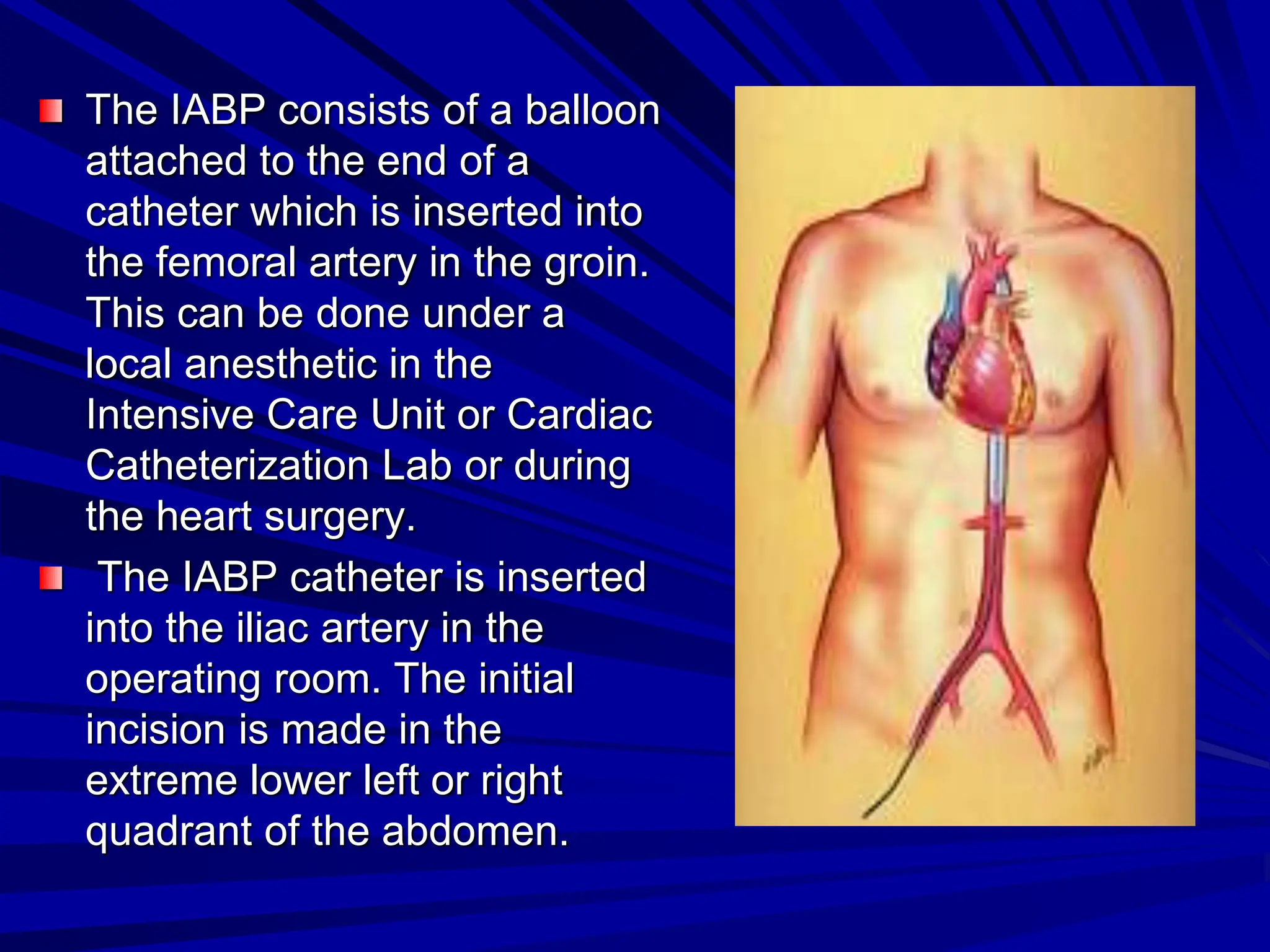
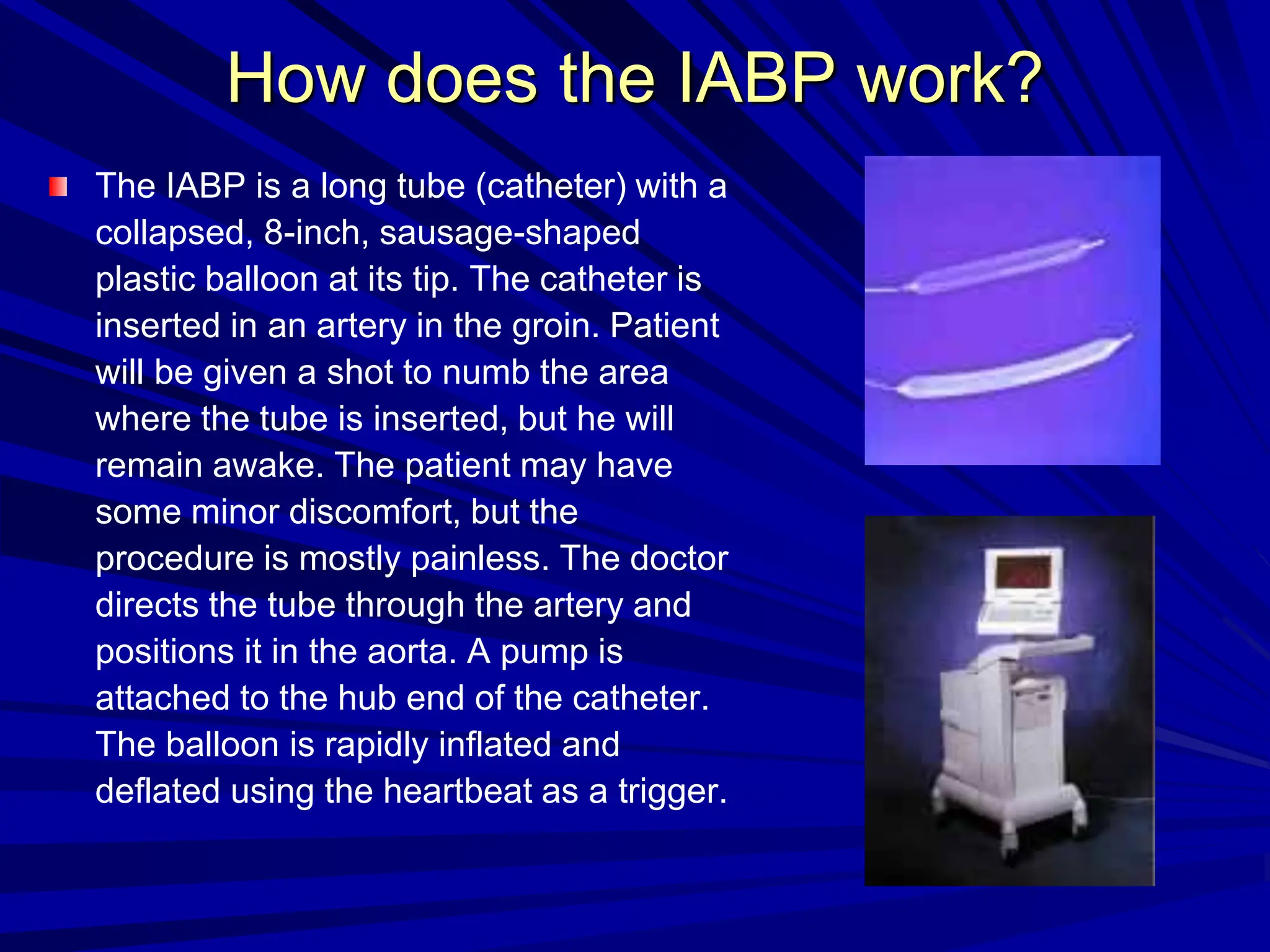
Intra-aortic Balloon Pump
(IABP)
content
Introduction.
Indications & Contraindications.
Basic mechanism.
Complications: 1- During insertion.
2- During use.
3- During or following
removal.
Nursing Interventions.
Introduction:-
The Intra-Aortic Balloon Pump (IABP) is a
mechanical device that increases blood
flow to the heart muscle and decreases
the heart’s workload through a process
called counter-pulsation.
The IABP is placed in the aorta, which is
the main artery that carries oxygen-rich
blood to the rest of the body.
The IABP consists of a balloon
attached to the end of a
catheter which is inserted into
the femoral artery in the groin.
This can be done under a
local anesthetic in the
Intensive Care Unit or Cardiac
Catheterization Lab or during
the heart surgery.
The IABP catheter is inserted
into the iliac artery in the
operating room. The initial
incision is made in the
extreme lower left or right
quadrant of the abdomen.
The balloon sits in the aorta
and opens and closes in
response to the hearts
contractions. After the heart
contracts and propels oxygen-
rich blood into the aorta, the
balloon rapidly opens up and
propels some of the oxygen-
rich blood back toward the
coronary arteries. Just before
the hearts next contraction, the
balloon rapidly deflates
creating a lower pressure in
the aorta so the heart does not
have to work as hard to pump
the blood out.
The primary goals of IABP treatment are
to increase myocardial oxygen supply and
decrease myocardial oxygen demand.
Secondary, improvement of cardiac output
(CO), ejection fraction (EF), an increase of
coronary perfusion pressure, systemic
perfusion and a decrease of heart rate,
pulmonary capillary wedge pressure and
systemic vascular resistance occur.
Basic mechanism
placed in the thoracic aorta
balloon inflated during
diastole, thus increasing
aortic pressure during
diastole and increasing
coronary blood flow
balloon deflated prior to
and during early left
ventricular ejection thus
reducing aortic pressure
and thus afterload
How does the IABP work?
The IABP is a long tube (catheter) with a
collapsed, 8-inch, sausage-shaped
plastic balloon at its tip. The catheter is
inserted in an artery in the groin. Patient
will be given a shot to numb the area
where the tube is inserted, but he will
remain awake. The patient may have
some minor discomfort, but the
procedure is mostly painless. The doctor
directs the tube through the artery and
positions it in the aorta. A pump is
attached to the hub end of the catheter.
The balloon is rapidly inflated and
deflated using the heartbeat as a trigger.
Indications
Heart failure.
Myocardial infarction.
Cardiogenic shock
Left ventricular failure.
If the heart cannot pump well enough to circulate the
blood.
To help the heart of severely ill people who are awaiting
a heart transplant.
To improve blood flow to the heart while you await
surgery or angioplasty for severe blockage of the heart
arteries
Acute heart attack during or after cardiac surgery to help
remove the patient from the heart-lung bypass machine
Contraindications
Aortic insufficiency
Aortic dissection
Prosthetic graft in thoracic aorta.
Complications
During insertion
1- Failure to advance catheter beyond ilio-
femoral system because of atherosclerotic
disease (most common complication).
2- Aortic dissection and arterial perforation.
During use
Limb ischemia most common. Sufficiently severe
to require removal of balloon in 11-27%
Sepsis. Relatively unusual but associated with
high mortality. Positive blood cultures require
prompt removal and culture of IAB and treatment
with antibiotics
Small perforation in balloon membrane may
allow small leak of blood into balloon lumen. Dry
helium desiccates the collected blood which
then forms a hard pellet which may prevent
removal of balloon except by surgical aortotomy.
During or following removal
haematoma
false aneurysm
AV fistula
The most common insertion site for the
IABP catheter is the femoral artery.
However, the IABP catheter has also been
inserted through indwelling supra-inguinal
bypass grafts, the ascending aorta, the
axillary artery, the subclavian artery, and
the common iliac artery.
The femoral approach is advantageous because
the catheter can be inserted percutaneously at
the patient's bedside.
Unfortunately, this approach requires the patient
to remain on bed rest with the head of the bed
elevated no more than 30 degrees. If prolonged,
circulatory support is needed, complications
associated with immobility can occur.
Complications that occur as a result of
prolonged bed rest, and immobility can be
avoided by inserting the IABP catheter through
the iliac artery, because patients are allowed to
sit, stand, and walk during counter-pulsation.
Thus, iliac placement of the IABP catheter helps
prevent deconditioning and the undesirable
consequences of immobility.
The iliac artery has a larger diameter than does
the femoral artery,
Nursing Interventions
The Nurse/Patient ratio for a patient with
an IABP is 1:1.
Nurses must ensure that the stay sutures
are intact. This observation is made at
least once a shift and before and after the
patient exercises
Nsg. intervention cont.
In order to prevent infection, the exit site of the
IABP catheter is cleansed with povidone-iodine
(Betadine) and alcohol and covered with a
sterile dressing every other day
During dressing changes, nurses must monitor
the exit site of the catheter for redness,
inflammation, and purulent drainage. Elevations
in patients' body temperature and white blood
cell count also may indicate infection.
Nsg. intervention cont.
Evaluate Cardiac Output within 1 hour of
any change in the assist interval, To
assess whether the change is tolerated.
Nurses should also observe patients for
bleeding complications.
Nsg. intervention cont.
Assess capillary refill and
pedal pulses q1h.
Assess capillary refill and
left radial pulse at the
start of each shift and
q1h.
Notify the physician of
any change from the
previous hour. Document
on flow sheet and nursing
note.
The balloon catheter or
thrombus can obstruct
flow to the distal
extremities.
If the balloon migrates too
high, it can obstruct flow
to the left subclavian
artery.
Nsg. intervention cont.
Maintain HOB 30
degrees.
Avoid hip flexion.
Raising the head of
the bed can shorten
the aorta and permit
migration of the IABP
catheter
Hip flexion can put
strain on the insertion
site and cause
vascular trauma.
Nsg. intervention cont.
Provide chest vibration to
promote secretion
clearance q4h - 6h and
prn.
Measure urinary output
q1h.
Immobility and sternal
pain predispose patient to
secretions and
atelectasis.
Decreased urine
production can occur as a
result of low cardiac
output, balloon placement
that is too low, prerenal
failure from a thrombus,
or hypotensive shock
conclusion
As the IABP inflates and deflates it allows an
increase in oxygen supply to the myocardium,
decreases workload of the left ventricle and
increases cardiac output and perfusion of the
vital organs. The device is designed to inflate
and deflate with each heart beat. The balloon
will inflate during diastole causing blood to be
forced back toward the extremities as well as
into the coronary arteries and main branches of
the aortic arch. Before systole the balloon will
deflate resulting in decreased pressure within
the aorta. This action result in less work for the
left ventricle, to expel blood during contraction.

Intra-aortic Balloon 00 and the Pump.ppt

  • 1.
  • 2.
    content Introduction. Indications & Contraindications. Basicmechanism. Complications: 1- During insertion. 2- During use. 3- During or following removal. Nursing Interventions.
  • 3.
    Introduction:- The Intra-Aortic BalloonPump (IABP) is a mechanical device that increases blood flow to the heart muscle and decreases the heart’s workload through a process called counter-pulsation. The IABP is placed in the aorta, which is the main artery that carries oxygen-rich blood to the rest of the body.
  • 4.
    The IABP consistsof a balloon attached to the end of a catheter which is inserted into the femoral artery in the groin. This can be done under a local anesthetic in the Intensive Care Unit or Cardiac Catheterization Lab or during the heart surgery. The IABP catheter is inserted into the iliac artery in the operating room. The initial incision is made in the extreme lower left or right quadrant of the abdomen.
  • 5.
    The balloon sitsin the aorta and opens and closes in response to the hearts contractions. After the heart contracts and propels oxygen- rich blood into the aorta, the balloon rapidly opens up and propels some of the oxygen- rich blood back toward the coronary arteries. Just before the hearts next contraction, the balloon rapidly deflates creating a lower pressure in the aorta so the heart does not have to work as hard to pump the blood out.
  • 6.
    The primary goalsof IABP treatment are to increase myocardial oxygen supply and decrease myocardial oxygen demand. Secondary, improvement of cardiac output (CO), ejection fraction (EF), an increase of coronary perfusion pressure, systemic perfusion and a decrease of heart rate, pulmonary capillary wedge pressure and systemic vascular resistance occur.
  • 7.
    Basic mechanism placed inthe thoracic aorta balloon inflated during diastole, thus increasing aortic pressure during diastole and increasing coronary blood flow balloon deflated prior to and during early left ventricular ejection thus reducing aortic pressure and thus afterload
  • 8.
    How does theIABP work? The IABP is a long tube (catheter) with a collapsed, 8-inch, sausage-shaped plastic balloon at its tip. The catheter is inserted in an artery in the groin. Patient will be given a shot to numb the area where the tube is inserted, but he will remain awake. The patient may have some minor discomfort, but the procedure is mostly painless. The doctor directs the tube through the artery and positions it in the aorta. A pump is attached to the hub end of the catheter. The balloon is rapidly inflated and deflated using the heartbeat as a trigger.
  • 9.
    Indications Heart failure. Myocardial infarction. Cardiogenicshock Left ventricular failure. If the heart cannot pump well enough to circulate the blood. To help the heart of severely ill people who are awaiting a heart transplant. To improve blood flow to the heart while you await surgery or angioplasty for severe blockage of the heart arteries Acute heart attack during or after cardiac surgery to help remove the patient from the heart-lung bypass machine
  • 10.
  • 11.
    Complications During insertion 1- Failureto advance catheter beyond ilio- femoral system because of atherosclerotic disease (most common complication). 2- Aortic dissection and arterial perforation.
  • 12.
    During use Limb ischemiamost common. Sufficiently severe to require removal of balloon in 11-27% Sepsis. Relatively unusual but associated with high mortality. Positive blood cultures require prompt removal and culture of IAB and treatment with antibiotics Small perforation in balloon membrane may allow small leak of blood into balloon lumen. Dry helium desiccates the collected blood which then forms a hard pellet which may prevent removal of balloon except by surgical aortotomy.
  • 13.
    During or followingremoval haematoma false aneurysm AV fistula
  • 14.
    The most commoninsertion site for the IABP catheter is the femoral artery. However, the IABP catheter has also been inserted through indwelling supra-inguinal bypass grafts, the ascending aorta, the axillary artery, the subclavian artery, and the common iliac artery.
  • 15.
    The femoral approachis advantageous because the catheter can be inserted percutaneously at the patient's bedside. Unfortunately, this approach requires the patient to remain on bed rest with the head of the bed elevated no more than 30 degrees. If prolonged, circulatory support is needed, complications associated with immobility can occur.
  • 16.
    Complications that occuras a result of prolonged bed rest, and immobility can be avoided by inserting the IABP catheter through the iliac artery, because patients are allowed to sit, stand, and walk during counter-pulsation. Thus, iliac placement of the IABP catheter helps prevent deconditioning and the undesirable consequences of immobility. The iliac artery has a larger diameter than does the femoral artery,
  • 17.
    Nursing Interventions The Nurse/Patientratio for a patient with an IABP is 1:1. Nurses must ensure that the stay sutures are intact. This observation is made at least once a shift and before and after the patient exercises
  • 18.
    Nsg. intervention cont. Inorder to prevent infection, the exit site of the IABP catheter is cleansed with povidone-iodine (Betadine) and alcohol and covered with a sterile dressing every other day During dressing changes, nurses must monitor the exit site of the catheter for redness, inflammation, and purulent drainage. Elevations in patients' body temperature and white blood cell count also may indicate infection.
  • 19.
    Nsg. intervention cont. EvaluateCardiac Output within 1 hour of any change in the assist interval, To assess whether the change is tolerated. Nurses should also observe patients for bleeding complications.
  • 20.
    Nsg. intervention cont. Assesscapillary refill and pedal pulses q1h. Assess capillary refill and left radial pulse at the start of each shift and q1h. Notify the physician of any change from the previous hour. Document on flow sheet and nursing note. The balloon catheter or thrombus can obstruct flow to the distal extremities. If the balloon migrates too high, it can obstruct flow to the left subclavian artery.
  • 21.
    Nsg. intervention cont. MaintainHOB 30 degrees. Avoid hip flexion. Raising the head of the bed can shorten the aorta and permit migration of the IABP catheter Hip flexion can put strain on the insertion site and cause vascular trauma.
  • 22.
    Nsg. intervention cont. Providechest vibration to promote secretion clearance q4h - 6h and prn. Measure urinary output q1h. Immobility and sternal pain predispose patient to secretions and atelectasis. Decreased urine production can occur as a result of low cardiac output, balloon placement that is too low, prerenal failure from a thrombus, or hypotensive shock
  • 23.
    conclusion As the IABPinflates and deflates it allows an increase in oxygen supply to the myocardium, decreases workload of the left ventricle and increases cardiac output and perfusion of the vital organs. The device is designed to inflate and deflate with each heart beat. The balloon will inflate during diastole causing blood to be forced back toward the extremities as well as into the coronary arteries and main branches of the aortic arch. Before systole the balloon will deflate resulting in decreased pressure within the aorta. This action result in less work for the left ventricle, to expel blood during contraction.