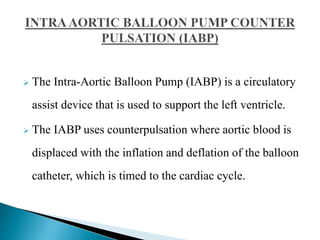
JOHNY WILBERT, M.SC[N]
LECTURER,
APOLLO INSTITUTE OF HOSPITAL
MANAGEMENT AND ALLIED SCIENCE
 The Intra-Aortic Balloon Pump (IABP) is a circulatory
assist device that is used to support the left ventricle.
 The IABP uses counterpulsation where aortic blood is
displaced with the inflation and deflation of the balloon
catheter, which is timed to the cardiac cycle.
 Severe aortic insufficiency
 Aortic or abdominal aneurism
 Severe peripheral vascular disease
The balloon catheter is inserted either percutaneously or surgically
by cutdown into the patients’ femoral artery.
 Connect to mains power to ensure the battery is preserved.
 Check the helium tank is open at the back of the pump.
 Ensure both an ECG and pressure trace can be obtained
from the patient on the screen of the IABP.
 The IABP can obtain a trigger, which stimulates the
pumping of the balloon.
 Frequency when first commencing pumping is on 1:1,
which means that for each heart beat the balloon, will
be inflated.
 To commence balloon pumping, inflation and deflation
points should be set at the midline and then once
pumping is established, timing should be reassessed.
 Connect the extension tubing to the balloon catheter
and on the balloon console at the back.
 Helium is used as it is easily dissolves in
blood than air and prevents the risk of air
emboli if the catheter ruptures.
The end of the balloon should be just distal (1-
2 cm) to the take-off of the left subclavian
artery
 Position should be confirmed by fluoroscopy
or chest x-ray
Trigger
Frequency
Augmentatio
LV contraction:
- Isovol. Contraction (b)
- maximal ejection (c)
LV relaxation:
- start of relaxation and
reduced
ejection (d)
- isovol.relaxation (e)
LV filling:
- LV filling , rapid phase (f)
- slow LV filling (g)
- atrial systole( a)
Inflation and deflation of the balloon
 Blood is displaced to the proximal aorta by inflation during
diastole.
 Aortic volume ( afterload) is reduced during systole through
vacuum effect created by rapid balloon deflation
 Decrease in SBP by 20 %
 Increase in aortic Diastolic Press. by 30 % ( raise coronary
blood flow)
 Increase in MAP
 Reduction of the HR by 20%
 Decrease in the mean PCWP by 20 %
 Elevation in the COP by 20%
First change from 1:1 to 1:2 augmentation
 Check the dicrotic notch
 See if augmentation starts at that point
This should produce a sharp “V” at inflation.
 Check if diastolic augmented wave is ›
systolic wave
Confirm if end diastolic wave following the
augmented wave is less than an non
augmented wave.
Is Deflation slope ok.
 Inflation of the IAB markedly after closure of the aortic
valve.
 Waveform Characteristics:
• Inflation of IAB after the dicrotic notch.
• Absence of sharp V.
• Sub optimal diastolic augmentation
Late deflation of the IAB during the diastolic phase.
 Waveform Characteristics:
• Assisted aortic end diastolic pressure may be equal to
the unassisted aortic end diastolic pressure.
• Rate of rise of assisted systole is prolonged.
• Diastolic augmentation may appear widened
Rounded balloon pressure waveform
- Loss of plateau resulting from a kink or
obstruction of shuttle gas
- Kink in the catheter tubing
- Improper IAB catheter position
- Sheath not being pulled back to allow
inflation of the IAB
- IAB is too large for the aorta
- IAB is not fully unwrapped
- H2O condensation in the external tubing
- due to a loose connection
- a leak in the IAB catheter
- H2O condensation in the external tubing
- a patient who is tachycardiac and febrile which causes
increased gas
diffusion through the IAB membrane
Anticoagulation-- maintain apTT at 50 to 70
seconds.
CXR daily – to R/O IAB migration.
 Check lower limb pulses - 2 hourly.
 Hip flexion is restricted, and the head of the
bed should not be elevated beyond 30°.
Never leave in standby by mode for more than 20
minutes
 Daily
 Haemoglobin (risk of bleeding or haemolysis)
 Platelet count (risk of thrombocytopenia)
 Renal function (risk of acute kidney injury
secondary to distal migration of IABP catheter)
 Wean off the IABP as early as possible as longer
duration is associated
 with higher incidence of limb complications
Timing of weaning:
 Patient should be stable for 12 – 24 hours
 Decrease inotropic support
 Decrease pump ratio
 From 1:1 to 1:2 or 1:3
 Decrease augmentation
 Monitor patient closely
 If patient becomes unstable, weaning should be
 Immediately discontinued
THANK YOU

IABP

  • 1.
    JOHNY WILBERT, M.SC[N] LECTURER, APOLLOINSTITUTE OF HOSPITAL MANAGEMENT AND ALLIED SCIENCE
  • 2.
     The Intra-AorticBalloon Pump (IABP) is a circulatory assist device that is used to support the left ventricle.  The IABP uses counterpulsation where aortic blood is displaced with the inflation and deflation of the balloon catheter, which is timed to the cardiac cycle.
  • 5.
     Severe aorticinsufficiency  Aortic or abdominal aneurism  Severe peripheral vascular disease
  • 6.
    The balloon catheteris inserted either percutaneously or surgically by cutdown into the patients’ femoral artery.
  • 7.
     Connect tomains power to ensure the battery is preserved.  Check the helium tank is open at the back of the pump.  Ensure both an ECG and pressure trace can be obtained from the patient on the screen of the IABP.  The IABP can obtain a trigger, which stimulates the pumping of the balloon.
  • 8.
     Frequency whenfirst commencing pumping is on 1:1, which means that for each heart beat the balloon, will be inflated.  To commence balloon pumping, inflation and deflation points should be set at the midline and then once pumping is established, timing should be reassessed.  Connect the extension tubing to the balloon catheter and on the balloon console at the back.
  • 9.
     Helium isused as it is easily dissolves in blood than air and prevents the risk of air emboli if the catheter ruptures.
  • 10.
    The end ofthe balloon should be just distal (1- 2 cm) to the take-off of the left subclavian artery  Position should be confirmed by fluoroscopy or chest x-ray
  • 11.
  • 12.
    LV contraction: - Isovol.Contraction (b) - maximal ejection (c) LV relaxation: - start of relaxation and reduced ejection (d) - isovol.relaxation (e) LV filling: - LV filling , rapid phase (f) - slow LV filling (g) - atrial systole( a)
  • 16.
    Inflation and deflationof the balloon  Blood is displaced to the proximal aorta by inflation during diastole.  Aortic volume ( afterload) is reduced during systole through vacuum effect created by rapid balloon deflation  Decrease in SBP by 20 %  Increase in aortic Diastolic Press. by 30 % ( raise coronary blood flow)
  • 17.
     Increase inMAP  Reduction of the HR by 20%  Decrease in the mean PCWP by 20 %  Elevation in the COP by 20%
  • 23.
    First change from1:1 to 1:2 augmentation  Check the dicrotic notch  See if augmentation starts at that point This should produce a sharp “V” at inflation.  Check if diastolic augmented wave is › systolic wave
  • 24.
    Confirm if enddiastolic wave following the augmented wave is less than an non augmented wave. Is Deflation slope ok.
  • 27.
     Inflation ofthe IAB markedly after closure of the aortic valve.  Waveform Characteristics: • Inflation of IAB after the dicrotic notch. • Absence of sharp V. • Sub optimal diastolic augmentation
  • 30.
    Late deflation ofthe IAB during the diastolic phase.  Waveform Characteristics: • Assisted aortic end diastolic pressure may be equal to the unassisted aortic end diastolic pressure. • Rate of rise of assisted systole is prolonged. • Diastolic augmentation may appear widened
  • 34.
    Rounded balloon pressurewaveform - Loss of plateau resulting from a kink or obstruction of shuttle gas - Kink in the catheter tubing - Improper IAB catheter position - Sheath not being pulled back to allow inflation of the IAB - IAB is too large for the aorta - IAB is not fully unwrapped - H2O condensation in the external tubing
  • 35.
    - due toa loose connection - a leak in the IAB catheter - H2O condensation in the external tubing - a patient who is tachycardiac and febrile which causes increased gas diffusion through the IAB membrane
  • 36.
    Anticoagulation-- maintain apTTat 50 to 70 seconds. CXR daily – to R/O IAB migration.  Check lower limb pulses - 2 hourly.  Hip flexion is restricted, and the head of the bed should not be elevated beyond 30°.
  • 37.
    Never leave instandby by mode for more than 20 minutes  Daily  Haemoglobin (risk of bleeding or haemolysis)  Platelet count (risk of thrombocytopenia)  Renal function (risk of acute kidney injury secondary to distal migration of IABP catheter)  Wean off the IABP as early as possible as longer duration is associated  with higher incidence of limb complications
  • 39.
    Timing of weaning: Patient should be stable for 12 – 24 hours  Decrease inotropic support  Decrease pump ratio  From 1:1 to 1:2 or 1:3  Decrease augmentation  Monitor patient closely  If patient becomes unstable, weaning should be  Immediately discontinued
  • 40.