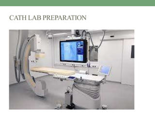
Cath lab preparation involves sterile setup of equipment and supplies. The sterile field must be carefully maintained to prevent infection. Patients are prepared by explaining procedures, identifying them, obtaining consent, and checking vitals. Emergency drugs and supplies are readied. Proper sterile technique includes scrubbing, gowning, gloving and draping. Procedures like angiograms, angioplasty, stent placement and temporary or permanent pacemakers require specific tray setup and patient preparation. Post-procedure care includes sheath removal, monitoring for bleeding, and discharge education on wound care and activity restrictions.
This document provides information about cardiac catheters and guidewires used in cardiac catheterization procedures. It discusses the history of cardiac catheters, ideal characteristics, parts of a catheter, materials used in construction, types of catheters including pigtail catheters, and features of guidewires. Characteristics such as size, stiffness, memory, and friction coefficient are compared for different catheter materials. The document also includes images and descriptions of specific catheters and guidewire tips.
The document summarizes key aspects of cardiac catheterization and hemodynamic data collection. It describes the normal cardiac cycle, pressure measurement systems, normal pressure waveforms, methods to measure cardiac output like thermodilution and Fick, how to evaluate valvular stenosis and regurgitation, determine vascular resistance and shunts. Specific details are provided on assessing aortic stenosis, mitral stenosis, right-sided valves and quantifying regurgitant fractions. Oxygen saturation analysis and Fick principles are outlined for shunt determinations.
Left ventricular angiography is used to assess global and regional left ventricular function and anatomy. It involves inserting a catheter into the left ventricle and injecting contrast dye to visualize the ventricle on x-ray imaging. The procedure provides key information on mitral valve function, ventricular shape and wall motion abnormalities, and congenital defects like VSD. LV volumes and ejection fraction are calculated from the images to quantify function. Regional wall motion is graded and correlated to coronary artery territories. Characteristic appearances are seen in conditions like cardiomyopathy, mitral regurgitation, and septal defects. Potential complications include arrhythmias and endocardial injury.
This document provides an overview of cardiac catheterization procedures. It discusses indications, contraindications, techniques, views obtained, and interpretation of pressure waveforms. Key points include that cardiac catheterization guides treatment decisions by measuring pressures, outputs, and obtaining images. It is now often used therapeutically for procedures like angioplasty and device closures. The document outlines patient preparation, access methods, catheters used, views obtained, and complications that can occur.
This document provides information about percutaneous transvenous mitral commissurotomy (PTMC), a procedure used to treat mitral stenosis. It discusses the stages and severity of mitral stenosis, indications and contraindications for PTMC, assessment of valve morphology, the PTMC procedure technique, instruments used, balloon size selection, post-procedure evaluation, complications, follow-up care, and long-term prognosis. PTMC is performed to improve the opening of a stenosed mitral valve by splitting the fused commissures using a balloon catheter, and is an important therapeutic option for treating symptomatic mitral stenosis.
The document discusses the history, anatomy, angiographic views, variations, and clinical relevance of coronary arteries. It provides a detailed overview of the typical anatomy and branches of the left main, left anterior descending, left circumflex, and right coronary arteries. It also describes common anatomical variations and anomalies seen in coronary arteries and their clinical implications. Angiographic classification methods for different coronary artery segments are presented.
BMV,PTMC,BALLOON MITRAL VALVOTOMY, BAL, VIRBHAN BALAI, DR VIRBHANDr Virbhan Balai
This document discusses balloon mitral valvuloplasty (BMV) and balloon aortic valvuloplasty (BAV). It describes the indications for BMV as symptomatic or asymptomatic severe mitral stenosis. The Inoue technique for BMV is explained in detail, including transseptal puncture and sequential balloon inflation. Complications of BMV include severe mitral regurgitation, mortality, and cardiac perforation. BAV was used historically but was abandoned due to high restenosis rates and no improvement in patient survival.
This document discusses hemodynamic principles and various cardiac pressures measured in the circulatory system. It begins by explaining how electrical activity leads to mechanical functions that generate pressure waves. It then discusses how to measure and interpret pressures in different parts of the heart including the aorta, pulmonary artery, right and left ventricles, and right atrium. Factors that influence pressures and common abnormalities are provided. Diagrams of normal pressure waveforms are displayed. The document concludes by defining pulmonary and systemic vascular resistances.
Diagnostic catheters for coronary angiography Aswin Rm
Overview of diagnostic catheters used in coronary angiography
Guide catheters not included
History of coronary catheters
Radial techniques and catheters
This document discusses vascular access during cardiac catheterization. It covers various access sites including femoral, radial, brachial and ulnar arteries as well as internal jugular, subclavian and femoral veins. Potential complications of vascular access like hematoma, pseudoaneurysm, retroperitoneal hemorrhage and arteriovenous fistula are described. Risk factors, diagnosis and management of these complications are provided. Prevention strategies to avoid vascular access complications are also mentioned.
1) The document discusses various techniques for balloon mitral valvuloplasty (BMV), including the history of the procedure and details of specific balloon designs.
2) Key balloons described are the Inoue balloon, Accura balloon, JOMIVA balloon, and the double balloon and multi-track techniques.
3) Complications of BMV discussed include balloon rupture and strategies to prevent it, such as avoiding overinflation and slow inflation to reduce rapid stretching of balloon layers.
This document discusses fractional flow reserve (FFR), a technique used during coronary catheterization to measure pressure differences across a coronary stenosis and determine if it is causing myocardial ischemia. An FFR value below 0.75 is considered functionally significant while a value above 0.80 rules out ischemia. FFR is useful for evaluating single-vessel disease, left main stenosis, tandem lesions, diffuse disease, grafts, and ostial lesions. Limitations include inability to assess plaque morphology.
The document discusses techniques for detecting intracardiac shunts including oximetry runs, indicator dilution curves, and angiography. It provides criteria for identifying left-to-right shunts using oxygen saturation step-ups in the oximetry run. Examples are given for detecting an atrial septal defect and ventricular septal defect. The ratio of pulmonary to systemic blood flow (Qp/Qs) is discussed as a measure of shunt magnitude.
Contrast echocardiography uses microbubble ultrasound contrast agents to improve image quality. These microbubbles remain in the intravascular space and allow for assessment of cardiac structure, function, and perfusion. Second generation contrast agents use an inert gas encapsulated by albumin or phospholipid shells. They interact with ultrasound by reflecting at fundamental frequencies and resonating to produce harmonic frequencies. Continuous infusion provides steady contrast levels needed for perfusion assessment. Contrast echocardiography is a non-invasive technique that improves evaluation of the heart.
Guide catheters in coronary interventionRohitWalse2
Guide catheters are essential for coronary interventions as they deliver hardware into the arteries. The document discusses the properties and types of guide catheters, highlighting how their structure provides support and torque control. It describes commonly used guide catheters like the Judkins, Amplatz and EBU catheters, noting what vessels each is best suited for. Specialty guide catheters for difficult anatomies are also reviewed. Proper guide selection and positioning are emphasized for coaxial engagement and optimal device delivery during interventions.
Raja Lahiri provides an overview of coronary angiography. Key points include:
- Coronary angiography is the current gold standard for visualizing the coronary arteries through X-ray imaging with contrast injection.
- The history of coronary angiography began in the 1920s-1940s with early experiments in cerebral and cardiac catheterization.
- Modern techniques involve accessing arteries typically through the femoral or radial arteries to insert a catheter for contrast injection into the coronary arteries under X-ray imaging.
- Multiple angiographic views are needed to visualize different segments of the left and right coronary arteries. Coronary angiography is used to evaluate coronary artery disease, graft patency, and left ventricular function.
Intravascular ultrasound (IVUS) uses sound waves to visualize the inside of arteries. There are two types of IVUS systems - mechanical systems using a rotating internal cable and solid-state systems using externally mounted transducers. Both produce 360-degree images with a resolution of 100-150 μm. IVUS is used to assess plaque, vessel dimensions, stent deployment, and more. It produces cross-sectional images showing the lumen, layers of the artery wall, and plaque composition and size. Measurements include diameters, areas, plaque burden, and indices of eccentricity. IVUS helps identify vulnerable plaque and has diagnostic and interventional applications.
Radial artery access ,complications and magementS S SRINIVASAN
1. Radial artery access is an alternative to femoral artery access for coronary procedures that offers advantages like lower bleeding risks and faster recovery times.
2. The radial artery anatomy and variations like tortuosity must be considered to select appropriate patients and techniques.
3. Radial artery puncture and navigation of wires and catheters through the arm requires specialized techniques to prevent complications like spasm or dissection.
4. Maintaining radial artery patency with anticoagulation, proper compression, and monitoring is important to prevent radial artery occlusion following the procedure.
This document discusses balloon aortic valvuloplasty (BAV) as a treatment for aortic stenosis. It can be performed via either a retrograde or antegrade approach. The retrograde approach involves crossing the aortic valve from the femoral artery, while the antegrade approach involves transseptal catheterization from the femoral vein. Key steps for both approaches include rapid ventricular pacing to stabilize the heart during balloon inflation. The goals of BAV are to increase the aortic valve area and reduce pressure gradients. Complications can include hypotension, aortic regurgitation, and embolization of calcium deposits. BAV provides symptomatic relief but is usually not curative, as restenosis may occur.
This document describes equipment, catheters, and basic intervals used in electrophysiology (EP) studies. It discusses radiographic tables, EP equipment like cardiac stimulators and mapping/ablation catheters. Patient preparation includes fasting, IV access, monitoring equipment. EP catheters come in different sizes and have electrodes for recording electrical activity. Basic intervals measured include P wave to atrial interval, atrial-His bundle interval, His-ventricular interval, and sinus node recovery time. Drive train stimulation with single, double, or triple extra stimuli is used. The document continues with further discussions of EP protocols, arrhythmias, ablation, and pre-excitation pathways.
1. Guide catheters provide support for advancing devices into coronary arteries and injecting contrast for visualization. Their selection depends on factors like coronary anatomy, aortic root size, and desired level of support.
2. Common guide catheters include the Judkins, Amplatz, and extra-backup guides. The Judkins provides balanced support while the Amplatz offers firm passive support. Long tip catheters provide coaxial support and manipulation.
3. Achieving proper coaxial alignment and maintaining backup support are important for device delivery and preventing complications. Catheter size, curves, and deep seating techniques impact the level of passive versus active support provided.
This document summarizes different devices used for closing ventricular septal defects (VSDs). It describes the common complications of VSD devices which are mostly minor, including embolization, arrhythmias, and conduction defects. Three types of Amplatzer devices are outlined - the muscular VSD device, asymmetric VSD occluder, and perimembranous VSD devices. Sizes and designs of each are provided. Results of post-myocardial infarction VSD closure show high residual leak rates. Finally, it briefly mentions some VSD devices manufactured in China including by Yatai and Lifetech, and introduces the novel NitOcclud VSD coil.
The document discusses drugs commonly used in cardiac catheterization laboratories. It describes the uses, mechanisms of action, dosages, and side effects of various drugs including lidocaine for local anesthesia, heparin and glycoprotein IIb/IIIa inhibitors for anticoagulation during procedures like percutaneous coronary intervention, nitrates like glyceryl trinitrate for vasodilation, inotropes like dopamine and dobutamine, antiarrhythmics like amiodarone, and contrast agents like iohexol. The document provides an overview of how these drugs are utilized during different cardiac procedures performed in cath labs.
Cardiac catheterization is useful for assessing left-to-right shunts through three main techniques: oximetry runs to detect oxygen saturation step-ups, indicator dye dilution to detect early recirculation of dye injected into the proximal chamber, and angiocardiography to directly visualize the anatomic site of the shunt. While oximetry is best to localize the shunt, dye dilution can detect smaller shunts and angiography confirms anatomy. Together these techniques allow diagnosis and quantification of left-to-right intracardiac shunts.
Coronary angiography is a diagnostic procedure that uses x-ray imaging and dye to visualize the coronary arteries and detect blockages. The technique was first performed in 1958 and involves inserting a catheter into the arteries and injecting dye so that blockages are highlighted on x-ray images. It remains the gold standard for detecting clinically significant coronary artery disease. Potential complications include bleeding, infection, and reactions to the dye, but it provides information on the location and severity of blockages when clinically suspected coronary artery disease needs to be diagnosed or bypass grafts evaluated. Careful patient preparation and monitoring during and after the procedure are important to minimize risks.
1. The document discusses proper specimen collection techniques and safety precautions. It covers specimen types including blood, urine, stool, and others.
2. Blood is the most common specimen collected, and can be obtained through venipuncture, arterial puncture, or skin puncture. Proper patient identification, site selection and preparation, and universal precautions are emphasized.
3. The document details the procedures for venipuncture and arterial puncture, including using evacuated tubes or syringes, order of draw, complications to watch for, and applying pressure after collection. Skin puncture for small volumes is also outlined. Proper handling and transport of all specimens to the lab is important for obtaining valid results.
Catheterization is the process of inserting a catheter into the urinary tract. It is used to drain urine from the bladder for various clinical reasons like surgery, inability to void, or monitoring urine output. The proper procedure involves preparing the patient and environment, selecting the correct catheter size and type, cleaning the perineal area aseptically, lubricating the catheter, and slowly inserting it into the urethra until urine flows or the catheter is at the proper depth. The balloon is then inflated to retain the catheter and the drainage bag is attached to collect urine. Catheterization must be done aseptically to prevent urinary tract infections.
Peripheral intravenous catheters are used to provide venous access for blood sampling, fluid administration, medications, and other purposes. They involve inserting a small gauge cannula into a superficial vein in the arm or hand. Central venous catheters are longer catheters placed into larger central veins to administer irritating or large volume substances. Proper techniques like using ultrasound guidance and the Seldinger technique aim to safely place the catheter and minimize complications like infection, bleeding, or injury to surrounding structures. Ongoing care of the insertion site and catheter is also important.
Cardiac catheterization is a test that uses a thin tube inserted into blood vessels to examine the heart. It can check for blockages in coronary arteries, assess heart function and valves, and diagnose congenital heart defects. The procedure involves inserting a catheter into the groin or wrist artery and guiding it to the heart while injecting dye for imaging. Potential risks include bleeding, infection, arrhythmias, and reactions to dye, though complications are rare. Post-procedure care involves monitoring for symptoms and restricting activity to allow puncture sites to heal.
The document discusses proper procedures for blood collection, including using universal safety precautions when handling needles and blood samples, selecting appropriate veins for venipuncture, collecting blood in tubes containing anticoagulants to prevent clotting, and taking care when collecting from babies or doing fingersticks to obtain small blood samples. Common anticoagulants added to blood collection tubes are EDTA, sodium citrate, heparin, and double oxalate, which work by binding calcium ions or inhibiting coagulation factors to prevent clotting.
Percutaneous transhepatic cholangiography (PTC) and percutaneous transhepatic biliary drainage (PTBD) are invasive radiological procedures used to image and drain the biliary tree. PTC involves inserting a needle into the liver under imaging guidance and injecting contrast dye to delineate the biliary anatomy. PTBD places a drainage catheter through the liver and into the blocked bile duct to relieve obstruction. Both procedures require imaging, sterile technique, antibiotics and monitoring for potential complications like bleeding, infection and bile leakage. They are used when less invasive options fail or are contraindicated for evaluating and treating biliary obstruction.
This document provides information about peripherally inserted central catheters (PICCs):
1. It discusses the benefits, risks, and characteristics of PICCs including catheter types, styles, sizes, and lengths.
2. It describes the PICC placement procedure and methods for verifying catheter tip location using chest x-ray or EKG tip positioning systems.
3. It outlines considerations for PICC assessment including patient complaints, new cardiac issues, extremity edema, catheter migration, and issues requiring consultation with the IV team.
4. It briefly mentions PICC line care including flushing procedures and discontinuing a PICC which requires a physician/provider order.
1) The document provides an overview of proper blood specimen collection procedures, including patient identification, types of blood draws, order of draw, tube requirements, and causes of rejected samples.
2) It emphasizes the importance of accurate patient identification and collection techniques to avoid issues like hemolysis that can impact test results and require redraws.
3) Key aspects of blood draws are outlined, including vein selection, cleaning the site, needle gauge, filling tubes, mixing, and disposal of sharps.
This document provides guidelines for collecting blood samples through three main methods: capillary, venous, and arterial puncture.
For capillary collection, the finger or earlobe is punctured with a lancet. For venous collection, a vein in the arm is accessed with a needle and syringe. Proper site selection, cleaning, tourniquet use, and needle insertion technique are described.
Arterial puncture is used to collect blood gases and is more difficult than venous puncture. The radial, brachial, or femoral arteries can be used. Precautions are outlined to minimize hemolysis and contamination across all collection methods.
1. The document provides an overview of phlebotomy basics including terminology, blood components, specimen collection and processing techniques, special patient populations, and quality control measures.
2. Key steps in phlebotomy are properly identifying the patient, selecting the appropriate vein and needle, performing the venipuncture, handling and labeling specimens correctly to avoid hemolysis or other issues.
3. Special considerations are discussed for elderly patients, babies, and those with IVs, recommending smaller needles, pressure for longer, and alternative collection sites as needed.
Peripheral angiography is a radiological procedure used to examine arteries and veins after injecting contrast media. It involves puncturing an artery such as the femoral artery using the Seldinger technique and threading a catheter over a guidewire to inject contrast media and obtain images. The procedure is used to diagnose and treat various vascular conditions. Precise positioning, sterile equipment and contrast injection are needed to obtain diagnostic images of the peripheral vasculature.
Neck angiography cect neck angiography carotid angiography
CT scan neck angiography
Carotid angiography useful for medical radiology students thank you process explain in simple language for more content like this presentation
This document provides information about the objectives and theory of phlebotomy. It discusses what phlebotomy is, the roles and responsibilities of phlebotomists, and related anatomy and physiology. It also covers important topics like professionalism, safety, equipment used, and procedures for collecting blood. Phlebotomists must properly identify patients, take safety precautions, position the patient, locate a vein, and collect blood samples while maintaining patient comfort and confidentiality.
This presentation include biliary anatomy ,indication, contraindication post op care of percutaneus transhepatic biliary drainage with important technique. and advantage and disadvantage of different technique. This is important for radiologist, radiographers, intervention radiologist radiology resident. Thanks
This document provides procedures for neonatal umbilical vessel catheterization. It describes:
1) Definitions and background information on when and where these procedures are performed and requirements for supervision.
2) Materials needed including catheter trays and additional items.
3) Steps for the procedure including patient preparation, umbilical arterial catheter insertion involving dilating the artery and advancing the catheter, and umbilical venous catheter insertion. Precautions are described.
This chapter discusses blood collection methods including capillary and venous blood collection. Capillary blood is collected via finger or heel prick and is used for small volume tests. Venous blood requires larger volumes and is collected from veins, usually in the arm. Proper safety precautions must be followed to prevent infection. The vacutainer method uses tubes of various anticoagulants while the syringe method involves manually filling tubes.
Similar to PPt Cath lab preparation (1) (1).pptx (20)
Anemia is a major health problem in India, especially among women. Some key points about anemia from the document include:
- Anemia is defined as a decrease in red blood cells or hemoglobin in the blood. It can be caused by blood loss, impaired red blood cell production, or increased red blood cell destruction.
- The document classifies anemias based on cause (hypo proliferative, hemorrhagic, hemolytic) and cell size (microcytic, normocytic, macrocytic). Common causes discussed are iron deficiency, B12/folate deficiency, aplastic anemia, and hemolytic anemia.
- Signs and symptoms of anemia
This document provides an overview of anemia for nursing students. It defines anemia, discusses its causes and types. It covers the pathophysiology, clinical manifestations, diagnostic evaluation and management of anemia. Nursing management focuses on improving nutrition, managing activity intolerance and improving tissue perfusion. The document aims to help nursing students understand anemia and how to care for patients with this condition.
The document discusses binary logistic regression. Some key points:
- Binary logistic regression predicts the probability of an outcome being 1 or 0 based on predictor variables. It addresses issues with ordinary least squares regression when the dependent variable is binary.
- The logistic regression model transforms the dependent variable using the logit function, ln(p/(1-p)), where p is the probability of an outcome being 1. This results in a linear relationship that can be modeled.
- Interpretation of coefficients is similar to ordinary least squares regression but focuses on odds ratios. A positive coefficient increases the odds of an outcome being 1, while a negative coefficient decreases the odds. The odds ratio indicates how much the odds change with a one-
Hypertension, or high blood pressure, is defined as a systolic blood pressure over 140 mm Hg or a diastolic blood pressure over 90 mm Hg. It has many risk factors including family history, age, gender, obesity, and substance abuse. If left untreated, it can lead to complications like heart attack, stroke, and kidney damage. The document discusses the types of hypertension, diagnostic tests, and medical and nursing management including lifestyle modifications and medications to control blood pressure.
This document summarizes key points about hypertension from Understanding Medical Surgical Nursing, 4th Edition by Linda S. Williams and Paula D. Hopper. It provides statistics on the incidence of hypertension, guidelines for taking blood pressure accurately, classifications of hypertension severity, risk factors, treatment options including lifestyle changes and medications, complications, hypertensive emergencies, and the importance of patient education for lifelong blood pressure control.
Hypertension, or high blood pressure, is defined as a systolic blood pressure over 140 mm Hg or a diastolic blood pressure over 90 mm Hg. It has many risk factors including family history, age, gender, obesity, and substance abuse. If left untreated, it can lead to complications like heart attack, stroke, and kidney damage. Treatment involves lifestyle modifications like diet changes and exercise as well as medication. Nurses educate patients on managing their condition, diet, medication compliance, and monitoring blood pressure at home.
Hypertension is high blood pressure that can lead to severe heart and other health problems if left untreated. It is often asymptomatic until advanced stages. Treatment may involve lifestyle changes like exercise and diet or medications to lower blood pressure. While those with hypertension can usually exercise moderately, untreated hypertension can impair exercise ability. Managing hypertension is important for reducing health risks in older adults.
Coronary angiography is a procedure that uses dye and x-rays to see how blood flows through the coronary arteries of the heart. It is the gold standard for evaluating coronary artery disease and can identify the location and severity of any blockages. A coronary angiogram involves inserting a catheter into the heart and injecting dye so that blockages are highlighted on x-ray images. Potential complications are usually minor but can include heart attack, stroke, or kidney injury from the dye. The results of the angiogram are used to determine if further procedures like angioplasty or bypass surgery are needed.
This document provides information on coronary angiography views and angiographic anatomy. It discusses the clinical divisions of the major coronary arteries and defines what constitutes significant coronary artery disease. Standard angiographic views are described for visualizing different segments of the left and right coronary arteries. Lesion classification systems and other angiogram interpretation elements like TIMI frame count are also summarized.
Coronary angiography remains the gold standard for detecting coronary artery disease. The technique was first performed in 1958 by Dr. Mason Sones at the Cleveland Clinic. Coronary angiography allows visualization of the coronary arteries, branches, and anomalies to precisely locate lesions. It remains an important diagnostic tool used to evaluate patients with suspected coronary artery disease. The procedure involves accessing the femoral artery and advancing a catheter into the heart to inject contrast and obtain images of the coronary arteries under fluoroscopy. Precise technique and monitoring are required to minimize risks of potential complications.
This document discusses vascular access during cardiac catheterization. It covers various topics related to arterial and venous access including common access sites, complications, risk factors, prevention of complications, and management of complications. Specific complications discussed in detail include hematoma, pseudoaneurysm, retroperitoneal hemorrhage. Treatment options for complications like ultrasound-guided compression, thrombin injection, endoluminal techniques are also summarized.
This document discusses congestive heart failure (CHF) and its nutrition management. Myocardial infarction can weaken the heart, limiting its ability to pump blood and removing fluid from the body. This causes a build up of fluid in the extremities and lungs. Nutrition is also impaired as the heart and lungs work harder to pump more fluid. Treatment includes diuretics to reduce fluid load and strengthen the heart. Nutrition therapy aims to reduce cardiac workload by limiting sodium and fluid intake to reduce fluid retention, and providing nutrient-dense foods and supplements if needed to support nutrition and weight status. Close monitoring is needed when providing nutrition support to avoid worsening heart failure.
Heart failure is a common clinical syndrome that can result from any structural or functional impairment of the ventricle that reduces its ability to fill or eject blood. It is the leading cause of hospitalization in adults over 65 years old. The document defines heart failure, discusses its key concepts like cardiac output and ejection fraction, classifications like NYHA and ACC/AHA stages, risk factors, pathophysiology including compensatory mechanisms and remodeling, symptoms, complications, diagnostic tests and emergency management.
This document discusses heart failure and its treatment with drugs. It begins by defining heart failure and listing the objectives of the lecture. It then covers cardiac physiology factors that influence cardiac output like preload, afterload, and contractility. The main drugs used to treat heart failure are also discussed - diuretics, ACE inhibitors, beta-blockers, vasodilators, and digitalis. Side effects and examples of drugs in each class are provided.
This document discusses myocardial infarction (MI), also known as a heart attack. It begins with an introduction defining MI as the death of heart muscle cells from loss of oxygen. It then provides details on the definition, causes, locations, and risk factors of MI. Modifiable risk factors include obesity, diabetes, smoking, and hypertension. The document outlines the pathophysiology of an MI, describing how reduced blood flow leads to cell death. It details the signs and symptoms, diagnostic tests, drug and surgical treatment options, and recent advances in MI management, including optimizing percutaneous coronary intervention outcomes and strategies to reduce reperfusion injury.
This document provides an overview of myocardial infarction (MI), also known as a heart attack. It defines MI as irreversible damage to the heart muscle caused by prolonged lack of oxygenated blood flow. The document outlines the types, epidemiology, causes, pathophysiology and clinical manifestations of MI. It also discusses the diagnostic criteria including cardiac enzymes, electrocardiogram changes and imaging tests. Finally, it summarizes the treatment approach for MI including both non-pharmacological and pharmacological management as well as revascularization procedures like angioplasty, stenting and bypass surgery.
This document discusses the analysis of a 12-lead EKG. It begins by describing the components that should be assessed, including rhythm, rate, axis, and grouped lead analysis. Specific abnormalities are then discussed in detail such as ST segment changes, bundle branch blocks, Q waves, and more. The overall goal is to systematically analyze all aspects of the 12-lead EKG to evaluate for any cardiac abnormalities.
Echocardiography uses ultrasound to generate images of cardiac structure and function and assess blood flow dynamics. Common laboratory tests for cardiovascular patients include complete blood count, electrolytes, renal function, liver function, lipid panel, and biomarkers like BNP and troponins. Modern cardiovascular imaging includes echocardiography, nuclear imaging like PET, cardiac magnetic resonance imaging, and computed tomography which provide information on structure, function, blood flow, and tissue characteristics.
These lecture slides, by Dr Sidra Arshad, offer a simplified look into the mechanisms involved in the regulation of respiration:
Learning objectives:
1. Describe the organisation of respiratory center
2. Describe the nervous control of inspiration and respiratory rhythm
3. Describe the functions of the dorsal and respiratory groups of neurons
4. Describe the influences of the Pneumotaxic and Apneustic centers
5. Explain the role of Hering-Breur inflation reflex in regulation of inspiration
6. Explain the role of central chemoreceptors in regulation of respiration
7. Explain the role of peripheral chemoreceptors in regulation of respiration
8. Explain the regulation of respiration during exercise
9. Integrate the respiratory regulatory mechanisms
10. Describe the Cheyne-Stokes breathing
Study Resources:
1. Chapter 42, Guyton and Hall Textbook of Medical Physiology, 14th edition
2. Chapter 36, Ganong’s Review of Medical Physiology, 26th edition
3. Chapter 13, Human Physiology by Lauralee Sherwood, 9th edition
Pictorial and detailed description of patellar instability with sign and symptoms and how to diagnose , what investigations you should go with and how to approach with treatment options . I have presented this slide in my 2nd year junior residency in orthopedics at LLRM medical college Meerut and got good reviews for it
After getting it read you will definitely understand the topic.
Know the difference between Endodontics and Orthodontics.Gokuldas Hospital
Your smile is beautiful.
Let’s be honest. Maintaining that beautiful smile is not an easy task. It is more than brushing and flossing. Sometimes, you might encounter dental issues that need special dental care. These issues can range anywhere from misalignment of the jaw to pain in the root of teeth.
Breast cancer: Post menopausal endocrine therapyDr. Sumit KUMAR
Breast cancer in postmenopausal women with hormone receptor-positive (HR+) status is a common and complex condition that necessitates a multifaceted approach to management. HR+ breast cancer means that the cancer cells grow in response to hormones such as estrogen and progesterone. This subtype is prevalent among postmenopausal women and typically exhibits a more indolent course compared to other forms of breast cancer, which allows for a variety of treatment options.
Diagnosis and Staging
The diagnosis of HR+ breast cancer begins with clinical evaluation, imaging, and biopsy. Imaging modalities such as mammography, ultrasound, and MRI help in assessing the extent of the disease. Histopathological examination and immunohistochemical staining of the biopsy sample confirm the diagnosis and hormone receptor status by identifying the presence of estrogen receptors (ER) and progesterone receptors (PR) on the tumor cells.
Staging involves determining the size of the tumor (T), the involvement of regional lymph nodes (N), and the presence of distant metastasis (M). The American Joint Committee on Cancer (AJCC) staging system is commonly used. Accurate staging is critical as it guides treatment decisions.
Treatment Options
Endocrine Therapy
Endocrine therapy is the cornerstone of treatment for HR+ breast cancer in postmenopausal women. The primary goal is to reduce the levels of estrogen or block its effects on cancer cells. Commonly used agents include:
Selective Estrogen Receptor Modulators (SERMs): Tamoxifen is a SERM that binds to estrogen receptors, blocking estrogen from stimulating breast cancer cells. It is effective but may have side effects such as increased risk of endometrial cancer and thromboembolic events.
Aromatase Inhibitors (AIs): These drugs, including anastrozole, letrozole, and exemestane, lower estrogen levels by inhibiting the aromatase enzyme, which converts androgens to estrogen in peripheral tissues. AIs are generally preferred in postmenopausal women due to their efficacy and safety profile compared to tamoxifen.
Selective Estrogen Receptor Downregulators (SERDs): Fulvestrant is a SERD that degrades estrogen receptors and is used in cases where resistance to other endocrine therapies develops.
Combination Therapies
Combining endocrine therapy with other treatments enhances efficacy. Examples include:
Endocrine Therapy with CDK4/6 Inhibitors: Palbociclib, ribociclib, and abemaciclib are CDK4/6 inhibitors that, when combined with endocrine therapy, significantly improve progression-free survival in advanced HR+ breast cancer.
Endocrine Therapy with mTOR Inhibitors: Everolimus, an mTOR inhibitor, can be added to endocrine therapy for patients who have developed resistance to aromatase inhibitors.
Chemotherapy
Chemotherapy is generally reserved for patients with high-risk features, such as large tumor size, high-grade histology, or extensive lymph node involvement. Regimens often include anthracyclines and taxanes.
Are you looking for a long-lasting solution to your missing tooth?
Dental implants are the most common type of method for replacing the missing tooth. Unlike dentures or bridges, implants are surgically placed in the jawbone. In layman’s terms, a dental implant is similar to the natural root of the tooth. It offers a stable foundation for the artificial tooth giving it the look, feel, and function similar to the natural tooth.
Summer is a time for fun in the sun, but the heat and humidity can also wreak havoc on your skin. From itchy rashes to unwanted pigmentation, several skin conditions become more prevalent during these warmer months.
Discover the benefits of homeopathic medicine for irregular periods with our guide on 5 common remedies. Learn how these natural treatments can help regulate menstrual cycles and improve overall menstrual health.
Visit Us: https://drdeepikashomeopathy.com/service/irregular-periods-treatment/
NAVIGATING THE HORIZONS OF TIME LAPSE EMBRYO MONITORING.pdfRahul Sen
Time-lapse embryo monitoring is an advanced imaging technique used in IVF to continuously observe embryo development. It captures high-resolution images at regular intervals, allowing embryologists to select the most viable embryos for transfer based on detailed growth patterns. This technology enhances embryo selection, potentially increasing pregnancy success rates.
- Video recording of this lecture in English language: https://youtu.be/Pt1nA32sdHQ
- Video recording of this lecture in Arabic language: https://youtu.be/uFdc9F0rlP0
- Link to download the book free: https://nephrotube.blogspot.com/p/nephrotube-nephrology-books.html
- Link to NephroTube website: www.NephroTube.com
- Link to NephroTube social media accounts: https://nephrotube.blogspot.com/p/join-nephrotube-on-social-media.html
4. Learning Objective
• At the end of this class student will be
• To identify different Cath lab. Sterile table Setup (sterile field),
Maintenance and Percutaneous Access, Sterile Draping.
5. Cath lab. preparation
Before receiving of patient to catheterization room:
• Arrange equipment’s properly.
• Crash trolley should be checked .
• Things need for emergency treatment should be kept ready eg.
difblilator infusion pump.
6. Emergency drugs preparation:
• Atropine1mL=1mg no dilution.
• Adrenaline 1mg /1mL will be dilute with 9ml N/S or D5W
• NTG 10mg/10ml (1mg/1ml), dilute with N/S 90 ml
• Amiodarone150mg/3ml loading dose 300mg (2amp) dilute with
20_30ml of D5W push over a minimum of 3 mints,
8. Con…
Upon receiving of patient
• Proper receiving of the patient will enhance the safety of
the patient.
• brief initial assessment of patient history performed by cath lab
nurse.
• proper identification of patient.
• Patient understand the procedure to be performed as written
consent by the physician.
9. Con…
• Allergen history of iodine shall be asked.
• Patient functional status and mobility.
• Patient psychological and emotional status.
• Check diagnostic test( ECG, Echo.)
• and laboratory result.(CBC, renal function, serology test)
• Undergarments to be removed before shifting the patient for
catheterization.
10. Surgical scrubbing procedure:
• Step 1: Remove all rings, watches and jewelers. Wet hands with
water.
• Step 2: Apply antiseptic solution again and spread over hands
and forearms.
• Step 3: Clean the hands again paying particular attention to
finger tips.
11. Con…
• Step 4: Thoroughly cleanse nails, fingers, inter digital spaces,
palms and back of hands. Wash each finger as if it has four
sides.
• Step 5: Scrub the wrists.
• Step 6: Once wrists have been scrubbed, scrub the forearm,
being sure to move from forearm towards elbow.
12. Con…
• Repeat the same process with the other forearm.
• Step 7: Thoroughly rinse hands and forearms being sure to hold
hands higher then elbows.
• Step 8: Allow excess water to drip off at elbows. Dry with a
sterile absorbent towel
13. Sterile Table Setup (sterile field), Maintenance and
Percutaneous Access, Sterile Draping.
• Arrange all necessary equipment in orderly manner before
starting the procedure.
14. Con…
It needs
• Clean person- clean person is designated to open and touch
non sterile items to maintain of the straile person .
• Sterile person - designated to handle all sterile instruments and
materials that will be used.
18. Con…
How to prepare a sterile field
1. Position all packages on tray away from your body.
2. Open the top flap of the package first open it away from you.
3. Then open the flap on the right side with right hand and the
flap on your left side with your left hand
4. Open the last flap towards your body.
20. Con…
Nursing alerts
• When dropping items in to a sterile fields hold the packaging
about three inch above the sterile tray.
21. Con…
Sterile Draping
• Sterile drape Should be placed on the patient , the furniture and
equipment to be included in the sterile field.
• Sterile drape Should be handled as little as possible.
• Never reach across the operating table to drape the opposite
side.
22. Con…
• Draping materials should be higher than the OR bed .
• draped from operative site to the periphery.
• Once the sterile drape is positioned , it should not be removed
because shifting the drape can compromise the sterility of the
field.
23. Con…
• If the drape contaminated , discard.
• If in doubt to sterility , consider it become contaminated.
26. Sterile Table Setup in Cardiac catheterization lab.
• A cardiac catheterization lab, also known as a “cardiac cath
lab,” is a special hospital room where doctors perform
minimally invasive tests and procedures to diagnose and treat
cardiovascular disease.
27. Con…
• Cardiac catheterization -is one of the invasive procedures used
to visualized the hearths chambers , valves and great vessels in
order to diagnose and treat disease related to abnormalities of
the coronary arteries.
28. Con…
• In Cardiac catheterization lab. Diagnostic and interventional
procedures done
• like : angiogram, angioplasty and implantation of pacemakers /
ICD,EP study and ablation.
29. Con…
Coronary angiogram (CAG):
• Coronary angiogram is a diagnostic invasive procedure to
visualize the coronary arteries under fluoroscopy by using a
contrast media.
31. Con…
Indications:
• IHD
• variant Angina.
• Following coronary artery bypass grafting.
• Stress test results that is very suggestive of myocardial
ischemia.
• Recurrent chest pain of undetermined cause.
32. Con…
Trolley Preparation ,a sterile trolley containing:
• A bowl containing antiseptic solution
• Sponge holding forceps to clean the patient.
• Sterile gauze pieces.
• Blade handles with Blade No 11.
33. Con…
• Artery forceps, Bowel with Heparin flush
• Needles and syringes 2% Lidocaine for local anesthesia
• Catheter introducer set (Dilator, sheath, guide wire, needle No
18).
• Sterile drapes
34. Con…
• Catheters 6F- JL 4,
• 6F – JR 4, Guide wire 0.038 – J- tip – 150cm
• IV Set ,Manifold ,Pressure line ,Inj. Heparin 3000 IU.
Preparation of the Patient:
• Explain the procedure to the patient.
• Make the Patient to lie down on supine position.
35. Con…
• Connect 4 lead ECG,BP and saturation probe.
• Clean the patient with antiseptic solution from umbilicus to
mid-thigh.
• Drape the patient with sterile drapes.
38. percutaneous trans luminal coronary angioplasty
(PTCA)
• To do percutaneous trans luminal coronary angioplasty.
• Implantation of inter coronary stents for relieving coronary
narrowing to relieve symptoms
40. Con…
Indications
• Patient with asymptotic ischemia or mild angina with one or
more significant lesions in one or two coronary artery lesions.
• Patient with moderate or severe symptoms with one or more
significant lesions in one or more coronary arteries.
41. Con…
Trolley preparation:
• Same to CAG +
• Guiding catheter
• Guide wire 0.038inch, J tip,150cm.
• Y-Connector with extension.
• Torque and introducing needle
42. Con…
• PTCA guide wire range from 0.014-0.038 inch
• PTCA Balloon and stent.
• Inflation device (in deflator)
• Diluted Contrast (1:3 ionic contrast).
46. PTMC: PercutaneousTrans luminal Mitral Commissurotomy
(Valvotomy).
• It is an interventional procedure in which mitral stenosis i.e.
relieved by the inflation of a balloon.
Indication:
• Severe Mitral Stenosis
A Sterile trolley containing:
• Same to CAG +
47. Con…
• Small bowl with diluted ionic contrast
• Undiluted contrast in 5ml syringe for septal puncture needle.
• Catheter introduces set (2 sheaths (6F & 7F) dilators 2 guide
wire,
• 3 way stopcock
• Patient drape
54. Temporary pacemaker
• an intervention that helps the heartbeat get back to a normal
pace if it has been temporarily out of rhythm.
• It consists of a trans venous catheter electrodes attached to an
external pulse generator.
55. Con…
Indication:
• Complete heart block with slow ventricular escape.
• Sinus bradycardia, asystole.
• Acute anterior myocardial infarction with complete heart block,
type II AV block.
• Malfunction of implanted pacemaker.
56. Con…
Trolley preparation:
• same to CAG +
• Pacing catheter
• Pacemaker generator [keep ready outside]
• Suturing materials to fix the pacing wire.
Preparing of the patient:
• same to CAG
58. Permanent pacemaker implantation
• Is an invasive procedure in which the electrode is inserted
intravenously and a pacemaker is implanted into a subcutaneous
packet.
Indication:
• Symptomatic bradyarrythmias
• Acquired AV block.
59. Con…
A sterile trolley containing:
• Sponge holding forceps.
• Bowl with antiseptic solution (Betadine iodine).
• A bowl with NS.
60. Con…
• One B. handles with No 10 Blade.
• Scissors – Mayo scissors / Straight scissors.
• Curved & Straight artery forceps.
• Needle holder
• Retractor
• Toothed & Noon toothed forceps.
62. Con…
• Vein dilator with peel away sheath
• Ventricular leads.
• Atrial leads
• Pulse generator
• Suturing material (silk and vicryl)
63. Con…
• Syringe and needles ,Lidocane
• Cautery pencil with cautery machine
• Antibiotic Inj. Gentamycin for subclavian pocket and
Pacemaker programmer.
64. Con…
Preparation of the Patient:
• Explain the procedure to the patient
• Connect ECG leads BP , saturation prove
• Clean the patient from the chin to till the umbilicus including hands and axilla.
• prepare femoral site for TPI [use betadine and
• Cover the patient with sterile drapes.
67. Patient education
Home care Patient permanent pacemaker instructions
• Avoid infection at the insertion site of the device.
• Leave the incision uncovered and observe it daily for redness,
increased swelling, and heat.
• Take temperature at same time each day; report any increase.
68. Con…
• Avoid wearing tight restrictive clothing that may cause friction
over the insertion site.
• Initially avoid soaking in the tub and lotion, creams, or powders
in the area of the device.
69. Con…
Adhere to activity restrictions.
• Restrict movement of arm until incision heals; do not raise arm
above head for 2 weeks.
• Avoid heavy lifting for a few weeks.
• Discuss safety of activities (eg, driving) with physician.
70. Con…
• Avoid contact sports.
• up to 2 to 3 weeks to resume normal activities, physical activity
does not usually have to be curtailed, with the exception of
contact sports
71. Con…
• Electromagnetic interference:
• Magnetic fields may deactivate the device,
• At security gates at airports, government buildings, or other
secured areas, show identification card .
72. Con…
• Some electrical devices, contain magnets (eg, cellular phones),
may interfere with the functioning of the cardiac device .
• if the electrical device is placed very close to it. place cellular
phone on opposite side of cardiac device.
73. Con…
Promote safety:
• Carry medical identification with physician’s name, type and
model number of the device,
• manufacturer’s name, and hospital where device was inserted.
74. Sheath removal
A tray containing:
• Inj. Atropine [1 ampule loaded in 2cc syringe]
• Sterile gauze pieces, IV set, IV F-NS
• Betadine solution
• Plaster
• Sterile gloves
75. Con…
Procedure:
• Explain the procedure to the patient.
• BP should be checked.
• Patient should be on cardiac monitor
• If Heart rate is below 50 beat/min iv fluid should be started and
Inj. Atropine .3mg iv should be administered.
76. Con…
• Feel the femoral pulse by using middle finger and index finger.
[1-2 inch above the puncture site]
• Ask the patient to hold the breath.
• Slowly loose the finger and remove the sheath.
• As soon as the sheath comes out, allow the 1-2 drop of blood to
flow out.
77. Con…
• Establish adequate hemostasis with 10 minute of manual
pressure.
• It will help to prevent puncture site complications.
• Compression should not be too forceful [It will occlude the flow
and risk femoral artery thrombosis.
78. Con…
• Check the distal pulse to confirm flow.
• Once hemostasis is established by manual compression,
• Mechanical device should not be used [sand bags, clamps].
79. Con…
PCI sheath removal:
• For PCI sheath should be taken out after 4 hrs
• After 10-15 minutes check the site for bleeding, hematoma and
oozing.
• To make sure that the bleeding is stopped, ask the patient to
cough once.
80. Con…
• Check the dorsalis pedis pulse.
• Clean the area with betadine solution.
• Prepare gauze roll and apply over the site.
• Put a tight pressure bandage over the site with plaster.
81. Con…
Instructions to the patient:
• Keep the affected leg straight fir 6 hours.
• Warm feeling, numbness over the site should be informed to the
allocated staff.
Instructions to the allocated nurse:
• Check the pedal pulse every 15 minutes for 3-4 hours.
• Check the dressed area for bleeding.
82. Patient education
• After discharge from the hospital for cardiac catheterization,
self-care include:
• For the next 24 hours, do not bend at the waist lift heavy
objects.
• Avoid tub baths, but shower as desired.
83. Con…
• Talk with your physician about when you may return to work,
drive, or resume strenuous activities.
• Bleeding, swelling, new bruising or pain from your puncture
site, temperature of 38.6 C (101.5 F) contact physcian.
84. Con…
If CAG test results show that patient have coronary artery
disease:
• Talk with patient about lifestyle changes to reduce risk for
further heart problems, such as quitting smoking, lowering
cholesterol level, initiating dietary changes, beginning an
exercise program, or losing weight.