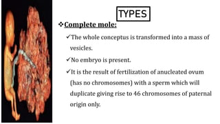
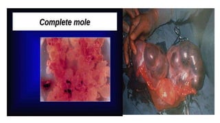
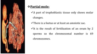
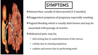
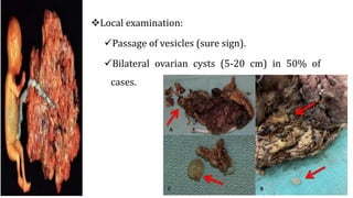
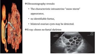
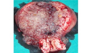
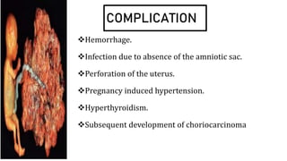
BY:
MS. LAMNUNNEM HAOKIP
SENIOR TUTOR/LECTURER
OBG NURSING
SSNSR, SU
DEFINITION
It is a benign neoplasm of the chorionic villi.
1:2000 pregnancies in United States and Europe,
but 10 times more in Asia. The incidence is higher
toward the beginning and more toward the end of
the childbearing period. It is 10 times more in
women over 45 years old
TYPES
Complete mole:
The whole conceptus is transformed into a mass of
vesicles.
No embryo is present.
It is the result of fertilization of anucleated ovum
(has no chromosomes) with a sperm which will
duplicate giving rise to 46 chromosomes of paternal
origin only.
Partial mole:
A part of trophoblastic tissue only shows molar
changes.
There is a foetus or at least an amniotic sac.
It is the result of fertilization of an ovum by 2
sperms so the chromosomal number is 69
chromosomes.
SYMPTOMS
Amenorrhea: usually of short period (2-3 months).
Exaggerated symptoms of pregnancy especially vomiting.
Vaginal bleeding which is usually dark brown and may be
associated with passage of vesicles.
Abdominal pain: may be,
• dull-aching due to rapid distension of the uterus,
• colicky due to starting expulsion,
• sudden and severe due to perforating mole
SIGNS
General examination:
Pre-eclampsia develops in 20% of cases, usually before
20 weeks’ gestation.
Hyperthyroidism develops in 10% of cases manifested
by enlarged thyroid gland, tachycardia and elevated
plasma thyroxin level.
Breast signs of pregnancy.
Abdominal examination:
The uterus is larger than the period of amenorrhea in
50% of cases, corresponds to it in 25% and smaller in
25% with inactive or dead mole.
The uterus is doughy in consistency.
Foetal parts and heart sound cannot be detected except
in partial mole.
Local examination:
Passage of vesicles (sure sign).
Bilateral ovarian cysts (5-20 cm) in 50% of
cases.
INVESTIGATIONS
Urine pregnancy test: is positive in high dilution. 1/200
is highly suggestive, 1/500 is surely diagnostic. In
normal pregnancy it is positive in dilutions up to 1/100.
Serum β-hCG level: is highly elevated (>100000
mIU/ml).
Ultrasonography reveals:
• The characteristic intrauterine "snow storm"
appearance,
• no identifiable foetus,
• bilateral ovarian cysts may be detected.
X-ray: shows no foetal skeleton
PRINCIPLES OF TREATMENT
• As soon as the diagnosis of vesicular mole is
established the uterus should be evacuated.
• The selected method depends on the size of the
uterus, whether partial expulsion has already occur
or not, the patient's age and fertility desire.
• Cross- matched blood should be available before
starting
TREATMENT
MEDICAL MANAGEMENT
Oxytocins and / or prostaglandins
may be used to encourage
expulsion of the mole but must
always be followed by surgical
evacuation.
SURGICAL MANAGEMENT
Suction Evacuation
It is carried out under general anesthesia, but not that which relax the
uterus as halothane as it may induce severe bleeding.
An infusion of 20 units oxytocin in 500 m1 of 5% glucose should be
maintained throughout the procedure.
Dilatation of the cervix is done up to a Hegar's number equal to the
period of amenorrhea in weeks e.g. No. 10 Hegar for 10 weeks’
amenorrhea. The suction canula used will be of the same size also.
A suction canula which may be metal or a disposable plastic
(preferred) is introduced into the uterine cavity.
The canula is connected to a suction pump adjusted at negative
pressure of 300-500 mmHg according to the duration of
pregnancy.
Although some recommended a gentle sharp curettage to the
uterus after evacuation, it is preferable to wait one week for fear of
uterine perforation
Hysterotomy
• It may be needed for evacuation of a large mole to
minimize and facilitate control of bleeding.
Hysterectomy
• It should be considered in women over 40 years who
have completed their family for fear of developing
choriocarcinoma
Early features suggesting residual molar tissue
include:
recurrent or persistent vaginal bleeding.
amenorrhea,
failure of uterine involution,
persistence of ovarian enlargement
NURSING MANAGEMENT
Assessment
Teach deep breathing techniques to alleviate the pain. Use
diversional activities if possible.
Check for abdominal pain, assess the abdominal area for signs
of internal bleeding(e.g. Cullen’s)
Assess for aspiration of fluids
Hygiene
Medication
Emotional support.
hCG level and follow-ups.
NURSING DIAGNOSIS
Deficit fluid volume related to heavy vaginal bleeding
secondary to hydatidiform mole as evidenced by blood
pressure level of or 90/70 mmHg, body weakness, less urine
output, pale and clammy skin.
Acute pain related to Hysterectomy/Hysterotomy as evidenced
by pain score of 10/10, verbalization of pelvic pain and
restlessness.
Risk for infection related to surgical incision.
Hopelessness related to loss of pregnancy as evidenced by
patient report of distressed.
COMPLICATION
Hemorrhage.
Infection due to absence of the amniotic sac.
Perforation of the uterus.
Pregnancy induced hypertension.
Hyperthyroidism.
Subsequent development of choriocarcinoma
BIBLIOGRAPHY
Annama Jocab, text book of comprehensive text book of ‘MIDWIFY
and GYNECOLOGY nursing ‘ JAYPEE publication 3rd edition page
no.285-287.
D.C. DUTTA text book of obsterical including perinatary and
contraception central publication 7th edition page no. 583-585.
Lily Podder, fundamentals of Midwifery and Obstetrical Nursing,
ELSEVIER publication. Page no. 280 – 285.
Mudaliar and Menon’s. Clinical Obstetrics, Universities Press 12th
Edition. Page no. 408 – 411.
HYDATIDIFORM MOLE.pptx

HYDATIDIFORM MOLE.pptx

  • 1.
    BY: MS. LAMNUNNEM HAOKIP SENIORTUTOR/LECTURER OBG NURSING SSNSR, SU
  • 2.
    DEFINITION It is abenign neoplasm of the chorionic villi. 1:2000 pregnancies in United States and Europe, but 10 times more in Asia. The incidence is higher toward the beginning and more toward the end of the childbearing period. It is 10 times more in women over 45 years old
  • 4.
    TYPES Complete mole: The wholeconceptus is transformed into a mass of vesicles. No embryo is present. It is the result of fertilization of anucleated ovum (has no chromosomes) with a sperm which will duplicate giving rise to 46 chromosomes of paternal origin only.
  • 6.
    Partial mole: A partof trophoblastic tissue only shows molar changes. There is a foetus or at least an amniotic sac. It is the result of fertilization of an ovum by 2 sperms so the chromosomal number is 69 chromosomes.
  • 8.
    SYMPTOMS Amenorrhea: usually ofshort period (2-3 months). Exaggerated symptoms of pregnancy especially vomiting. Vaginal bleeding which is usually dark brown and may be associated with passage of vesicles. Abdominal pain: may be, • dull-aching due to rapid distension of the uterus, • colicky due to starting expulsion, • sudden and severe due to perforating mole
  • 9.
    SIGNS General examination: Pre-eclampsia developsin 20% of cases, usually before 20 weeks’ gestation. Hyperthyroidism develops in 10% of cases manifested by enlarged thyroid gland, tachycardia and elevated plasma thyroxin level. Breast signs of pregnancy.
  • 10.
    Abdominal examination: The uterusis larger than the period of amenorrhea in 50% of cases, corresponds to it in 25% and smaller in 25% with inactive or dead mole. The uterus is doughy in consistency. Foetal parts and heart sound cannot be detected except in partial mole.
  • 11.
    Local examination: Passage ofvesicles (sure sign). Bilateral ovarian cysts (5-20 cm) in 50% of cases.
  • 12.
    INVESTIGATIONS Urine pregnancy test:is positive in high dilution. 1/200 is highly suggestive, 1/500 is surely diagnostic. In normal pregnancy it is positive in dilutions up to 1/100. Serum β-hCG level: is highly elevated (>100000 mIU/ml).
  • 13.
    Ultrasonography reveals: • Thecharacteristic intrauterine "snow storm" appearance, • no identifiable foetus, • bilateral ovarian cysts may be detected. X-ray: shows no foetal skeleton
  • 14.
    PRINCIPLES OF TREATMENT •As soon as the diagnosis of vesicular mole is established the uterus should be evacuated. • The selected method depends on the size of the uterus, whether partial expulsion has already occur or not, the patient's age and fertility desire. • Cross- matched blood should be available before starting
  • 15.
    TREATMENT MEDICAL MANAGEMENT Oxytocins and/ or prostaglandins may be used to encourage expulsion of the mole but must always be followed by surgical evacuation.
  • 16.
    SURGICAL MANAGEMENT Suction Evacuation Itis carried out under general anesthesia, but not that which relax the uterus as halothane as it may induce severe bleeding. An infusion of 20 units oxytocin in 500 m1 of 5% glucose should be maintained throughout the procedure. Dilatation of the cervix is done up to a Hegar's number equal to the period of amenorrhea in weeks e.g. No. 10 Hegar for 10 weeks’ amenorrhea. The suction canula used will be of the same size also.
  • 18.
    A suction canulawhich may be metal or a disposable plastic (preferred) is introduced into the uterine cavity. The canula is connected to a suction pump adjusted at negative pressure of 300-500 mmHg according to the duration of pregnancy. Although some recommended a gentle sharp curettage to the uterus after evacuation, it is preferable to wait one week for fear of uterine perforation
  • 20.
    Hysterotomy • It maybe needed for evacuation of a large mole to minimize and facilitate control of bleeding. Hysterectomy • It should be considered in women over 40 years who have completed their family for fear of developing choriocarcinoma
  • 22.
    Early features suggestingresidual molar tissue include: recurrent or persistent vaginal bleeding. amenorrhea, failure of uterine involution, persistence of ovarian enlargement
  • 23.
    NURSING MANAGEMENT Assessment Teach deepbreathing techniques to alleviate the pain. Use diversional activities if possible. Check for abdominal pain, assess the abdominal area for signs of internal bleeding(e.g. Cullen’s) Assess for aspiration of fluids Hygiene Medication Emotional support. hCG level and follow-ups.
  • 24.
    NURSING DIAGNOSIS Deficit fluidvolume related to heavy vaginal bleeding secondary to hydatidiform mole as evidenced by blood pressure level of or 90/70 mmHg, body weakness, less urine output, pale and clammy skin. Acute pain related to Hysterectomy/Hysterotomy as evidenced by pain score of 10/10, verbalization of pelvic pain and restlessness. Risk for infection related to surgical incision. Hopelessness related to loss of pregnancy as evidenced by patient report of distressed.
  • 25.
    COMPLICATION Hemorrhage. Infection due toabsence of the amniotic sac. Perforation of the uterus. Pregnancy induced hypertension. Hyperthyroidism. Subsequent development of choriocarcinoma
  • 26.
    BIBLIOGRAPHY Annama Jocab, textbook of comprehensive text book of ‘MIDWIFY and GYNECOLOGY nursing ‘ JAYPEE publication 3rd edition page no.285-287. D.C. DUTTA text book of obsterical including perinatary and contraception central publication 7th edition page no. 583-585. Lily Podder, fundamentals of Midwifery and Obstetrical Nursing, ELSEVIER publication. Page no. 280 – 285. Mudaliar and Menon’s. Clinical Obstetrics, Universities Press 12th Edition. Page no. 408 – 411.