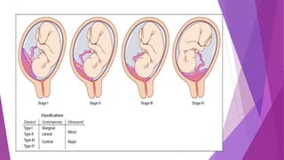
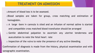
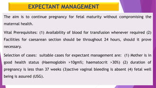
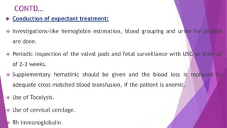
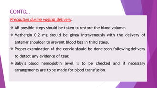
PLACENTA PREVIA
PRESENTED BY
LIPI MONDAL
M.SC NURSING STUDENT
MEANING
When the placenta is implanted partially or
completely over the lower uterine segment (over
and adjacent to the internal OS) it is called
Placenta Previa. It is one type of antepartum
hemorrhage.
ETIOLOGY
DROPPING DOWN THEORY
PERSISTENT CHORIONIC
ACTIVITY
DEFECTIVE DECIDUA
BIG SURFACE OF PLACENTA
TYPES OF PLACENTA PREVIA
CLINICAL FEATURES
Abdominal examination:
 The size of the uterus is proportionate of the period of gestation.
 The uterus feels relaxed, soft and elastic.
 Persistence of malpresentation like breech or transverse or
unstable lie is more frequent.
 Persistent displacement of the fetal head is very suggestive.
 Slowing of the fetal heart rate on pressing the head down into the
pelvis which soon recovers promptly as the pressure is released is
suggestive presence of low lying placenta (Stallworth's sign).
CONTD…
Valval inspection: Only inspection is to be done to note whether
the bleeding is still occurring or has ceased.
Character of the blood – Bright red or dark colored
Amount of blood loss - Assessed from the blood stained
clothing. In placenta previa, the blood is bright red as the
bleeding occurs from the separated utero-placental sinuses close
to the cervical opening and escape out immediately.
Vaginal examination must not be done outside the operation
theatre in the hospital, as it can provoke further separation of
placenta.
DIAGNOSIS
I.LOCALISATION OF PLACENTA
(PLACENTOGRAPHY)
 Sonography
1. Transabdominal ultrasound (TAS)
2. Transvaginal ultrasound (TVS)
3. Trans perineal ultrasound
4. Color Doppler flow study
 Magnetic resonance imaging (MRI)
II. CLINICAL
1. By internal examination
(double set up
examination)
2. Direct visualization during
caesarean section
3. Examination of the
placenta following vaginal
delivery
MANAGEMENT
PREVENTION:
-Adequate antenatal care to improve the health status of women and
correction of anemia.
-Antenatal diagnosis of low lying placenta at 20 weeks with routine ultra
sound needs repeat ultra sound examination at 34 weeks to confirm the
diagnosis.
-Significance of “Warning hemorrhage “ should not be ignored.
-Color flow Doppler USG in Placenta Previa is indicated to detect any
placenta accrete.
ADMISSION TO HOSPITAL
All cases of APH, even if the bleeding is slight or absent by the time
the patient reaches the hospital, should be admitted.
The reasons are:
(1) All the cases of APH should be regarded as due to Placenta Previa
unless proved otherwise.
(2) (2) The bleeding may recur sooner or later and none can predict
when it recurs and how much she will bleed.
TREATMENT ON ADMISSION
oAmount of blood loss is to be assessed.
oBlood samples are taken for group, cross matching and estimation of
hemoglobin.
o A large –bore iv cannula is sited and an infusion of normal saline is started
and compatible cross matched blood transfusion should be arranged
o Gentle abdominal palpation to ascertain any uterine tenderness and
auscultation to note the fetal heart rate
o Inspection of the vulva to note the presence of any active bleeding.
Confirmation of diagnosis is made from the history, physical examination and with
sonographic examination.
EXPECTANT MANAGEMENT
The aim is to continue pregnancy for fetal maturity without compromising the
maternal health.
Vital Prerequisites: (1) Availability of blood for transfusion whenever required (2)
Facilities for caesarean section should be throughout 24 hours, should it prove
necessary.
Selection of cases: suitable cases for expectant management are: (1) Mother is in
good health status (Haemoglobin >10gm%; haematocrit >30%) (2) duration of
pregnancy is less than 37 weeks (3)active vaginal bleeding is absent (4) fetal well
being is assured (USG).
CONTD…
 Conduction of expectant treatment:
 Investigations-like hemoglobin estimation, blood grouping and urine for protein
are done.
 Periodic inspection of the valval pads and fetal surveillance with USG at interval
of 2-3 weeks.
 Supplementary hematinic should be given and the blood loss is replaced by
adequate cross matched blood transfusion, if the patient is anemic.
 Use of Tocolysis.
 Use of cervical cerclage.
 Rh immunoglobulin.
ACTIVE MANAGEMENT
The indications of definitive management (delivery) are:
 bleeding occurs at or after 37 weeks of pregnancy
 Patient is in labor.
 Patient is in exsanguinated state on admission.
 Bleeding is continuing & in moderate degree.
 Baby is dead or known to be congenitally deformed.
ACTIVE MANAGEMENT
A.Caesarean delivery is done for all women with sonographic evidence of Placenta
Previa where placental edge is within 2cm from the internal os.
B. Vaginal delivery may be considered where placenta edge is clearly 2-3 cm away
from the internal cervical os (based on sonography)
Contraindications of vaginal examination are:
 Patient in exsanguinated state & diagnosed cases of major degree of Placenta
Previa confirmed by ultrasonography.
 Associated complicating factors such as malpresentation, elderly primigravida,
pregnancy with history of previous caesarean section, contracted pelvis etc.
Low Rupture of Membranes.
CONTD…
Precaution during vaginal delivery:
 All possible steps should be taken to restore the blood volume.
 Methergin 0.2 mg should be given intravenously with the delivery of
anterior shoulder to prevent blood loss in third stage.
 Proper examination of the cervix should be done soon following delivery
to detect any evidence of tear.
 Baby’s blood hemoglobin level is to be checked and if necessary
arrangements are to be made for blood transfusion.
BIBLIOGRAPHY
Teacher’s reference:
 Jacob A. “Mannual Midwifery and gynaecological Nursing”. New delhi,jaypee
brothers:2012, pg:381-3.
 Mckinney ES, James SR, Murray SS, Ashwill JW. “Matrnal child
Nursing”.canada, Elsevier: 2009,pg:509-30.
 Dutta’s DC. “Text book of obstetrics”. west Bengal, India: New Central Book of
Agency:2011,pg :444-6.
Student’s reference:
 Salhan S. “Text book of Obstetrics”. New Delhi, India: jaypee
brothers:2014,pg:509-13.
 Saili A, Bhat S, Shenoi A, “ obstetrics principles and practice”. New delhi,
jaypee, 3rd edition;2005.
Pg:439-54.

Placenta previa

  • 1.
    PLACENTA PREVIA PRESENTED BY LIPIMONDAL M.SC NURSING STUDENT
  • 2.
    MEANING When the placentais implanted partially or completely over the lower uterine segment (over and adjacent to the internal OS) it is called Placenta Previa. It is one type of antepartum hemorrhage.
  • 3.
    ETIOLOGY DROPPING DOWN THEORY PERSISTENTCHORIONIC ACTIVITY DEFECTIVE DECIDUA BIG SURFACE OF PLACENTA
  • 4.
  • 6.
    CLINICAL FEATURES Abdominal examination: The size of the uterus is proportionate of the period of gestation.  The uterus feels relaxed, soft and elastic.  Persistence of malpresentation like breech or transverse or unstable lie is more frequent.  Persistent displacement of the fetal head is very suggestive.  Slowing of the fetal heart rate on pressing the head down into the pelvis which soon recovers promptly as the pressure is released is suggestive presence of low lying placenta (Stallworth's sign).
  • 7.
    CONTD… Valval inspection: Onlyinspection is to be done to note whether the bleeding is still occurring or has ceased. Character of the blood – Bright red or dark colored Amount of blood loss - Assessed from the blood stained clothing. In placenta previa, the blood is bright red as the bleeding occurs from the separated utero-placental sinuses close to the cervical opening and escape out immediately. Vaginal examination must not be done outside the operation theatre in the hospital, as it can provoke further separation of placenta.
  • 8.
    DIAGNOSIS I.LOCALISATION OF PLACENTA (PLACENTOGRAPHY) Sonography 1. Transabdominal ultrasound (TAS) 2. Transvaginal ultrasound (TVS) 3. Trans perineal ultrasound 4. Color Doppler flow study  Magnetic resonance imaging (MRI) II. CLINICAL 1. By internal examination (double set up examination) 2. Direct visualization during caesarean section 3. Examination of the placenta following vaginal delivery
  • 9.
    MANAGEMENT PREVENTION: -Adequate antenatal careto improve the health status of women and correction of anemia. -Antenatal diagnosis of low lying placenta at 20 weeks with routine ultra sound needs repeat ultra sound examination at 34 weeks to confirm the diagnosis. -Significance of “Warning hemorrhage “ should not be ignored. -Color flow Doppler USG in Placenta Previa is indicated to detect any placenta accrete.
  • 10.
    ADMISSION TO HOSPITAL Allcases of APH, even if the bleeding is slight or absent by the time the patient reaches the hospital, should be admitted. The reasons are: (1) All the cases of APH should be regarded as due to Placenta Previa unless proved otherwise. (2) (2) The bleeding may recur sooner or later and none can predict when it recurs and how much she will bleed.
  • 11.
    TREATMENT ON ADMISSION oAmountof blood loss is to be assessed. oBlood samples are taken for group, cross matching and estimation of hemoglobin. o A large –bore iv cannula is sited and an infusion of normal saline is started and compatible cross matched blood transfusion should be arranged o Gentle abdominal palpation to ascertain any uterine tenderness and auscultation to note the fetal heart rate o Inspection of the vulva to note the presence of any active bleeding. Confirmation of diagnosis is made from the history, physical examination and with sonographic examination.
  • 12.
    EXPECTANT MANAGEMENT The aimis to continue pregnancy for fetal maturity without compromising the maternal health. Vital Prerequisites: (1) Availability of blood for transfusion whenever required (2) Facilities for caesarean section should be throughout 24 hours, should it prove necessary. Selection of cases: suitable cases for expectant management are: (1) Mother is in good health status (Haemoglobin >10gm%; haematocrit >30%) (2) duration of pregnancy is less than 37 weeks (3)active vaginal bleeding is absent (4) fetal well being is assured (USG).
  • 13.
    CONTD…  Conduction ofexpectant treatment:  Investigations-like hemoglobin estimation, blood grouping and urine for protein are done.  Periodic inspection of the valval pads and fetal surveillance with USG at interval of 2-3 weeks.  Supplementary hematinic should be given and the blood loss is replaced by adequate cross matched blood transfusion, if the patient is anemic.  Use of Tocolysis.  Use of cervical cerclage.  Rh immunoglobulin.
  • 14.
    ACTIVE MANAGEMENT The indicationsof definitive management (delivery) are:  bleeding occurs at or after 37 weeks of pregnancy  Patient is in labor.  Patient is in exsanguinated state on admission.  Bleeding is continuing & in moderate degree.  Baby is dead or known to be congenitally deformed.
  • 15.
    ACTIVE MANAGEMENT A.Caesarean deliveryis done for all women with sonographic evidence of Placenta Previa where placental edge is within 2cm from the internal os. B. Vaginal delivery may be considered where placenta edge is clearly 2-3 cm away from the internal cervical os (based on sonography) Contraindications of vaginal examination are:  Patient in exsanguinated state & diagnosed cases of major degree of Placenta Previa confirmed by ultrasonography.  Associated complicating factors such as malpresentation, elderly primigravida, pregnancy with history of previous caesarean section, contracted pelvis etc. Low Rupture of Membranes.
  • 16.
    CONTD… Precaution during vaginaldelivery:  All possible steps should be taken to restore the blood volume.  Methergin 0.2 mg should be given intravenously with the delivery of anterior shoulder to prevent blood loss in third stage.  Proper examination of the cervix should be done soon following delivery to detect any evidence of tear.  Baby’s blood hemoglobin level is to be checked and if necessary arrangements are to be made for blood transfusion.
  • 18.
    BIBLIOGRAPHY Teacher’s reference:  JacobA. “Mannual Midwifery and gynaecological Nursing”. New delhi,jaypee brothers:2012, pg:381-3.  Mckinney ES, James SR, Murray SS, Ashwill JW. “Matrnal child Nursing”.canada, Elsevier: 2009,pg:509-30.  Dutta’s DC. “Text book of obstetrics”. west Bengal, India: New Central Book of Agency:2011,pg :444-6. Student’s reference:  Salhan S. “Text book of Obstetrics”. New Delhi, India: jaypee brothers:2014,pg:509-13.  Saili A, Bhat S, Shenoi A, “ obstetrics principles and practice”. New delhi, jaypee, 3rd edition;2005. Pg:439-54.