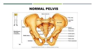
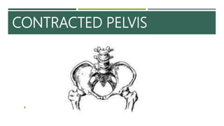
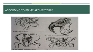
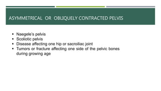
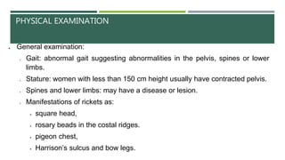
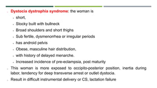
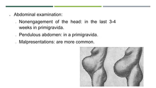
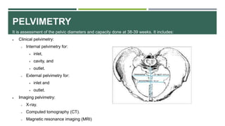
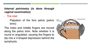
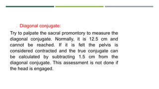
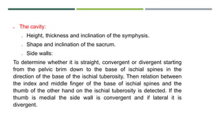
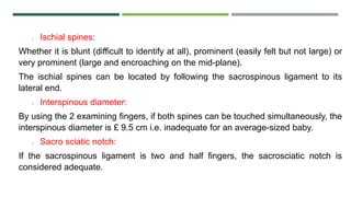
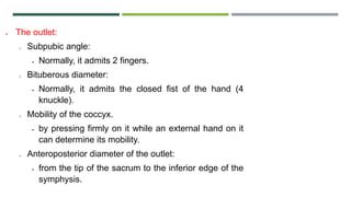
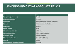
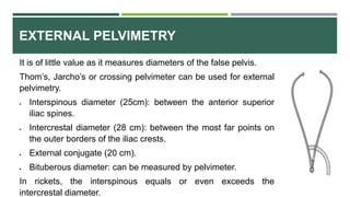
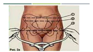
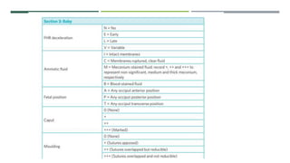
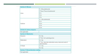
SlideShare a Scribd company logo
Contracted pelvis
and
Dystocia
Neethu S.S.
Second year MSc Nursing
Govt. College of Nursing
Kozhikode.
NORMAL PELVIS
CONTRACTED PELVIS

DEFINITION
Anatomically contracted pelvis is defined as one where the
essential diameters of one or more planes are shortened by 0.5cm.
Obstetrically, it is a state in which there is alteration in size or
shape of the pelvis of sufficient degree as to alter the normal
mechanism of labor in an average size baby.
Factors influencing the size and shape of the pelvis
 Developmental factor: hereditary or congenital.
 Racial factor.
 Nutritional factor: malnutrition results in small pelvis.
 Sexual factor: as excessive androgen may produce android pelvis.
 Metabolic factor: as rickets and osteomalacia.
 Trauma, diseases or tumors of the bony pelvis, legs or spines
ETIOLOGY OF CONTRACTED PELVIS
 Causes in the pelvis
 Developmental (congenital):
o Small gynecoid pelvis (generally contracted pelvis).
o Small android pelvis.
o Small anthropoid pelvis.
o Small platypelloid pelvis (simple flat pelvis).
o Naegele’s pelvis: absence of one sacral ala.
o Robert’s pelvis: absence of both sacral alae.
o High assimilation pelvis: The sacrum is composed of 6 vertebrae.
o Low assimilation pelvis: The sacrum is composed of 4 vertebrae.
o Split pelvis: splitted symphysis pubis.
 Metabolic:
o Rickets.
o Osteomalacia (triradiate pelvic brim).
 Traumatic: as fractures.
 Neoplastic: as osteoma.
Causes in the spine
 Lumbar kyphosis.
 Lumbar scoliosis.
 Spondylolisthesis: The 5th lumbar vertebra with the above vertebral
column is pushed forward while the promontory is pushed backwards
and the tip of the sacrum is pushed forwards leading to outlet
contraction.
 Coccygeal deformity
Causes in the lower limbs
 Dislocation of one or both femurs.
 Atrophy of one or both lower limbs.
 Hip joint disease
CLASSIFICATION OF CONTRACTED PELVIS
 According to degree of contracture
 Minor degree: The true conjugate is 9-10 cm. It corresponds to
minor disproportion.
 Moderate degree: The true conjugate is 8-9 cm. It corresponds to
moderate disproportion.
 Severe degree: The true conjugate is 6-8 cm. It corresponds to
marked disproportion.
 Extreme degree: The true conjugate is less than 6 cm. Vaginal
delivery is impossible even after craniotomy as the bimastoid
diameter (7.5 cm) is not crushed. Also known as absolutely
contracted pelvis.
ACCORDING TO PELVIC ARCHITECTURE
RACHITIC FLAT PELVIS
 Rickets in early childhood cause bones to remain soft and unossified.
 Inlet
Sacral promontory is pushed downwards and forwards producing a reniform
shape Short APD
 Cavity
Sacrum is flat and tilted backwards
Sharp angulation at sacrococcygeal joint
 Outlet
Widened transverse diameter and pubic arch
OSTEOMALACIC PELVIS
 Caused by softening of the pubic bones
 Due to deficiency of calcium, vitamin D and lack of exposure to
sunrays
 The promontory is pushed downwards and forwards and the
lateral pelvic walls are pushed inwards causing the anterior wall to
form a beak
 Triradiate shape of inlet
 Approximation of 2 ischial tuberosities
 Markedly shortened sacrum
 Coccyx is pushed forward
ASYMMETRICAL OR OBLIQUELY CONTRACTED PELVIS
 Naegele’s pelvis
 Scoliotic pelvis
 Disease affecting one hip or sacroiliac joint
 Tumors or fracture affecting one side of the pelvic bones
during growing age
Naegele’s pelvis
 Extremely rare
 Due to arrested development of one ala of the
sacrum
 It can be
i. Congenital: associated with urinary tract of the
same side
ii. Acquired: osteitis of sacroiliac joint
 Pelvis is obliquely contracted at all levels but more
marked in the outlet
 Straight iliopectineal line on the affected side
SCOLIOSIS
 Acetabulam is pushed inwards on the
weight bearing side
 Contraction of one of the oblique
diameters
ROBERT’S PELVIS ( TRANSVERSELY CONTRACTED PELVIS)
 Ala of both the sides
are absent
 Sacrum is fused
with innominate
bones
KYPHOTIC PELVIS
 Developed secondary to the kyphotic changes of the vertebral column.
 sacrum is tilted backwards in the upper part and forwards in the lower part,
 it is narrow and straight
 APD is increased at the inlet but is decreased at the outlet
 Narrow suprapubic angle
 Extreme funneling of the pelvis
 Pendulous abdomen
PELVIS AEQUABILITER JUSTO MINOR
 Characterized by general reduction of all
diameters; equally shortened usually by
1-2cm
 Occurs in short. Also occurs in women
with massive skeletal bones and
developed muscles, the pelvis has
masculine features such as narrow
sacrum, narrow pubic outlet (funnel-
shaped)
RARE FORMS OF CONTRACTED PELVIS
 Otto’s pelvis – develop as result of inflammatory
process in the hip or knee
 Beaked (rostrate) pelvis – under development of
both sacral wings
 Spondylolithetic pelvis – formed due to partial
dislocation of last lumbar vertebra in front of 1st sacral
vertebra
DIAGNOSIS OF CONTRACTED PELVIS
 History
 Rickets: is expected if there is a history of delayed walking and dentition.
 Osteomalacia
 Tuberculosis of the pelvic joints or spines
 poliomyelitis
 Trauma or diseases: of the pelvis, spines or lower limbs.
 Bad obstetric history: e.g. prolonged labour ended by;
o difficult forceps,
o caesarean section or
o still birth.
o Weight of the baby, evidence of maternal injuries such as complete perineal tear,
vesicovaginal or rectovaginal fistula.
PHYSICAL EXAMINATION
 General examination:
o Gait: abnormal gait suggesting abnormalities in the pelvis, spines or lower
limbs.
o Stature: women with less than 150 cm height usually have contracted pelvis.
o Spines and lower limbs: may have a disease or lesion.
o Manifestations of rickets as:
 square head,
 rosary beads in the costal ridges.
 pigeon chest,
 Harrison’s sulcus and bow legs.
o Dystocia dystrophia syndrome: the woman is
 short,
 Stocky built with bullneck
 Broad shoulders and short thighs
 Sub fertile, dysmenorrhea or irregular periods
 has android pelvis
 Obese, masculine hair distribution,
 with history of delayed menarche.
 Increased incidence of pre-eclampsia, post maturity
o This woman is more exposed to occipito-posterior position, inertia during
labor, tendency for deep transverse arrest or outlet dystocia.
o Result in difficult instrumental delivery or CS, lactation failure
 Abdominal examination:
o Nonengagement of the head: in the last 3-4
weeks in primigravida.
o Pendulous abdomen: in a primigravida.
o Malpresentations: are more common.
PELVIMETRY
It is assessment of the pelvic diameters and capacity done at 38-39 weeks. It includes:
 Clinical pelvimetry:
o Internal pelvimetry for:
 inlet,
 cavity, and
 outlet.
o External pelvimetry for:
 inlet and
 outlet.
 Imaging pelvimetry:
o X-ray.
o Computed tomography (CT).
o Magnetic resonance imaging (MRI)
Internal pelvimetry (is done through
vaginal examination)
 The inlet:
o Palpation of the fore pelvis (pelvic
brim):
The index and middle fingers are moved
along the pelvic brim. Note whether it is
round or angulated, causing the fingers to
dip into a V-shaped depression behind the
symphysis.
o Diagonal conjugate:
Try to palpate the sacral promontory to measure the
diagonal conjugate. Normally, it is 12.5 cm and
cannot be reached. If it is felt the pelvis is
considered contracted and the true conjugate can
be calculated by subtracting 1.5 cm from the
diagonal conjugate. This assessment is not done if
the head is engaged.
 The cavity:
o Height, thickness and inclination of the symphysis.
o Shape and inclination of the sacrum.
o Side walls:
To determine whether it is straight, convergent or divergent starting
from the pelvic brim down to the base of ischial spines in the
direction of the base of the ischial tuberosity. Then relation between
the index and middle finger of the base of ischial spines and the
thumb of the other hand on the ischial tuberosity is detected. If the
thumb is medial the side wall is convergent and if lateral it is
divergent.
o Ischial spines:
Whether it is blunt (difficult to identify at all), prominent (easily felt but not large) or
very prominent (large and encroaching on the mid-plane).
The ischial spines can be located by following the sacrospinous ligament to its
lateral end.
o Interspinous diameter:
By using the 2 examining fingers, if both spines can be touched simultaneously, the
interspinous diameter is £ 9.5 cm i.e. inadequate for an average-sized baby.
o Sacro sciatic notch:
If the sacrospinous ligament is two and half fingers, the sacrosciatic notch is
considered adequate.
 The outlet:
o Subpubic angle:
 Normally, it admits 2 fingers.
o Bituberous diameter:
 Normally, it admits the closed fist of the hand (4
knuckle).
o Mobility of the coccyx.
 by pressing firmly on it while an external hand on it
can determine its mobility.
o Anteroposterior diameter of the outlet:
 from the tip of the sacrum to the inferior edge of the
symphysis.
Data Finding
Forepelvis (pelvic brim)
Diagonal conjugate
Symphysis
Sacrum
Side walls
Ischial spines
Interspinous diameter
Sacrosciatic notch
Subpubic angle
Bituberous diameter
Coccyx
Anterposterior diameter of outlet
Round.
≥11.5 cm.
Average thickness, parallel to sacrum.
Hollow, average inclination.
Straight.
Blunt.
≥10.0 cm.
2.5 -3 finger - breadths.
2finger - breadths.
4 knuckles >8.0 cm).
Mobile.
≥11.0 cm.
FINDINGS INDICATING ADEQUATE PELVIS:
EXTERNAL PELVIMETRY
It is of little value as it measures diameters of the false pelvis.
Thom’s, Jarcho’s or crossing pelvimeter can be used for external
pelvimetry.
 Interspinous diameter (25cm): between the anterior superior
iliac spines.
 Intercrestal diameter (28 cm): between the most far points on
the outer borders of the iliac crests.
 External conjugate (20 cm).
 Bituberous diameter: can be measured by pelvimeter.
In rickets, the interspinous equals or even exceeds the
intercrestal diameter.
RADIOLOGICAL PELVIMETRY
It is indicated mainly in borderline pelvic contraction.
 Lateral view: The patient stands with the X-ray tube on one side and the film cassette on the
opposite side.
o It is the most important view as it shows the anteroposterior diameters of the pelvis, angle
of inclination of the brim, width of sacro sciatic notch, curvature of the sacrum and
cephalo-pelvic relationship.
 Inlet view: The patient sits on the film cassette and leans backwards so that the plane of the
pelvic brim becomes parallel to the film.
 Outlet view: The patient sits on the film cassette and leans forwards.
EFFECT OF CONTRACTED PELVIS
 On pregnancy
I. More chance of incarceration of the retroverted gravid uterus in
flat pelvis.
II. Abdomen becomes pendulous in multigravida
III.Malpresentations
IV.Unstable lie
 On labor
Increased incidence of:
1. Early rupture of membranes
2. Cord prolapse
3. Slow cervical dilatation
4. Prolonged labor
5. Obstructed labor with exhaustion, dehydration, ketoacidosis and
sepsis
6. Operative interference
7. Shock
8. Postpartum hemorrhage and sepsis
 Maternal injuries
 Fetal hazards
due to trauma and asphyxia
MECHANISM OF LABOR IN CONTRACTED PELVIS
 Generally in contracted pelvis all the diameters in the different planes are
shortened. So there is difficulty from beginning to the end.
 In the flat pelvis, the head finds difficulty in negotiating the brim and once it
passes through the brim, there is no difficulty in the cavity and outlet.
 The head negotiates the brim by the following mechanism:
 The head engages with the sagittal suture in transverse diameter.
 Head remains deflexed and engagement is delayed.
 If the APD is too short, the occiput is mobilized to the same side to occupy the sacral
bay. The biparietal diameter is thus placed in the sacrocotyloid diameter and the
narrow bitemporal diameter is placed in the narrow conjugate. If lateral mobilization
is not possible, there is a chance of extension of the head leading to brow or face
presentation.
 Engagement occurs by exaggerated parietal presentation so that the super- sub
parietal diameter, instead of the biparietal diameter passes through the pelvic brim.
 Moulding may be extreme and often there is an indentation or even a fracture of one
parietal bone. However, the caput that forms is not big.
 Once the head negotiates the brim, there is no difficulty in the cavity and outlet and
normal mechanism follows.
MANAGEMENT OF CONTRACTED PELVIS
Ascertain the degree of disproportion
 Minor inlet contraction: spontaneous delivery
 Moderate and severe degrees:
Induction of labor
Elective cesarean section at term
 Trial labor
INDUCTION OF LABOR
 Induction 2-3 weeks prior to the EDC may be considered only in
cases with minor to moderate degrees of pelvic contraction.
 2-3 weeks before the date in selected multigravida with previous
history of difficult vaginal delivery.
ELECTIVE CESAREAN SECTION AT TERM
 INDICATIONS:
 Major degree of inlet contraction
 Moderate degree of inlet contraction
associated with outlet contraction or
complicating factors like elderly
primigravida, malpresentation, post-
cesarean pregnancy.
 Ascertain maturity of fetus before planning.
TRIAL LABOR
 It is the conduction of spontaneous labor in
moderate degree of cephalopelvic disproportion,
in an institution under supervision with watchful
expectancy, hoping for a vaginal delivery.
AIM
Aims at avoiding an unnecessary cesarean section and at delivering a healthy
baby.
CONTRAINDICATIONS
 Associated mid pelvic and outlet contraction
 Presence of complicating factors like primigarvida,
malpresentation, post maturity, post caesarean pregnancy, pre
eclampsia, medical disorders like heart disease, diabetes, TB etc
 Lack of facilities for caesarean section round the clock
CONDUCTION OF TRIAL LABOR
 Prefers spontaneous labor, induce only if labor does not start even
after due date
 NPO, maintain hydration by IVF, adequate analgesics
 Maintain partograph
 Maternal and fetal monitoring
 In failure to progress: amniotomy+ oxytocin after cervix is 3cm
 Pelvic examination after membranes are ruptured
FAVOURABLE FACTORS
 Flat pelvis better than android
 Vertex
 Degree of contraction: minor
 Intact membranes till full dilatation
 Good uterine contraction
 Emotional stability of woman
UNFAVOURABLE FEATURES
 Appearance of abnormal uterine contraction
 Cervical < 1cm/hour
 Arrest of cervical dilation and no descent of fetal head in spite of
oxytocin therapy
 Early rupture of membranes
 Formation of caput and evidence of excessive moulding
 Fetal distress
HOW LONG TRIAL TO BE CONTINUED
 Termination of trial
 Spontaneous delivery with or without episiotomy
 Forceps/ ventouse: difficult forceps delivery is to be avoided
Caesarean section
SUCCESSFUL TRIAL
 A healthy baby is born vaginally, spontaneously or by forceps or
ventouse with the mother in good condition.
 Delivery by cesarean section or delivery of a dead baby,
spontaneously or by craniotomy, is called failure of trial labor.
ADVANTAGES OF TRIAL LABOR
 It eliminates unnecessary cesarean section electively decided
upon
 It eliminates injudicious use of premature induction of labor with its
antecedent hazards
 A successful trial ensures the women a good future obstetrics
DISADVANTAGES OF TRIAL LABOR
 Test of disproportion remains unproven when cesarean delivery is
done due to fetal distress or uterine dysfunction
 Increased perinatal morbidity or mortality due to asphyxia or
intracranial hemorrhage
 Increased maternal morbidity
 Increased psychological morbidity
NURSING MANAGEMENT
 Check vitals every 4 hourly
 Monitor both contraction and fetus continuously
 Report immediately the sign of fetal distress
 Position the mother in ways to increase the pelvic diameter such as sitting or
squatting which increase the outlet diameter and also aid in fetal descent
 Assess the fetus for hypoxia
 Provide support to the client and the family members in coping with stress of
a complicated labor
COMPLICATIONS OF CONTRACTED PELVIS
 Maternal:
o During pregnancy:
 Incarcerated retroverted gravid uterus.
 Malpresentations.
 Pendulous abdomen.
 Nonengagement.
 Pyelonephritis especially in high assimilation
pelvis due to more compression of the ureter.
o During labour:
 Inertia, slow cervical dilatation and prolonged labor.
 Premature rupture of membranes and cord prolapse.
 Obstructed labor and rupture uterus.
 Necrotic genito-urinary fistula.
 Injury to pelvic joints or nerves from difficult forceps
delivery.
 Postpartum hemorrhage.
 Fetal:
o Intracranial hemorrhage.
o Asphyxia.
o Fracture skull.
o Nerve injuries.
o Intra-amniotic infection.
DYSTOCIA
 Dystocia refers to the abnormal progress of labor.
 The labor is longer, more painful, or abnormal because of
problems with the mechanics of labor, powers, passageway,
passenger, or psyche.
 Dystocia is the most common indication for primary cesarean
section, accounting for 50% of surgical deliveries.
CAUSES
4 P’s
1.Powers:
 uterine contractions that are not sufficiently strong to
cause cervical dilatation and effacement
 Voluntary pushing combined with uterine contractions not
be sufficient to cause descent and expulsion of the fetus.
2.Passageway:
 Variations in the size and shape of the bony pelvis like
contracture of the pelvic diameter
 Abnormalities of the reproductive tract like immature pelvic
size or deformities
3. Passenger
 Malpresentation
 Malposition
 Unusual size
 Abnormal development of the fetus
4.Psyche
Maternal factors:
 Anxiety
 Lack of preparation
 fear
PROBLEMS WITH THE POWER
 Problems with the powers of labor involve the forces of
labor, uterine contractions, and bearing down efforts.
 Dysfunctional labor is a term commonly used to describe
abnormal uterine contractions that interfere with normal
progress of labor.
TYPES OF UTERINE DYSFUNCTION
1.Hyper tonic contraction pattern
2.Hypotonic contraction pattern
HYPERTONIC CONTRACTION PATTERN
 Involves a distortion of the pressure gradient .
 The midsegment may contract with more force than the fundus, or there could
be complete asynchronism of the impulses originating in each cornu.
 Increased frequency or elevated resting tone > 15 mmHg
 Occurs during latent phase.
 Ineffective in accomplishing dilatation
 Increased uterine tone result in maternal discomfort
 Contraction described as ‘colicky’ and extremely painful
 Uterus tender to palpate even between contractions.
MANAGEMENT
 Rest
 Administration of fluids to maintain hydration and electrolyte balance
 Inj. Morphine 10-15 mg IM to inhibit abnormal excitability
 Short acting barbiturates
 Oxytocin is contraindicated as it may cause even greater resting tension.
 90% resume normal labor when the sedation is disappeared.
 CS if the contractions remain uncoordinated and ineffective even after rest
and with evident signs of fetal distress.
HYPOTONIC CONTRACTION PATTERN
 Occurs in approximately 4% of all labor
 Uterine contractions are less frequent, no basal tone and their
slight rise in pressure is insufficient to dilate the cervix at
satisfactory rate.
 Occurs in active phase
 Contractions also may become hypotonic during second stage
 It is a pattern of uterine activity that is less than the adequate
labor pattern
MANAGEMENT
 Timely diagnosis
 Vaginal examinations every 4th hourly
 Rest and fluids
 enema
 Augmentation of contraction by amniotomy or
 Oxytocin administration
 ARM
ACTIVE PHASE DISORDERS
I. Protraction disorders
II.Arrest disorders
PROTRACTION DISORDERS
 Characterized by a slower than normal rate of cervical dilation and by delayed
descent of the fetal head in the active phase of labor.
 Cervical dilation < 1 cm/hr in nulliparous
 Cervical dilation <2 cm/hr in multiparas
 Treated by supportive fluids, reassurance and minimum sedation.
ARREST DISORDERS
1. Prolonged deceleration phase: > 3 hrs in nullipara and > 1 hr in a multipara
2. Secondary arrest of dilation: no progress in cervical dilation occurs for > 2
hours
3. Arrest of descent: fetal head dose not descent for > 1 hr in nulliparous and >
0.5 hours for a multipara.
4. Failure of descent: no descent during first stage, deceleration phase or active
phase.
 It may occur following protraction disorders or when a normally progressing
labor suddenly stops
 Frequently associated with CPD.
PROBLEMS WITH EXPULSIVE FORCES
I. Inadequate voluntary expulsive forces
II.Pathologic retraction ring
III.Constriction ring
INADEQUATE VOLUNTARY EXPULSIVE FORCES
Affected by
 Anesthesia or heavy sedation
 Fatigue or intensification of pain during pushing
 Rarely physical problems such as spinal cord injury
 Management related to the cause.
 Appropriate encouragement, support, instruction and positioning.
PATHOLOGIC RING AND CONSTRICTION RING
 Pathologic retraction ring or Bandl’s ring is an exaggeration of the normal
physiologic retraction ring which occurs at the junction of the upper and lower
uterine segments.
 Uterus above the ring becomes thicker, lower uterine segment thins out and
rupture unless the obstruction is relieved or delivery is accomplished by
cesarean section.
 Constriction ring usually conform to a depression in the fetus such as the
neck or abdomen . The area pf spasm is thick, but the lower uterine segment
does not become stretched or thinned out.
 Managed by CS
CLASSIFICATION OF DYSTOCIA
1. PELVIC DYSTOCIA
This occurs when there is a significant shortening of the internal diameters of the
bony pelvis.
2. SOFT TISSUE DYSTOCIA
This is caused by an obstruction of the birth passage by an anatomic abnormality
other than that of bony pelvis.
Those abnormalities may be tumor, injuries that prevent dilatation, and
congenital anomalies ( bicornuate uterus)
3.FETAL DYSTOCIA
This refers to conditions that involve the passenger that can delay and complicate the
process of labor.
It may be excessive size of fetus, fetal anomaly( hydrocephalus, conjoined twins, gross
ascites) or fetal malpresentations.
4.UTERINE DYSTOCIA
This is an abnormality of the contractile pattern of the uterine muscles that prevents
normal progress in labor.
The contractions may be too weak, too short, or too infrequent.
Labor may also be extremely forceful, rapid or traumatic.
NURSING MANAGEMENT
ASSESSMENT
 condition of the fetus
 FHR and baseline variability
 Signs of fetal distress: meconium stained amniotic fluid, increased fetal activity
 Maternal vital signs
 Urine checked for acetone
 Intake and output
 Contractions: frequency, strength, duration
 Cervical dilation and effacement
DIAGNOSIS
 Acute pain related to intense uterine contractions
 Fatigue related to prolonged labor
 Anxiety related to unexpected length of labor
 Fear related to uncertainty of outcome
 Knowledge deficit related to dystocia, treatment and care
 Ineffective individual coping related to fatigue and fear
 Risk for infection related to prolonged rupture of membranes
 Risk for fluid volume deficit related to increased insensible fluid loss during
prolonged labor
INTERVENTIONS
 Emotional support
 Repeated reinforcement of the explanations
 Encourage feedback
 Comfort measures to promote relaxation
 Sponge bath, soothing back rubs
 Changes in position:
 Diversional activities
 Companionship
 Emptying of bladder
 Enema
 Maintain partograph
LABOR CARE GUIDE
 The LCG has been designed for the care of women and their babies during labour and
childbirth. It includes assessments and observations that are essential for the care of all
pregnant women, regardless of their risk status.
 Documentation on the LCG of the well-being of the woman and her baby as well as
progression of labour should be initiated when the woman enters active phase of the first
stage of labour (5 cm or more cervical dilatation), regardless of her parity and membranes
status.
STRUCTURE OF LCG
 The LCG has seven sections, which were adapted from the previous
partograph design. The sections are as follows :
 1. Identifying information and labour characteristics at admission
 2. Supportive care
 3. Care of the baby
 4. Care of the woman
 5. Labour progress
 6. Medication
 7. Shared decision-making
RELATED STUDIES
 Study of anthropometric measurements to predict contracted pelvis
 Deepika N, Arun kumar
 International journal of clinical obstetrics and gynecology, 2019; 3(1).07-11
 This cohort study is done to know the efficacy of using maternal height, foot length, external
pelvic measurements, sacral rhomboid dimensions as predictors of CP. 1000 uncomplicated
primigravid are selected.
 Found that CPD was present in 123 women. In univariant analysis, maternal height, foot
length, biacromial diameters were found to be associated with CP. Smaller dimensions of
sacral rhomboid are promising screening parameters for contracted pelvis which can be used
in community to pick up high risk primigravid women.
Thank you

More Related Content

What's hot

Hydatidiform (vesicular) mole
Hydatidiform (vesicular) moleHydatidiform (vesicular) mole
Hydatidiform (vesicular) moleraj kumar
 
Preterm labour
Preterm labourPreterm labour
Preterm labour
Shaells Joshi
 
Version..
Version..Version..
D&E procedure
D&E procedure D&E procedure
D&E procedure
farranajwa
 
multiple pregnancy
multiple pregnancymultiple pregnancy
multiple pregnancy
Snehlata Parashar
 
Non stress test
Non stress testNon stress test
Non stress test
preetishukla38
 
Destructive operations
Destructive operationsDestructive operations
Destructive operations
Neethu Satheesan
 
Ventouse or vaccum delivery
Ventouse or vaccum deliveryVentouse or vaccum delivery
Ventouse or vaccum delivery
Priyanka Gohil
 
Puerperium
PuerperiumPuerperium
Puerperium
priya saxena
 
Forceps delivery
Forceps deliveryForceps delivery
Forceps deliveryraj kumar
 
Cpd & contracted pelvis
Cpd & contracted pelvisCpd & contracted pelvis
Cpd & contracted pelvis
SREEVIDYA UMMADISETTI
 
forceps delivery
 forceps delivery forceps delivery
forceps delivery
Saima Habeeb
 
Second stage of labor
Second stage of laborSecond stage of labor
Second stage of labor
DR MUKESH SAH
 
Placenta abnormalities
Placenta abnormalitiesPlacenta abnormalities
Placenta abnormalities
Abhilasha verma
 
Preconceptional care
Preconceptional carePreconceptional care
Preconceptional care
Santosh Kumari
 
Female pelvis
Female pelvisFemale pelvis
Female pelvis
Krupa Meet Patel
 
Brow presentation
Brow presentationBrow presentation
Brow presentationraj kumar
 

What's hot (20)

Placenta examination
Placenta examinationPlacenta examination
Placenta examination
 
Hydatidiform (vesicular) mole
Hydatidiform (vesicular) moleHydatidiform (vesicular) mole
Hydatidiform (vesicular) mole
 
Preterm labour
Preterm labourPreterm labour
Preterm labour
 
Version..
Version..Version..
Version..
 
D&E procedure
D&E procedure D&E procedure
D&E procedure
 
multiple pregnancy
multiple pregnancymultiple pregnancy
multiple pregnancy
 
Non stress test
Non stress testNon stress test
Non stress test
 
Destructive operations
Destructive operationsDestructive operations
Destructive operations
 
Ventouse or vaccum delivery
Ventouse or vaccum deliveryVentouse or vaccum delivery
Ventouse or vaccum delivery
 
Puerperium
PuerperiumPuerperium
Puerperium
 
Forceps delivery
Forceps deliveryForceps delivery
Forceps delivery
 
Cpd & contracted pelvis
Cpd & contracted pelvisCpd & contracted pelvis
Cpd & contracted pelvis
 
forceps delivery
 forceps delivery forceps delivery
forceps delivery
 
Second stage of labor
Second stage of laborSecond stage of labor
Second stage of labor
 
Placenta abnormalities
Placenta abnormalitiesPlacenta abnormalities
Placenta abnormalities
 
Cpd
CpdCpd
Cpd
 
Female pelvis ppt
Female pelvis pptFemale pelvis ppt
Female pelvis ppt
 
Preconceptional care
Preconceptional carePreconceptional care
Preconceptional care
 
Female pelvis
Female pelvisFemale pelvis
Female pelvis
 
Brow presentation
Brow presentationBrow presentation
Brow presentation
 

Similar to Contracted pelvis

Contracted pelvis (diagnosis and treatment)
Contracted pelvis (diagnosis and treatment)Contracted pelvis (diagnosis and treatment)
Contracted pelvis (diagnosis and treatment)fidaey48
 
Contracted Pelvis
Contracted PelvisContracted Pelvis
Contracted Pelvis
CoMed
 
cpd-190209143022.pdf
cpd-190209143022.pdfcpd-190209143022.pdf
cpd-190209143022.pdf
Ashraf Shaik
 
Contracted pelvis
Contracted pelvisContracted pelvis
Contracted pelvis
Kanchan Mehra
 
Contracted pelvis.PPT
Contracted pelvis.PPTContracted pelvis.PPT
Contracted pelvis.PPT
Simrannkauur
 
CPD.pptx
CPD.pptxCPD.pptx
CPD.pptx
annupanchal6
 
CPD.pptx
CPD.pptxCPD.pptx
CPD.pptx
MrsP6
 
UcNABpQPT2Oog9ho916.pptx
UcNABpQPT2Oog9ho916.pptxUcNABpQPT2Oog9ho916.pptx
UcNABpQPT2Oog9ho916.pptx
IslamSaeed19
 
f920Fe566ePpQKKc699.pptx
f920Fe566ePpQKKc699.pptxf920Fe566ePpQKKc699.pptx
f920Fe566ePpQKKc699.pptx
IslamSaeed19
 
CONGENITAL ANOMALIES ANDNORMAL SKELETAL VARIENTS.pptx a.pptx final ppt.pptx
CONGENITAL ANOMALIES ANDNORMAL SKELETAL VARIENTS.pptx a.pptx  final ppt.pptxCONGENITAL ANOMALIES ANDNORMAL SKELETAL VARIENTS.pptx a.pptx  final ppt.pptx
CONGENITAL ANOMALIES ANDNORMAL SKELETAL VARIENTS.pptx a.pptx final ppt.pptx
yashwanthnaik8
 
Developmental dysplasia of the hip
Developmental dysplasia of the hip Developmental dysplasia of the hip
Developmental dysplasia of the hip
Diaa Srahin
 
Developmental dysplasia of the hip
Developmental dysplasia of the hipDevelopmental dysplasia of the hip
Developmental dysplasia of the hip
Abhishek Chaturvedi
 
pediatric hip dioerders
pediatric hip dioerderspediatric hip dioerders
pediatric hip dioerders
Lukman Al Nomani
 
Cpd and contracted pelvis
Cpd and contracted pelvisCpd and contracted pelvis
Cpd and contracted pelvis
Shaells Joshi
 
Developmental dysplasia hip
Developmental dysplasia hipDevelopmental dysplasia hip
Developmental dysplasia hip
vedant bansal
 
cpdandcontractedpelvis-140305095223-phpapp01.pptx
cpdandcontractedpelvis-140305095223-phpapp01.pptxcpdandcontractedpelvis-140305095223-phpapp01.pptx
cpdandcontractedpelvis-140305095223-phpapp01.pptx
vijaymala00
 
Abnormal labour process and management for nursing students
Abnormal labour process and management for nursing studentsAbnormal labour process and management for nursing students
Abnormal labour process and management for nursing students
brownmunde108
 
Obg contracted pelvis and other pelvic deformations
Obg contracted pelvis and other pelvic deformationsObg contracted pelvis and other pelvic deformations
Obg contracted pelvis and other pelvic deformations
Beema3
 
maternalpelvis-160311112407.pdf
maternalpelvis-160311112407.pdfmaternalpelvis-160311112407.pdf
maternalpelvis-160311112407.pdf
MeetPatel288087
 

Similar to Contracted pelvis (20)

Contracted pelvis (diagnosis and treatment)
Contracted pelvis (diagnosis and treatment)Contracted pelvis (diagnosis and treatment)
Contracted pelvis (diagnosis and treatment)
 
Contracted Pelvis
Contracted PelvisContracted Pelvis
Contracted Pelvis
 
cpd-190209143022.pdf
cpd-190209143022.pdfcpd-190209143022.pdf
cpd-190209143022.pdf
 
Contracted pelvis
Contracted pelvisContracted pelvis
Contracted pelvis
 
Contracted pelvis.PPT
Contracted pelvis.PPTContracted pelvis.PPT
Contracted pelvis.PPT
 
CPD.pptx
CPD.pptxCPD.pptx
CPD.pptx
 
Coxa vara
Coxa varaCoxa vara
Coxa vara
 
CPD.pptx
CPD.pptxCPD.pptx
CPD.pptx
 
UcNABpQPT2Oog9ho916.pptx
UcNABpQPT2Oog9ho916.pptxUcNABpQPT2Oog9ho916.pptx
UcNABpQPT2Oog9ho916.pptx
 
f920Fe566ePpQKKc699.pptx
f920Fe566ePpQKKc699.pptxf920Fe566ePpQKKc699.pptx
f920Fe566ePpQKKc699.pptx
 
CONGENITAL ANOMALIES ANDNORMAL SKELETAL VARIENTS.pptx a.pptx final ppt.pptx
CONGENITAL ANOMALIES ANDNORMAL SKELETAL VARIENTS.pptx a.pptx  final ppt.pptxCONGENITAL ANOMALIES ANDNORMAL SKELETAL VARIENTS.pptx a.pptx  final ppt.pptx
CONGENITAL ANOMALIES ANDNORMAL SKELETAL VARIENTS.pptx a.pptx final ppt.pptx
 
Developmental dysplasia of the hip
Developmental dysplasia of the hip Developmental dysplasia of the hip
Developmental dysplasia of the hip
 
Developmental dysplasia of the hip
Developmental dysplasia of the hipDevelopmental dysplasia of the hip
Developmental dysplasia of the hip
 
pediatric hip dioerders
pediatric hip dioerderspediatric hip dioerders
pediatric hip dioerders
 
Cpd and contracted pelvis
Cpd and contracted pelvisCpd and contracted pelvis
Cpd and contracted pelvis
 
Developmental dysplasia hip
Developmental dysplasia hipDevelopmental dysplasia hip
Developmental dysplasia hip
 
cpdandcontractedpelvis-140305095223-phpapp01.pptx
cpdandcontractedpelvis-140305095223-phpapp01.pptxcpdandcontractedpelvis-140305095223-phpapp01.pptx
cpdandcontractedpelvis-140305095223-phpapp01.pptx
 
Abnormal labour process and management for nursing students
Abnormal labour process and management for nursing studentsAbnormal labour process and management for nursing students
Abnormal labour process and management for nursing students
 
Obg contracted pelvis and other pelvic deformations
Obg contracted pelvis and other pelvic deformationsObg contracted pelvis and other pelvic deformations
Obg contracted pelvis and other pelvic deformations
 
maternalpelvis-160311112407.pdf
maternalpelvis-160311112407.pdfmaternalpelvis-160311112407.pdf
maternalpelvis-160311112407.pdf
 

More from Neethu Satheesan

Unstable lie
Unstable lieUnstable lie
Unstable lie
Neethu Satheesan
 
Breast complications
Breast complicationsBreast complications
Breast complications
Neethu Satheesan
 
Collective bargaining
Collective bargainingCollective bargaining
Collective bargaining
Neethu Satheesan
 
Vaginitis
VaginitisVaginitis
Vaginitis
Neethu Satheesan
 
Assessment of postnatal women
Assessment of postnatal womenAssessment of postnatal women
Assessment of postnatal women
Neethu Satheesan
 
Total parenteral nutrition
Total parenteral nutritionTotal parenteral nutrition
Total parenteral nutrition
Neethu Satheesan
 

More from Neethu Satheesan (6)

Unstable lie
Unstable lieUnstable lie
Unstable lie
 
Breast complications
Breast complicationsBreast complications
Breast complications
 
Collective bargaining
Collective bargainingCollective bargaining
Collective bargaining
 
Vaginitis
VaginitisVaginitis
Vaginitis
 
Assessment of postnatal women
Assessment of postnatal womenAssessment of postnatal women
Assessment of postnatal women
 
Total parenteral nutrition
Total parenteral nutritionTotal parenteral nutrition
Total parenteral nutrition
 

Recently uploaded

Hemodialysis: Chapter 4, Dialysate Circuit - Dr.Gawad
Hemodialysis: Chapter 4, Dialysate Circuit - Dr.GawadHemodialysis: Chapter 4, Dialysate Circuit - Dr.Gawad
Hemodialysis: Chapter 4, Dialysate Circuit - Dr.Gawad
NephroTube - Dr.Gawad
 
Netter's Atlas of Human Anatomy 7.ed.pdf
Netter's Atlas of Human Anatomy 7.ed.pdfNetter's Atlas of Human Anatomy 7.ed.pdf
Netter's Atlas of Human Anatomy 7.ed.pdf
BrissaOrtiz3
 
Local Advanced Lung Cancer: Artificial Intelligence, Synergetics, Complex Sys...
Local Advanced Lung Cancer: Artificial Intelligence, Synergetics, Complex Sys...Local Advanced Lung Cancer: Artificial Intelligence, Synergetics, Complex Sys...
Local Advanced Lung Cancer: Artificial Intelligence, Synergetics, Complex Sys...
Oleg Kshivets
 
Pharynx and Clinical Correlations BY Dr.Rabia Inam Gandapore.pptx
Pharynx and Clinical Correlations BY Dr.Rabia Inam Gandapore.pptxPharynx and Clinical Correlations BY Dr.Rabia Inam Gandapore.pptx
Pharynx and Clinical Correlations BY Dr.Rabia Inam Gandapore.pptx
Dr. Rabia Inam Gandapore
 
SURGICAL ANATOMY OF THE RETROPERITONEUM, ADRENALS, KIDNEYS AND URETERS.pptx
SURGICAL ANATOMY OF THE RETROPERITONEUM, ADRENALS, KIDNEYS AND URETERS.pptxSURGICAL ANATOMY OF THE RETROPERITONEUM, ADRENALS, KIDNEYS AND URETERS.pptx
SURGICAL ANATOMY OF THE RETROPERITONEUM, ADRENALS, KIDNEYS AND URETERS.pptx
Bright Chipili
 
Knee anatomy and clinical tests 2024.pdf
Knee anatomy and clinical tests 2024.pdfKnee anatomy and clinical tests 2024.pdf
Knee anatomy and clinical tests 2024.pdf
vimalpl1234
 
basicmodesofventilation2022-220313203758.pdf
basicmodesofventilation2022-220313203758.pdfbasicmodesofventilation2022-220313203758.pdf
basicmodesofventilation2022-220313203758.pdf
aljamhori teaching hospital
 
Tom Selleck Health: A Comprehensive Look at the Iconic Actor’s Wellness Journey
Tom Selleck Health: A Comprehensive Look at the Iconic Actor’s Wellness JourneyTom Selleck Health: A Comprehensive Look at the Iconic Actor’s Wellness Journey
Tom Selleck Health: A Comprehensive Look at the Iconic Actor’s Wellness Journey
greendigital
 
263778731218 Abortion Clinic /Pills In Harare ,
263778731218 Abortion Clinic /Pills In Harare ,263778731218 Abortion Clinic /Pills In Harare ,
263778731218 Abortion Clinic /Pills In Harare ,
sisternakatoto
 
Sex determination from mandible pelvis and skull
Sex determination from mandible pelvis and skullSex determination from mandible pelvis and skull
Sex determination from mandible pelvis and skull
ShashankRoodkee
 
Basavarajeeyam - Ayurvedic heritage book of Andhra pradesh
Basavarajeeyam - Ayurvedic heritage book of Andhra pradeshBasavarajeeyam - Ayurvedic heritage book of Andhra pradesh
Basavarajeeyam - Ayurvedic heritage book of Andhra pradesh
Dr. Madduru Muni Haritha
 
Integrating Ayurveda into Parkinson’s Management: A Holistic Approach
Integrating Ayurveda into Parkinson’s Management: A Holistic ApproachIntegrating Ayurveda into Parkinson’s Management: A Holistic Approach
Integrating Ayurveda into Parkinson’s Management: A Holistic Approach
Ayurveda ForAll
 
Non-respiratory Functions of the Lungs.pdf
Non-respiratory Functions of the Lungs.pdfNon-respiratory Functions of the Lungs.pdf
Non-respiratory Functions of the Lungs.pdf
MedicoseAcademics
 
Pictures of Superficial & Deep Fascia.ppt.pdf
Pictures of Superficial & Deep Fascia.ppt.pdfPictures of Superficial & Deep Fascia.ppt.pdf
Pictures of Superficial & Deep Fascia.ppt.pdf
Dr. Rabia Inam Gandapore
 
micro teaching on communication m.sc nursing.pdf
micro teaching on communication m.sc nursing.pdfmicro teaching on communication m.sc nursing.pdf
micro teaching on communication m.sc nursing.pdf
Anurag Sharma
 
The Electrocardiogram - Physiologic Principles
The Electrocardiogram - Physiologic PrinciplesThe Electrocardiogram - Physiologic Principles
The Electrocardiogram - Physiologic Principles
MedicoseAcademics
 
Identification and nursing management of congenital malformations .pptx
Identification and nursing management of congenital malformations .pptxIdentification and nursing management of congenital malformations .pptx
Identification and nursing management of congenital malformations .pptx
MGM SCHOOL/COLLEGE OF NURSING
 
Best Ayurvedic medicine for Gas and Indigestion
Best Ayurvedic medicine for Gas and IndigestionBest Ayurvedic medicine for Gas and Indigestion
Best Ayurvedic medicine for Gas and Indigestion
SwastikAyurveda
 
Colonic and anorectal physiology with surgical implications
Colonic and anorectal physiology with surgical implicationsColonic and anorectal physiology with surgical implications
Colonic and anorectal physiology with surgical implications
Dr Maria Tamanna
 
Light House Retreats: Plant Medicine Retreat Europe
Light House Retreats: Plant Medicine Retreat EuropeLight House Retreats: Plant Medicine Retreat Europe
Light House Retreats: Plant Medicine Retreat Europe
Lighthouse Retreat
 

Recently uploaded (20)

Hemodialysis: Chapter 4, Dialysate Circuit - Dr.Gawad
Hemodialysis: Chapter 4, Dialysate Circuit - Dr.GawadHemodialysis: Chapter 4, Dialysate Circuit - Dr.Gawad
Hemodialysis: Chapter 4, Dialysate Circuit - Dr.Gawad
 
Netter's Atlas of Human Anatomy 7.ed.pdf
Netter's Atlas of Human Anatomy 7.ed.pdfNetter's Atlas of Human Anatomy 7.ed.pdf
Netter's Atlas of Human Anatomy 7.ed.pdf
 
Local Advanced Lung Cancer: Artificial Intelligence, Synergetics, Complex Sys...
Local Advanced Lung Cancer: Artificial Intelligence, Synergetics, Complex Sys...Local Advanced Lung Cancer: Artificial Intelligence, Synergetics, Complex Sys...
Local Advanced Lung Cancer: Artificial Intelligence, Synergetics, Complex Sys...
 
Pharynx and Clinical Correlations BY Dr.Rabia Inam Gandapore.pptx
Pharynx and Clinical Correlations BY Dr.Rabia Inam Gandapore.pptxPharynx and Clinical Correlations BY Dr.Rabia Inam Gandapore.pptx
Pharynx and Clinical Correlations BY Dr.Rabia Inam Gandapore.pptx
 
SURGICAL ANATOMY OF THE RETROPERITONEUM, ADRENALS, KIDNEYS AND URETERS.pptx
SURGICAL ANATOMY OF THE RETROPERITONEUM, ADRENALS, KIDNEYS AND URETERS.pptxSURGICAL ANATOMY OF THE RETROPERITONEUM, ADRENALS, KIDNEYS AND URETERS.pptx
SURGICAL ANATOMY OF THE RETROPERITONEUM, ADRENALS, KIDNEYS AND URETERS.pptx
 
Knee anatomy and clinical tests 2024.pdf
Knee anatomy and clinical tests 2024.pdfKnee anatomy and clinical tests 2024.pdf
Knee anatomy and clinical tests 2024.pdf
 
basicmodesofventilation2022-220313203758.pdf
basicmodesofventilation2022-220313203758.pdfbasicmodesofventilation2022-220313203758.pdf
basicmodesofventilation2022-220313203758.pdf
 
Tom Selleck Health: A Comprehensive Look at the Iconic Actor’s Wellness Journey
Tom Selleck Health: A Comprehensive Look at the Iconic Actor’s Wellness JourneyTom Selleck Health: A Comprehensive Look at the Iconic Actor’s Wellness Journey
Tom Selleck Health: A Comprehensive Look at the Iconic Actor’s Wellness Journey
 
263778731218 Abortion Clinic /Pills In Harare ,
263778731218 Abortion Clinic /Pills In Harare ,263778731218 Abortion Clinic /Pills In Harare ,
263778731218 Abortion Clinic /Pills In Harare ,
 
Sex determination from mandible pelvis and skull
Sex determination from mandible pelvis and skullSex determination from mandible pelvis and skull
Sex determination from mandible pelvis and skull
 
Basavarajeeyam - Ayurvedic heritage book of Andhra pradesh
Basavarajeeyam - Ayurvedic heritage book of Andhra pradeshBasavarajeeyam - Ayurvedic heritage book of Andhra pradesh
Basavarajeeyam - Ayurvedic heritage book of Andhra pradesh
 
Integrating Ayurveda into Parkinson’s Management: A Holistic Approach
Integrating Ayurveda into Parkinson’s Management: A Holistic ApproachIntegrating Ayurveda into Parkinson’s Management: A Holistic Approach
Integrating Ayurveda into Parkinson’s Management: A Holistic Approach
 
Non-respiratory Functions of the Lungs.pdf
Non-respiratory Functions of the Lungs.pdfNon-respiratory Functions of the Lungs.pdf
Non-respiratory Functions of the Lungs.pdf
 
Pictures of Superficial & Deep Fascia.ppt.pdf
Pictures of Superficial & Deep Fascia.ppt.pdfPictures of Superficial & Deep Fascia.ppt.pdf
Pictures of Superficial & Deep Fascia.ppt.pdf
 
micro teaching on communication m.sc nursing.pdf
micro teaching on communication m.sc nursing.pdfmicro teaching on communication m.sc nursing.pdf
micro teaching on communication m.sc nursing.pdf
 
The Electrocardiogram - Physiologic Principles
The Electrocardiogram - Physiologic PrinciplesThe Electrocardiogram - Physiologic Principles
The Electrocardiogram - Physiologic Principles
 
Identification and nursing management of congenital malformations .pptx
Identification and nursing management of congenital malformations .pptxIdentification and nursing management of congenital malformations .pptx
Identification and nursing management of congenital malformations .pptx
 
Best Ayurvedic medicine for Gas and Indigestion
Best Ayurvedic medicine for Gas and IndigestionBest Ayurvedic medicine for Gas and Indigestion
Best Ayurvedic medicine for Gas and Indigestion
 
Colonic and anorectal physiology with surgical implications
Colonic and anorectal physiology with surgical implicationsColonic and anorectal physiology with surgical implications
Colonic and anorectal physiology with surgical implications
 
Light House Retreats: Plant Medicine Retreat Europe
Light House Retreats: Plant Medicine Retreat EuropeLight House Retreats: Plant Medicine Retreat Europe
Light House Retreats: Plant Medicine Retreat Europe
 

Contracted pelvis

  • 1. Contracted pelvis and Dystocia Neethu S.S. Second year MSc Nursing Govt. College of Nursing Kozhikode.
  • 4. DEFINITION Anatomically contracted pelvis is defined as one where the essential diameters of one or more planes are shortened by 0.5cm. Obstetrically, it is a state in which there is alteration in size or shape of the pelvis of sufficient degree as to alter the normal mechanism of labor in an average size baby.
  • 5. Factors influencing the size and shape of the pelvis  Developmental factor: hereditary or congenital.  Racial factor.  Nutritional factor: malnutrition results in small pelvis.  Sexual factor: as excessive androgen may produce android pelvis.  Metabolic factor: as rickets and osteomalacia.  Trauma, diseases or tumors of the bony pelvis, legs or spines
  • 6. ETIOLOGY OF CONTRACTED PELVIS  Causes in the pelvis  Developmental (congenital): o Small gynecoid pelvis (generally contracted pelvis). o Small android pelvis. o Small anthropoid pelvis. o Small platypelloid pelvis (simple flat pelvis). o Naegele’s pelvis: absence of one sacral ala.
  • 7. o Robert’s pelvis: absence of both sacral alae. o High assimilation pelvis: The sacrum is composed of 6 vertebrae. o Low assimilation pelvis: The sacrum is composed of 4 vertebrae. o Split pelvis: splitted symphysis pubis.  Metabolic: o Rickets. o Osteomalacia (triradiate pelvic brim).  Traumatic: as fractures.  Neoplastic: as osteoma.
  • 8. Causes in the spine  Lumbar kyphosis.  Lumbar scoliosis.  Spondylolisthesis: The 5th lumbar vertebra with the above vertebral column is pushed forward while the promontory is pushed backwards and the tip of the sacrum is pushed forwards leading to outlet contraction.  Coccygeal deformity
  • 9. Causes in the lower limbs  Dislocation of one or both femurs.  Atrophy of one or both lower limbs.  Hip joint disease
  • 10. CLASSIFICATION OF CONTRACTED PELVIS  According to degree of contracture  Minor degree: The true conjugate is 9-10 cm. It corresponds to minor disproportion.  Moderate degree: The true conjugate is 8-9 cm. It corresponds to moderate disproportion.  Severe degree: The true conjugate is 6-8 cm. It corresponds to marked disproportion.  Extreme degree: The true conjugate is less than 6 cm. Vaginal delivery is impossible even after craniotomy as the bimastoid diameter (7.5 cm) is not crushed. Also known as absolutely contracted pelvis.
  • 11. ACCORDING TO PELVIC ARCHITECTURE
  • 12. RACHITIC FLAT PELVIS  Rickets in early childhood cause bones to remain soft and unossified.  Inlet Sacral promontory is pushed downwards and forwards producing a reniform shape Short APD  Cavity Sacrum is flat and tilted backwards Sharp angulation at sacrococcygeal joint  Outlet Widened transverse diameter and pubic arch
  • 13. OSTEOMALACIC PELVIS  Caused by softening of the pubic bones  Due to deficiency of calcium, vitamin D and lack of exposure to sunrays  The promontory is pushed downwards and forwards and the lateral pelvic walls are pushed inwards causing the anterior wall to form a beak  Triradiate shape of inlet  Approximation of 2 ischial tuberosities  Markedly shortened sacrum  Coccyx is pushed forward
  • 14. ASYMMETRICAL OR OBLIQUELY CONTRACTED PELVIS  Naegele’s pelvis  Scoliotic pelvis  Disease affecting one hip or sacroiliac joint  Tumors or fracture affecting one side of the pelvic bones during growing age
  • 15. Naegele’s pelvis  Extremely rare  Due to arrested development of one ala of the sacrum  It can be i. Congenital: associated with urinary tract of the same side ii. Acquired: osteitis of sacroiliac joint  Pelvis is obliquely contracted at all levels but more marked in the outlet  Straight iliopectineal line on the affected side
  • 16. SCOLIOSIS  Acetabulam is pushed inwards on the weight bearing side  Contraction of one of the oblique diameters
  • 17. ROBERT’S PELVIS ( TRANSVERSELY CONTRACTED PELVIS)  Ala of both the sides are absent  Sacrum is fused with innominate bones
  • 18. KYPHOTIC PELVIS  Developed secondary to the kyphotic changes of the vertebral column.  sacrum is tilted backwards in the upper part and forwards in the lower part,  it is narrow and straight  APD is increased at the inlet but is decreased at the outlet  Narrow suprapubic angle  Extreme funneling of the pelvis  Pendulous abdomen
  • 19. PELVIS AEQUABILITER JUSTO MINOR  Characterized by general reduction of all diameters; equally shortened usually by 1-2cm  Occurs in short. Also occurs in women with massive skeletal bones and developed muscles, the pelvis has masculine features such as narrow sacrum, narrow pubic outlet (funnel- shaped)
  • 20. RARE FORMS OF CONTRACTED PELVIS  Otto’s pelvis – develop as result of inflammatory process in the hip or knee  Beaked (rostrate) pelvis – under development of both sacral wings  Spondylolithetic pelvis – formed due to partial dislocation of last lumbar vertebra in front of 1st sacral vertebra
  • 21. DIAGNOSIS OF CONTRACTED PELVIS  History  Rickets: is expected if there is a history of delayed walking and dentition.  Osteomalacia  Tuberculosis of the pelvic joints or spines  poliomyelitis  Trauma or diseases: of the pelvis, spines or lower limbs.  Bad obstetric history: e.g. prolonged labour ended by; o difficult forceps, o caesarean section or o still birth. o Weight of the baby, evidence of maternal injuries such as complete perineal tear, vesicovaginal or rectovaginal fistula.
  • 22. PHYSICAL EXAMINATION  General examination: o Gait: abnormal gait suggesting abnormalities in the pelvis, spines or lower limbs. o Stature: women with less than 150 cm height usually have contracted pelvis. o Spines and lower limbs: may have a disease or lesion. o Manifestations of rickets as:  square head,  rosary beads in the costal ridges.  pigeon chest,  Harrison’s sulcus and bow legs.
  • 23. o Dystocia dystrophia syndrome: the woman is  short,  Stocky built with bullneck  Broad shoulders and short thighs  Sub fertile, dysmenorrhea or irregular periods  has android pelvis  Obese, masculine hair distribution,  with history of delayed menarche.  Increased incidence of pre-eclampsia, post maturity o This woman is more exposed to occipito-posterior position, inertia during labor, tendency for deep transverse arrest or outlet dystocia. o Result in difficult instrumental delivery or CS, lactation failure
  • 24.  Abdominal examination: o Nonengagement of the head: in the last 3-4 weeks in primigravida. o Pendulous abdomen: in a primigravida. o Malpresentations: are more common.
  • 25. PELVIMETRY It is assessment of the pelvic diameters and capacity done at 38-39 weeks. It includes:  Clinical pelvimetry: o Internal pelvimetry for:  inlet,  cavity, and  outlet. o External pelvimetry for:  inlet and  outlet.  Imaging pelvimetry: o X-ray. o Computed tomography (CT). o Magnetic resonance imaging (MRI)
  • 26. Internal pelvimetry (is done through vaginal examination)  The inlet: o Palpation of the fore pelvis (pelvic brim): The index and middle fingers are moved along the pelvic brim. Note whether it is round or angulated, causing the fingers to dip into a V-shaped depression behind the symphysis.
  • 27. o Diagonal conjugate: Try to palpate the sacral promontory to measure the diagonal conjugate. Normally, it is 12.5 cm and cannot be reached. If it is felt the pelvis is considered contracted and the true conjugate can be calculated by subtracting 1.5 cm from the diagonal conjugate. This assessment is not done if the head is engaged.
  • 28.  The cavity: o Height, thickness and inclination of the symphysis. o Shape and inclination of the sacrum. o Side walls: To determine whether it is straight, convergent or divergent starting from the pelvic brim down to the base of ischial spines in the direction of the base of the ischial tuberosity. Then relation between the index and middle finger of the base of ischial spines and the thumb of the other hand on the ischial tuberosity is detected. If the thumb is medial the side wall is convergent and if lateral it is divergent.
  • 29. o Ischial spines: Whether it is blunt (difficult to identify at all), prominent (easily felt but not large) or very prominent (large and encroaching on the mid-plane). The ischial spines can be located by following the sacrospinous ligament to its lateral end. o Interspinous diameter: By using the 2 examining fingers, if both spines can be touched simultaneously, the interspinous diameter is £ 9.5 cm i.e. inadequate for an average-sized baby. o Sacro sciatic notch: If the sacrospinous ligament is two and half fingers, the sacrosciatic notch is considered adequate.
  • 30.  The outlet: o Subpubic angle:  Normally, it admits 2 fingers. o Bituberous diameter:  Normally, it admits the closed fist of the hand (4 knuckle). o Mobility of the coccyx.  by pressing firmly on it while an external hand on it can determine its mobility. o Anteroposterior diameter of the outlet:  from the tip of the sacrum to the inferior edge of the symphysis.
  • 31. Data Finding Forepelvis (pelvic brim) Diagonal conjugate Symphysis Sacrum Side walls Ischial spines Interspinous diameter Sacrosciatic notch Subpubic angle Bituberous diameter Coccyx Anterposterior diameter of outlet Round. ≥11.5 cm. Average thickness, parallel to sacrum. Hollow, average inclination. Straight. Blunt. ≥10.0 cm. 2.5 -3 finger - breadths. 2finger - breadths. 4 knuckles >8.0 cm). Mobile. ≥11.0 cm. FINDINGS INDICATING ADEQUATE PELVIS:
  • 32. EXTERNAL PELVIMETRY It is of little value as it measures diameters of the false pelvis. Thom’s, Jarcho’s or crossing pelvimeter can be used for external pelvimetry.  Interspinous diameter (25cm): between the anterior superior iliac spines.  Intercrestal diameter (28 cm): between the most far points on the outer borders of the iliac crests.  External conjugate (20 cm).  Bituberous diameter: can be measured by pelvimeter. In rickets, the interspinous equals or even exceeds the intercrestal diameter.
  • 33.
  • 34. RADIOLOGICAL PELVIMETRY It is indicated mainly in borderline pelvic contraction.  Lateral view: The patient stands with the X-ray tube on one side and the film cassette on the opposite side. o It is the most important view as it shows the anteroposterior diameters of the pelvis, angle of inclination of the brim, width of sacro sciatic notch, curvature of the sacrum and cephalo-pelvic relationship.  Inlet view: The patient sits on the film cassette and leans backwards so that the plane of the pelvic brim becomes parallel to the film.  Outlet view: The patient sits on the film cassette and leans forwards.
  • 35. EFFECT OF CONTRACTED PELVIS  On pregnancy I. More chance of incarceration of the retroverted gravid uterus in flat pelvis. II. Abdomen becomes pendulous in multigravida III.Malpresentations IV.Unstable lie
  • 36.  On labor Increased incidence of: 1. Early rupture of membranes 2. Cord prolapse 3. Slow cervical dilatation 4. Prolonged labor 5. Obstructed labor with exhaustion, dehydration, ketoacidosis and sepsis 6. Operative interference 7. Shock 8. Postpartum hemorrhage and sepsis
  • 37.  Maternal injuries  Fetal hazards due to trauma and asphyxia
  • 38. MECHANISM OF LABOR IN CONTRACTED PELVIS  Generally in contracted pelvis all the diameters in the different planes are shortened. So there is difficulty from beginning to the end.  In the flat pelvis, the head finds difficulty in negotiating the brim and once it passes through the brim, there is no difficulty in the cavity and outlet.  The head negotiates the brim by the following mechanism:  The head engages with the sagittal suture in transverse diameter.  Head remains deflexed and engagement is delayed.
  • 39.  If the APD is too short, the occiput is mobilized to the same side to occupy the sacral bay. The biparietal diameter is thus placed in the sacrocotyloid diameter and the narrow bitemporal diameter is placed in the narrow conjugate. If lateral mobilization is not possible, there is a chance of extension of the head leading to brow or face presentation.  Engagement occurs by exaggerated parietal presentation so that the super- sub parietal diameter, instead of the biparietal diameter passes through the pelvic brim.  Moulding may be extreme and often there is an indentation or even a fracture of one parietal bone. However, the caput that forms is not big.  Once the head negotiates the brim, there is no difficulty in the cavity and outlet and normal mechanism follows.
  • 40. MANAGEMENT OF CONTRACTED PELVIS Ascertain the degree of disproportion  Minor inlet contraction: spontaneous delivery  Moderate and severe degrees: Induction of labor Elective cesarean section at term  Trial labor
  • 41. INDUCTION OF LABOR  Induction 2-3 weeks prior to the EDC may be considered only in cases with minor to moderate degrees of pelvic contraction.  2-3 weeks before the date in selected multigravida with previous history of difficult vaginal delivery.
  • 42. ELECTIVE CESAREAN SECTION AT TERM  INDICATIONS:  Major degree of inlet contraction  Moderate degree of inlet contraction associated with outlet contraction or complicating factors like elderly primigravida, malpresentation, post- cesarean pregnancy.  Ascertain maturity of fetus before planning.
  • 43. TRIAL LABOR  It is the conduction of spontaneous labor in moderate degree of cephalopelvic disproportion, in an institution under supervision with watchful expectancy, hoping for a vaginal delivery.
  • 44. AIM Aims at avoiding an unnecessary cesarean section and at delivering a healthy baby.
  • 45. CONTRAINDICATIONS  Associated mid pelvic and outlet contraction  Presence of complicating factors like primigarvida, malpresentation, post maturity, post caesarean pregnancy, pre eclampsia, medical disorders like heart disease, diabetes, TB etc  Lack of facilities for caesarean section round the clock
  • 46. CONDUCTION OF TRIAL LABOR  Prefers spontaneous labor, induce only if labor does not start even after due date  NPO, maintain hydration by IVF, adequate analgesics  Maintain partograph  Maternal and fetal monitoring  In failure to progress: amniotomy+ oxytocin after cervix is 3cm  Pelvic examination after membranes are ruptured
  • 47. FAVOURABLE FACTORS  Flat pelvis better than android  Vertex  Degree of contraction: minor  Intact membranes till full dilatation  Good uterine contraction  Emotional stability of woman
  • 48. UNFAVOURABLE FEATURES  Appearance of abnormal uterine contraction  Cervical < 1cm/hour  Arrest of cervical dilation and no descent of fetal head in spite of oxytocin therapy  Early rupture of membranes  Formation of caput and evidence of excessive moulding  Fetal distress
  • 49. HOW LONG TRIAL TO BE CONTINUED  Termination of trial  Spontaneous delivery with or without episiotomy  Forceps/ ventouse: difficult forceps delivery is to be avoided Caesarean section
  • 50. SUCCESSFUL TRIAL  A healthy baby is born vaginally, spontaneously or by forceps or ventouse with the mother in good condition.  Delivery by cesarean section or delivery of a dead baby, spontaneously or by craniotomy, is called failure of trial labor.
  • 51. ADVANTAGES OF TRIAL LABOR  It eliminates unnecessary cesarean section electively decided upon  It eliminates injudicious use of premature induction of labor with its antecedent hazards  A successful trial ensures the women a good future obstetrics
  • 52. DISADVANTAGES OF TRIAL LABOR  Test of disproportion remains unproven when cesarean delivery is done due to fetal distress or uterine dysfunction  Increased perinatal morbidity or mortality due to asphyxia or intracranial hemorrhage  Increased maternal morbidity  Increased psychological morbidity
  • 53. NURSING MANAGEMENT  Check vitals every 4 hourly  Monitor both contraction and fetus continuously  Report immediately the sign of fetal distress  Position the mother in ways to increase the pelvic diameter such as sitting or squatting which increase the outlet diameter and also aid in fetal descent  Assess the fetus for hypoxia  Provide support to the client and the family members in coping with stress of a complicated labor
  • 54. COMPLICATIONS OF CONTRACTED PELVIS  Maternal: o During pregnancy:  Incarcerated retroverted gravid uterus.  Malpresentations.  Pendulous abdomen.  Nonengagement.  Pyelonephritis especially in high assimilation pelvis due to more compression of the ureter.
  • 55. o During labour:  Inertia, slow cervical dilatation and prolonged labor.  Premature rupture of membranes and cord prolapse.  Obstructed labor and rupture uterus.  Necrotic genito-urinary fistula.  Injury to pelvic joints or nerves from difficult forceps delivery.  Postpartum hemorrhage.
  • 56.  Fetal: o Intracranial hemorrhage. o Asphyxia. o Fracture skull. o Nerve injuries. o Intra-amniotic infection.
  • 57. DYSTOCIA  Dystocia refers to the abnormal progress of labor.  The labor is longer, more painful, or abnormal because of problems with the mechanics of labor, powers, passageway, passenger, or psyche.  Dystocia is the most common indication for primary cesarean section, accounting for 50% of surgical deliveries.
  • 58. CAUSES 4 P’s 1.Powers:  uterine contractions that are not sufficiently strong to cause cervical dilatation and effacement  Voluntary pushing combined with uterine contractions not be sufficient to cause descent and expulsion of the fetus.
  • 59. 2.Passageway:  Variations in the size and shape of the bony pelvis like contracture of the pelvic diameter  Abnormalities of the reproductive tract like immature pelvic size or deformities
  • 60. 3. Passenger  Malpresentation  Malposition  Unusual size  Abnormal development of the fetus
  • 61. 4.Psyche Maternal factors:  Anxiety  Lack of preparation  fear
  • 62. PROBLEMS WITH THE POWER  Problems with the powers of labor involve the forces of labor, uterine contractions, and bearing down efforts.  Dysfunctional labor is a term commonly used to describe abnormal uterine contractions that interfere with normal progress of labor.
  • 63. TYPES OF UTERINE DYSFUNCTION 1.Hyper tonic contraction pattern 2.Hypotonic contraction pattern
  • 64. HYPERTONIC CONTRACTION PATTERN  Involves a distortion of the pressure gradient .  The midsegment may contract with more force than the fundus, or there could be complete asynchronism of the impulses originating in each cornu.  Increased frequency or elevated resting tone > 15 mmHg  Occurs during latent phase.  Ineffective in accomplishing dilatation  Increased uterine tone result in maternal discomfort  Contraction described as ‘colicky’ and extremely painful  Uterus tender to palpate even between contractions.
  • 65. MANAGEMENT  Rest  Administration of fluids to maintain hydration and electrolyte balance  Inj. Morphine 10-15 mg IM to inhibit abnormal excitability  Short acting barbiturates  Oxytocin is contraindicated as it may cause even greater resting tension.  90% resume normal labor when the sedation is disappeared.  CS if the contractions remain uncoordinated and ineffective even after rest and with evident signs of fetal distress.
  • 66. HYPOTONIC CONTRACTION PATTERN  Occurs in approximately 4% of all labor  Uterine contractions are less frequent, no basal tone and their slight rise in pressure is insufficient to dilate the cervix at satisfactory rate.  Occurs in active phase  Contractions also may become hypotonic during second stage  It is a pattern of uterine activity that is less than the adequate labor pattern
  • 67. MANAGEMENT  Timely diagnosis  Vaginal examinations every 4th hourly  Rest and fluids  enema  Augmentation of contraction by amniotomy or  Oxytocin administration  ARM
  • 68. ACTIVE PHASE DISORDERS I. Protraction disorders II.Arrest disorders
  • 69. PROTRACTION DISORDERS  Characterized by a slower than normal rate of cervical dilation and by delayed descent of the fetal head in the active phase of labor.  Cervical dilation < 1 cm/hr in nulliparous  Cervical dilation <2 cm/hr in multiparas  Treated by supportive fluids, reassurance and minimum sedation.
  • 70. ARREST DISORDERS 1. Prolonged deceleration phase: > 3 hrs in nullipara and > 1 hr in a multipara 2. Secondary arrest of dilation: no progress in cervical dilation occurs for > 2 hours 3. Arrest of descent: fetal head dose not descent for > 1 hr in nulliparous and > 0.5 hours for a multipara. 4. Failure of descent: no descent during first stage, deceleration phase or active phase.  It may occur following protraction disorders or when a normally progressing labor suddenly stops  Frequently associated with CPD.
  • 71. PROBLEMS WITH EXPULSIVE FORCES I. Inadequate voluntary expulsive forces II.Pathologic retraction ring III.Constriction ring
  • 72. INADEQUATE VOLUNTARY EXPULSIVE FORCES Affected by  Anesthesia or heavy sedation  Fatigue or intensification of pain during pushing  Rarely physical problems such as spinal cord injury  Management related to the cause.  Appropriate encouragement, support, instruction and positioning.
  • 73. PATHOLOGIC RING AND CONSTRICTION RING  Pathologic retraction ring or Bandl’s ring is an exaggeration of the normal physiologic retraction ring which occurs at the junction of the upper and lower uterine segments.  Uterus above the ring becomes thicker, lower uterine segment thins out and rupture unless the obstruction is relieved or delivery is accomplished by cesarean section.  Constriction ring usually conform to a depression in the fetus such as the neck or abdomen . The area pf spasm is thick, but the lower uterine segment does not become stretched or thinned out.  Managed by CS
  • 74. CLASSIFICATION OF DYSTOCIA 1. PELVIC DYSTOCIA This occurs when there is a significant shortening of the internal diameters of the bony pelvis. 2. SOFT TISSUE DYSTOCIA This is caused by an obstruction of the birth passage by an anatomic abnormality other than that of bony pelvis. Those abnormalities may be tumor, injuries that prevent dilatation, and congenital anomalies ( bicornuate uterus)
  • 75. 3.FETAL DYSTOCIA This refers to conditions that involve the passenger that can delay and complicate the process of labor. It may be excessive size of fetus, fetal anomaly( hydrocephalus, conjoined twins, gross ascites) or fetal malpresentations. 4.UTERINE DYSTOCIA This is an abnormality of the contractile pattern of the uterine muscles that prevents normal progress in labor. The contractions may be too weak, too short, or too infrequent. Labor may also be extremely forceful, rapid or traumatic.
  • 76. NURSING MANAGEMENT ASSESSMENT  condition of the fetus  FHR and baseline variability  Signs of fetal distress: meconium stained amniotic fluid, increased fetal activity  Maternal vital signs  Urine checked for acetone  Intake and output  Contractions: frequency, strength, duration  Cervical dilation and effacement
  • 77. DIAGNOSIS  Acute pain related to intense uterine contractions  Fatigue related to prolonged labor  Anxiety related to unexpected length of labor  Fear related to uncertainty of outcome  Knowledge deficit related to dystocia, treatment and care  Ineffective individual coping related to fatigue and fear  Risk for infection related to prolonged rupture of membranes  Risk for fluid volume deficit related to increased insensible fluid loss during prolonged labor
  • 78. INTERVENTIONS  Emotional support  Repeated reinforcement of the explanations  Encourage feedback  Comfort measures to promote relaxation  Sponge bath, soothing back rubs  Changes in position:  Diversional activities  Companionship  Emptying of bladder  Enema  Maintain partograph
  • 79. LABOR CARE GUIDE  The LCG has been designed for the care of women and their babies during labour and childbirth. It includes assessments and observations that are essential for the care of all pregnant women, regardless of their risk status.  Documentation on the LCG of the well-being of the woman and her baby as well as progression of labour should be initiated when the woman enters active phase of the first stage of labour (5 cm or more cervical dilatation), regardless of her parity and membranes status.
  • 80. STRUCTURE OF LCG  The LCG has seven sections, which were adapted from the previous partograph design. The sections are as follows :  1. Identifying information and labour characteristics at admission  2. Supportive care  3. Care of the baby  4. Care of the woman  5. Labour progress  6. Medication  7. Shared decision-making
  • 81.
  • 82.
  • 83.
  • 84.
  • 85.
  • 86. RELATED STUDIES  Study of anthropometric measurements to predict contracted pelvis  Deepika N, Arun kumar  International journal of clinical obstetrics and gynecology, 2019; 3(1).07-11  This cohort study is done to know the efficacy of using maternal height, foot length, external pelvic measurements, sacral rhomboid dimensions as predictors of CP. 1000 uncomplicated primigravid are selected.  Found that CPD was present in 123 women. In univariant analysis, maternal height, foot length, biacromial diameters were found to be associated with CP. Smaller dimensions of sacral rhomboid are promising screening parameters for contracted pelvis which can be used in community to pick up high risk primigravid women.