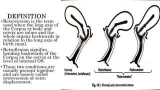
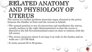
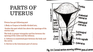
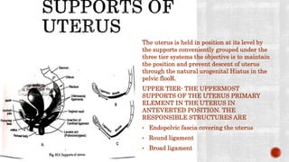
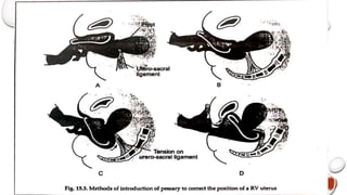
Susmita Haldar
Sister tutor
School of nursing Asia foundation
The uterus is not a fixed organ. Minor variations in
position in direction occur in constantly with changes
in poster with standing with full bladder or loaded
rectum. Only when the uterus rest habitually in a
position beyond the limit of normal variation it
should be called displacement of uterus.
Retroversion is present in about 15 to 20
person of normal women
 Retroversion is the term
used when the long axis of
the Corpus or body and
cervix are inline and the
whole organs backwards in
relation to the long axis of
birth canal.
 Retroflexion signifies
bending backwards of the
Corpus on the cervix at the
level of internal OS.
 These two conditions are
usually present together
and are loosely called
retroversion or retro
displacement.
The uterus is a hollow pyriform muscular organ situated in the pelvis
between the bladder in front and the rectum in behind
 The normal position is one of anteversion and anteflexion the uterus,
usually inclines to the right (dextrorotation) so that the cervix is
directed to the left (levorotation)and comes in close to relation with the
left ureter.
 The uterus measures about 8 cm long 5 cm wide at the fundus and its
walls are 1.25 CM thick
 It waits around 50 to 80 grams.
Uterus has got following part
1.Body or Corpus is furthEr divided into,
fundus the part which lies above the openings of the
uterine tube
The body is proper triangular and lies between the
opening of the tubes and the isthmus.
2. Isthmus is constricted part between the body and
the cervix
3. Service is the lowermost part of uterus
The uterus is held in position at its level by
the supports conveniently grouped under the
three tier systems the objective is to maintain
the position and prevent descent of uterus
through the natural urogenital Hiatus in the
pelvic flooR.
UPPER TIER- THE UPPERMOST
SUPPORTS OF THE UTERUS PRIMARY
ELEMENT IN THE UTERUS IN
ANTEVERTED POSITION. THE
RESPONSIBLE STRUCTURES ARE
• Endopelvic fascia covering the uterus
• Round ligament
• Broad ligament
 Middle tier constitutes the strongest support of uterus
1. Paracervical ring is the coller of fibroelastic connective tissue encircling the
supravaginal cervix. It is connected with the pubo cervical ligaments And physical
vaginal septum anteriorly cardinal ligaments laterally and Ritu vaginal septum
posteriorly. It’s stabilizer service at the level of interspinous diameter along the
other ligaments.
2. Pelvic cellular tissues and the in endopelvic fascia consists of connective tissue and
smooth muscles . the blood vessels nerves supplying the uterus bladder and vagina
pass through it from the lateral pelvic wall. As they passed the pelvic cellular
tissues condense surrounding them and give them direct support to the viscera.
Inferior tier give
indirect support to
the the Uterus. The
support is
principally given by
the pelvic floor
muscles.
Supports of anterior vaginal wall
1. Positional support in the erect foster the vagina makes an angle of 45 degree to
the horizontal. Normal vaginal axis is horizontal in the upper two third and
vertical in the lower third
2. the vagina is insured by strong condensation of pelvic cellular tissue called
endopelvic fascia.
Supports of posterior vaginal wall
1. Endopelvic fascia sheath covering the vagina and the rectuM.
2. Attachment of the uterus after ligament to the lateral wall of the vault
Conveniently 3° are described
1.First degree- the fundus is vertical and
pointing towards sacral promontory
2.Second degree-The fundus lies in the sacral
hollow but not below the internal os.
3.Third degree- the fun does lies below the level
of internal OS
Developmental
Retro displacement is quite
common in foetuses And young
children.
Due to developmental defect
there is lack of tone to uterine
muscles.
The infantile position is
retained.
This is often associated with
short vagina with shallow
anterior vaginal fornix.
Acquired
 Puerperal – the stretch ligaments caused by child
but fail to keep the uterus in its normal position. A
subinvoluted bulky uterus agree with this condition
 Prolapse- retroversion is usually implicated in the
pathophysiology of prolapse which is mechanically
caused by traction following cystocele
 Tumor- fibroid either in anterior or posterior wall
produces heaviness to uterus and hence it falls
behind
 Pelvic adhesions- additions either inflammatory
operative or due to pelvic endometriosis pull the
uterus posteriorly.
This condition is classified either as mobile
and fixed or uncomplicated and complicated
by pelvic diseases
Symptoms
Mobile retroverted uterus is quite common and almost remains asymptomatic. However following
symptoms can be seen
 Chronic premenstrual pelvic pain- is due to verikos cities in broad ligament produced by the
kinks.
 Backache
 Dyspareunia- deep dyspareunia may be due to direct thurst by the penis against the retroflexed
uterus for the prolapsed ovaries lying in the pouch of Douglas. Similar pain is reproduced by
pressing with examining fingers mein confirm its reality.
 Infertility- in third degree retroversion the external cost is away from the seminal pool at the
posterior fornix during coitus or it may be operated by the anterior vaginal wall. Associated
under development of uterus may also be our contributing factor.
The physician should however think twice before declaring the patient
to the the fact that the particular symptoms is related to backward
position of the womb. This applies specially to baccha chronic pelvic
pain or dyspareunia.
In such cases a Hodge Smith pessary may be placed inside for about 3
months after correcting the uterine position to anteversion.
If the symptoms disappear during the period and recover back after its
removal it may be concluded that the symptoms are due to retroverted
uterus.
This is known as PESSARY TEST.
Signs
1.Bimanual examination reveals
 The Cervix is directed upwards and forwards
 The body of the uterus is felt through the posterior fornix
 It is found continuous with the cervix and it moves when the cervix is
pressed up.
 The size of uterus is difficult to assess at times
2.Speculum examination reveals
 The cervix comes in view much easily and the external OS points forwards
3.Rectal examination
 Helps to confirm the diagnosis.
 Menstrual abnormalities like
 Menorrhagia
 congestive dysmenorrhea
 Chronic pelvic pain
 Dyspareunia
 When the uterus is fixed on mobile can be elicited by attempting to
replace 8 by moving the service backwards and pushing the fundus
upwards
 Rectal examination may be more effective
 Retroversion has got practically no adverse effect either
on fertility or early pregnancy wastage.
 In pregnancy the spontaneous correction usually occurs
by 12 to 14 weeks.
 Why the cause of infertility is mainly mechanical cause of
repeated pregnancy e waste May be due to disturbance in
uterine vascularity are due to thurst during intercourse
specially in abortion prone women.
1. A complete composite examination includes inspection and palpation of vagina
rectum after vaginal or even under anaesthesia may be required to arrive a
correct diagnosis
2. General examination details including BMI signs of myopathy on neuropathy
features of chronic airway disease or any abdominal mass should be done
3. Pelvic examination in both dorsal and standing positions
4. Bimanual examination
5. Speculum examination
6. Rectal examination
7. Pessary test to assess correct position.
Preventive:
The following guidelines are of help during the weeks after abortion or childbirth
1. To empty the bladder regularly
2. To increase the tone of pelvic muscles by regular exercise
3. To encourage lying in prone position for half to 1 hour once or twice daily between
2 to 4 weeks of postpartum period.
Corrective treatment:
PESSARY
Pessary less commonly used in present day gynaecological practice. This may be indicated
 For pessary test
 In subinvolution of uterus
 In pregnancy when spontaneous correction to anteversion fails by 12 week
Usually Hodge Smith pessary is used.
Pessary acts by stretching the uterosacral ligaments so as to pull the cervix backwards.
Surgical correction is indicated in
1.Cases where the ‘ pessary test’ is positive indicating that the symptoms are due to retroversion.
2.Fixed retroverted uterus producing symptoms like backache and dyspareunia
The principle of surgical correction is
 Ventrosuspension of uterus bye implicating the round ligament of both sides of extra peritoneal
early to the undersurface of anterior rectus sheath.
 This will pull the uterus forwards and maintains it permanently in the same position.
ANSWER THE FOLLOWING
QUESTIONS✍️
1.Fill in the blanks with suitable words. 1x4 = 4
1. Usually ____________ Pessary is used for treatment of retroverted uterus.
2. Surgical correction of retroverted uterus is ______________
3. Painful coitus is termed as _________________
4. In pregnancy spontaneous correction of position of uterus takes place by ________weeks.
2.State the following sentences are true or false. ½ x 2 =1
1. Emptying bladder prior to examination does not make any change in diagnosis.
2. Retroversion of uterus has got no adverse effect on fertility or early pregnancy.
3.Write Short note on Retroversion of uterus. ( Homework) 5
4 + 1+ 5 = 10
Thank you ❤️

Retroverted uterus

  • 1.
    Susmita Haldar Sister tutor Schoolof nursing Asia foundation
  • 2.
    The uterus isnot a fixed organ. Minor variations in position in direction occur in constantly with changes in poster with standing with full bladder or loaded rectum. Only when the uterus rest habitually in a position beyond the limit of normal variation it should be called displacement of uterus.
  • 3.
    Retroversion is presentin about 15 to 20 person of normal women
  • 4.
     Retroversion isthe term used when the long axis of the Corpus or body and cervix are inline and the whole organs backwards in relation to the long axis of birth canal.  Retroflexion signifies bending backwards of the Corpus on the cervix at the level of internal OS.  These two conditions are usually present together and are loosely called retroversion or retro displacement.
  • 5.
    The uterus isa hollow pyriform muscular organ situated in the pelvis between the bladder in front and the rectum in behind  The normal position is one of anteversion and anteflexion the uterus, usually inclines to the right (dextrorotation) so that the cervix is directed to the left (levorotation)and comes in close to relation with the left ureter.  The uterus measures about 8 cm long 5 cm wide at the fundus and its walls are 1.25 CM thick  It waits around 50 to 80 grams.
  • 6.
    Uterus has gotfollowing part 1.Body or Corpus is furthEr divided into, fundus the part which lies above the openings of the uterine tube The body is proper triangular and lies between the opening of the tubes and the isthmus. 2. Isthmus is constricted part between the body and the cervix 3. Service is the lowermost part of uterus
  • 7.
    The uterus isheld in position at its level by the supports conveniently grouped under the three tier systems the objective is to maintain the position and prevent descent of uterus through the natural urogenital Hiatus in the pelvic flooR. UPPER TIER- THE UPPERMOST SUPPORTS OF THE UTERUS PRIMARY ELEMENT IN THE UTERUS IN ANTEVERTED POSITION. THE RESPONSIBLE STRUCTURES ARE • Endopelvic fascia covering the uterus • Round ligament • Broad ligament
  • 8.
     Middle tierconstitutes the strongest support of uterus 1. Paracervical ring is the coller of fibroelastic connective tissue encircling the supravaginal cervix. It is connected with the pubo cervical ligaments And physical vaginal septum anteriorly cardinal ligaments laterally and Ritu vaginal septum posteriorly. It’s stabilizer service at the level of interspinous diameter along the other ligaments. 2. Pelvic cellular tissues and the in endopelvic fascia consists of connective tissue and smooth muscles . the blood vessels nerves supplying the uterus bladder and vagina pass through it from the lateral pelvic wall. As they passed the pelvic cellular tissues condense surrounding them and give them direct support to the viscera.
  • 9.
    Inferior tier give indirectsupport to the the Uterus. The support is principally given by the pelvic floor muscles.
  • 10.
    Supports of anteriorvaginal wall 1. Positional support in the erect foster the vagina makes an angle of 45 degree to the horizontal. Normal vaginal axis is horizontal in the upper two third and vertical in the lower third 2. the vagina is insured by strong condensation of pelvic cellular tissue called endopelvic fascia. Supports of posterior vaginal wall 1. Endopelvic fascia sheath covering the vagina and the rectuM. 2. Attachment of the uterus after ligament to the lateral wall of the vault
  • 11.
    Conveniently 3° aredescribed 1.First degree- the fundus is vertical and pointing towards sacral promontory 2.Second degree-The fundus lies in the sacral hollow but not below the internal os. 3.Third degree- the fun does lies below the level of internal OS
  • 13.
    Developmental Retro displacement isquite common in foetuses And young children. Due to developmental defect there is lack of tone to uterine muscles. The infantile position is retained. This is often associated with short vagina with shallow anterior vaginal fornix. Acquired  Puerperal – the stretch ligaments caused by child but fail to keep the uterus in its normal position. A subinvoluted bulky uterus agree with this condition  Prolapse- retroversion is usually implicated in the pathophysiology of prolapse which is mechanically caused by traction following cystocele  Tumor- fibroid either in anterior or posterior wall produces heaviness to uterus and hence it falls behind  Pelvic adhesions- additions either inflammatory operative or due to pelvic endometriosis pull the uterus posteriorly.
  • 14.
    This condition isclassified either as mobile and fixed or uncomplicated and complicated by pelvic diseases
  • 15.
    Symptoms Mobile retroverted uterusis quite common and almost remains asymptomatic. However following symptoms can be seen  Chronic premenstrual pelvic pain- is due to verikos cities in broad ligament produced by the kinks.  Backache  Dyspareunia- deep dyspareunia may be due to direct thurst by the penis against the retroflexed uterus for the prolapsed ovaries lying in the pouch of Douglas. Similar pain is reproduced by pressing with examining fingers mein confirm its reality.  Infertility- in third degree retroversion the external cost is away from the seminal pool at the posterior fornix during coitus or it may be operated by the anterior vaginal wall. Associated under development of uterus may also be our contributing factor.
  • 16.
    The physician shouldhowever think twice before declaring the patient to the the fact that the particular symptoms is related to backward position of the womb. This applies specially to baccha chronic pelvic pain or dyspareunia. In such cases a Hodge Smith pessary may be placed inside for about 3 months after correcting the uterine position to anteversion. If the symptoms disappear during the period and recover back after its removal it may be concluded that the symptoms are due to retroverted uterus. This is known as PESSARY TEST.
  • 17.
    Signs 1.Bimanual examination reveals The Cervix is directed upwards and forwards  The body of the uterus is felt through the posterior fornix  It is found continuous with the cervix and it moves when the cervix is pressed up.  The size of uterus is difficult to assess at times 2.Speculum examination reveals  The cervix comes in view much easily and the external OS points forwards 3.Rectal examination  Helps to confirm the diagnosis.
  • 18.
     Menstrual abnormalitieslike  Menorrhagia  congestive dysmenorrhea  Chronic pelvic pain  Dyspareunia  When the uterus is fixed on mobile can be elicited by attempting to replace 8 by moving the service backwards and pushing the fundus upwards  Rectal examination may be more effective
  • 19.
     Retroversion hasgot practically no adverse effect either on fertility or early pregnancy wastage.  In pregnancy the spontaneous correction usually occurs by 12 to 14 weeks.  Why the cause of infertility is mainly mechanical cause of repeated pregnancy e waste May be due to disturbance in uterine vascularity are due to thurst during intercourse specially in abortion prone women.
  • 20.
    1. A completecomposite examination includes inspection and palpation of vagina rectum after vaginal or even under anaesthesia may be required to arrive a correct diagnosis 2. General examination details including BMI signs of myopathy on neuropathy features of chronic airway disease or any abdominal mass should be done 3. Pelvic examination in both dorsal and standing positions 4. Bimanual examination 5. Speculum examination 6. Rectal examination 7. Pessary test to assess correct position.
  • 21.
    Preventive: The following guidelinesare of help during the weeks after abortion or childbirth 1. To empty the bladder regularly 2. To increase the tone of pelvic muscles by regular exercise 3. To encourage lying in prone position for half to 1 hour once or twice daily between 2 to 4 weeks of postpartum period.
  • 22.
    Corrective treatment: PESSARY Pessary lesscommonly used in present day gynaecological practice. This may be indicated  For pessary test  In subinvolution of uterus  In pregnancy when spontaneous correction to anteversion fails by 12 week Usually Hodge Smith pessary is used. Pessary acts by stretching the uterosacral ligaments so as to pull the cervix backwards.
  • 24.
    Surgical correction isindicated in 1.Cases where the ‘ pessary test’ is positive indicating that the symptoms are due to retroversion. 2.Fixed retroverted uterus producing symptoms like backache and dyspareunia The principle of surgical correction is  Ventrosuspension of uterus bye implicating the round ligament of both sides of extra peritoneal early to the undersurface of anterior rectus sheath.  This will pull the uterus forwards and maintains it permanently in the same position.
  • 25.
    ANSWER THE FOLLOWING QUESTIONS✍️ 1.Fillin the blanks with suitable words. 1x4 = 4 1. Usually ____________ Pessary is used for treatment of retroverted uterus. 2. Surgical correction of retroverted uterus is ______________ 3. Painful coitus is termed as _________________ 4. In pregnancy spontaneous correction of position of uterus takes place by ________weeks. 2.State the following sentences are true or false. ½ x 2 =1 1. Emptying bladder prior to examination does not make any change in diagnosis. 2. Retroversion of uterus has got no adverse effect on fertility or early pregnancy. 3.Write Short note on Retroversion of uterus. ( Homework) 5 4 + 1+ 5 = 10
  • 26.