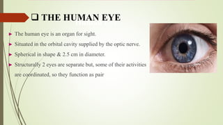
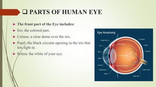
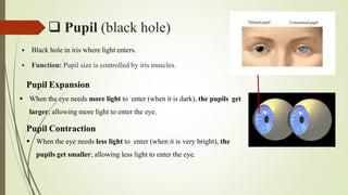
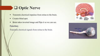
STRUCTURE
&
FUNCTION OF THE EYE
Prepared by
Mr. Vinayak Raju Bodhankar
M. Pharm. (QA)
Human Eye
► The human eye is an organ for sight.
► Situated in the orbital cavity supplied by the optic nerve.
► Spherical in shape & 2.5 cm in diameter.
► Structurally 2 eyes are separate but, some of their activities
are coordinated, so they function as pair
 THE HUMAN EYE
 STRUCTURE
There are 3 layers of tissue in the wall of eye
1. Outer fibrous layer: Sclera & cornea
2. Middle vascular layer: Choroid, ciliary body & iris
3. Inner nervous tissue layer: Retina
 PARTS OF HUMAN EYE
► The front part of the Eye includes:
► Iris: the colored part.
► Cornea: a clear dome over the iris.
► Pupil: the black circular opening in the iris that
lets light in.
► Sclera: the white of your eye.
 SCLERA & CORNEA
CORNEA
 Transparent covering of the front of the eye.
 Allows for the passage of light into the eye and functions as a fixed lens.
Function:
Allows for the passage of light into the eye and it also focuses the light on retina.
SCLERA
 A tough white skin (made of tissue) that covers all of the eyeball except the cornea.
Function: Supports eyeball and provides attachment for muscles.
 Iris, Choroid & Ciliary body
IRIS
 Colored part of eye.
 Lying behind the cornea in front of lens.
 It is a circular body composed of pigment cells & two layers of smooth muscle fibres.
 Controls the amount of light entering the eye.
CHOROID
 Lines the inner surface of sclera.
 Rich in blood vessel & is deep chocolate brown in colour.
 Light enters the eye through pupil, stimulate the nerve endings in retina & then absorbed by choroid.
CILIARY BODY
 It is the anterior continuation of the choroid.
 Consist of ciliary muscle & secretory epithelial cells.
 Contraction & relaxation of ciliary muscle changes the thickness of the lens.
 Retina
 Innermost layer of the wall of the eye.
 Layer is highly sensitive to the light.
 Contains light-receptive cells (rods and cones)
 Converts light to electrical signals
 Pupil (black hole)
 Black hole in iris where light enters.
 Function: Pupil size is controlled by iris muscles.
Pupil Expansion
 When the eye needs more light to enter (when it is dark), the pupils get
larger; allowing more light to enter the eye.
Pupil Contraction
 When the eye needs less light to enter (when it is very bright), the
pupils get smaller; allowing less light to enter the eye.
 Optic Nerve
 Transmits electrical impulses from retina to the brain.
 Creates blind spot.
 Brain takes inverted image and flips it so we can see.
Function:
Transmits electrical signals from retina to the brain.
 Functions
► The eye has many parts that must work together to produce clear vision:
► The sclera, or white part of the eye, protects the eyeball.
► The pupil, or black dot at the center of the eye, is a hole through which light can
enter the eye.
► The iris, or colored part of the eye, surrounds the pupil.

Human Eye

  • 1.
    STRUCTURE & FUNCTION OF THEEYE Prepared by Mr. Vinayak Raju Bodhankar M. Pharm. (QA)
  • 2.
    Human Eye ► Thehuman eye is an organ for sight. ► Situated in the orbital cavity supplied by the optic nerve. ► Spherical in shape & 2.5 cm in diameter. ► Structurally 2 eyes are separate but, some of their activities are coordinated, so they function as pair  THE HUMAN EYE
  • 3.
     STRUCTURE There are3 layers of tissue in the wall of eye 1. Outer fibrous layer: Sclera & cornea 2. Middle vascular layer: Choroid, ciliary body & iris 3. Inner nervous tissue layer: Retina
  • 4.
     PARTS OFHUMAN EYE ► The front part of the Eye includes: ► Iris: the colored part. ► Cornea: a clear dome over the iris. ► Pupil: the black circular opening in the iris that lets light in. ► Sclera: the white of your eye.
  • 5.
     SCLERA &CORNEA CORNEA  Transparent covering of the front of the eye.  Allows for the passage of light into the eye and functions as a fixed lens. Function: Allows for the passage of light into the eye and it also focuses the light on retina. SCLERA  A tough white skin (made of tissue) that covers all of the eyeball except the cornea. Function: Supports eyeball and provides attachment for muscles.
  • 6.
     Iris, Choroid& Ciliary body IRIS  Colored part of eye.  Lying behind the cornea in front of lens.  It is a circular body composed of pigment cells & two layers of smooth muscle fibres.  Controls the amount of light entering the eye. CHOROID  Lines the inner surface of sclera.  Rich in blood vessel & is deep chocolate brown in colour.  Light enters the eye through pupil, stimulate the nerve endings in retina & then absorbed by choroid. CILIARY BODY  It is the anterior continuation of the choroid.  Consist of ciliary muscle & secretory epithelial cells.  Contraction & relaxation of ciliary muscle changes the thickness of the lens.
  • 7.
     Retina  Innermostlayer of the wall of the eye.  Layer is highly sensitive to the light.  Contains light-receptive cells (rods and cones)  Converts light to electrical signals
  • 8.
     Pupil (blackhole)  Black hole in iris where light enters.  Function: Pupil size is controlled by iris muscles. Pupil Expansion  When the eye needs more light to enter (when it is dark), the pupils get larger; allowing more light to enter the eye. Pupil Contraction  When the eye needs less light to enter (when it is very bright), the pupils get smaller; allowing less light to enter the eye.
  • 9.
     Optic Nerve Transmits electrical impulses from retina to the brain.  Creates blind spot.  Brain takes inverted image and flips it so we can see. Function: Transmits electrical signals from retina to the brain.
  • 10.
     Functions ► Theeye has many parts that must work together to produce clear vision: ► The sclera, or white part of the eye, protects the eyeball. ► The pupil, or black dot at the center of the eye, is a hole through which light can enter the eye. ► The iris, or colored part of the eye, surrounds the pupil.