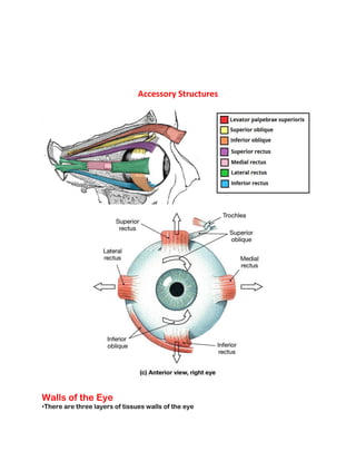
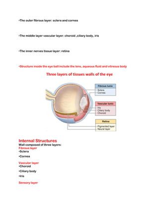
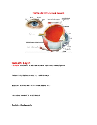
The Eye & Vision
SHAHINA BANO
Objectives
•At the end of this session students will be able to:
•Describe eye in terms of:
•Structure related to vision
• Accessory structures
•Discuss image formation by describing refraction, accommodation and constriction of the pupil.
•Explain the process of dark adaptation and light adaptation.
Vision
•Vision is extremely important to human survival, More than half the sensory receptors in the human
body are located in the eyes.
• A large part of cerebral cortex is devoted to processing visual information.
Vision
•Vision is extremely important to human survival, More than half the sensory receptors in the human
body are located in the eyes.
• A large part of cerebral cortex is devoted to processing visual information.
Accessory Structures of the Eye
•Eyelidsshade the eye during sleep, protect the eye from excessive light and spreading the
lubrication over the eye
•Eye browshelp to protect eye ball from foreign substance
•Eye lashesproject from the border of each eyelid
•Lacrimal(tearing)apparatus produces and drains tear
•Extrinsic eye musclesattached to the outside of the eyeball and help the eyes to move
in all directions of sight
Tear Gland
Lacrimal glands produce tears Tears are dilute salt solution Contain lysozyme to kill bacteria Drain into
nasal cavity
Accessory Structures
Walls of the Eye
•There are three layers of tissues walls of the eye
•The outer fibrous layer: sclera and cornea
•The middle layer vascular layer: choroid ,ciliary body, iris
•The inner nerves tissue layer: retina
•Structure inside the eye ball include the lens, aqueous fluid and vitreous body
Three layers of tissues walls of the eye
Internal Structures
Wall composed of three layers:
Fibrous layer
•Sclera
•Cornea
Vascular layer
•Choroid
•Ciliary body
•Iris
Sensory layer
•Retina
•Pigmented layer (prevent light from scattering)
•Neural layer (photoreceptors)
Fibrous Layer
•Sclera: thick, white connective tissue (“white of the eye”)
•For protection & attachment.
•Posterior and laterally of the eye ball and is continuous anteriorly with transparent
cornea.
•Maintain the shape of eye.
•Anteriorly sclera continues as a clear transparent epithelial membrane.
Fibrous Layer
•Cornea: clear “window” through which light enters eye
•Anterior portion of eye.
•many nerve endings (pain fibers), easily repairs itself.
•Avascular, so only tissue in the body that can be transplanted without fear of rejection.
•Helps focus entering light rays.
•Connective tissue with thin layer of epithelium; unusually regular fiber pattern.
Fibrous Layer Sclera & Cornea
Vascular Layer
•Choroid: blood-rich nutritive tunic that contains a dark pigment
•Prevents light from scattering inside the eye
•Modified anteriorly to form ciliary body & iris
•Produces melanin to absorb light
•Contains blood vessels
Vascular Layer
•Ciliary body: Two smooth muscle structures to which the lens is attached with the
ciliary zonula (ligament).
•Forms internal ring around front of eye (muscle fibers & ligaments).
•Iris: pigmented, has an opening called the pupil through which light passes.
Choroid
Ciliary Body
Sensory Layer Retina
•Retina : two layers which extends anteriorly only to the ciliary body.
•Composed of pigment cells that absorb light and prevent light from scattering inside
the light.
•Act as phagocytes to remove dead or damaged receptor cells.
•Store vitamin A needed for vision.
Sensory Layer Retina
•Neural layer: contains millions of receptor cells.
•Photoreceptors (rods & cones): respond to light, bipolar neurons.
•70% of sensory receptors are in the eyes.
•Rods: more sensitive in low light, gives general outline, seen as black and white, 120
millions
•Cones: less sensitive in low light, sharp picture, color , 6 millions
Sensory Layer Retina
•Electrical impulse leaves the retina via the optic nerve & nerve impulses are
transmitted to the optic cortex which results in vision.
•Fovea: all cones, sharpest vision (visual acuity).
•Optic disc (“blind spot”) – where optic nerve leaves eyeball.
Retina
Sensory Layer Retina
•Night blindness: fewer working rods
(lack of vitamin A).
•Day blindness: lack of working cones.
Lens
•Lies directly behind iris & pupil.
•Focuses light entering the eye on the retina (changes shape to focus).
•Held upright in the eye by a suspensory ligament (ciliary zonule) attached to ciliary
body.
•Epithelial cells (cytoplasm is transparent part)
Lens
Aqueous humor
•Anterior to the lens, clear watery fluid
•Fills space between cornea and lens
•Maintain shape of eye ball and supply oxygen and nutrient to lens and cornea
Vitreous humor
•Reinforces
•eyeball internally
•Posterior to lens
•Maintain shape of eye ball and keeps the retina attached to the choroid
So how do you actually see?
•Retina – receives and translates light message into nerve impulses
•Contains photoreceptors called rods and cones
•Messages leave photoreceptors and pass to optic nerve, then to occipital lobe of brain
for translation.
•Fovea Centralis- Region of the retina that produces the sharpest
Blind Spot
•Optic nerve is covered by photoreceptors
•Except for optic disc (where optic nerve leaves eyeball)
Physiology of Vision
•Light rays are bent (refracted) as light encounters the cornea, aqueous humor, lens,
and vitreous humor.
•Refractory power of the lens can be changed by changing its shape (ciliary body
controls shape of lens).
•Accommodation: Ability of eye to focus for close objects (less than 20 ft. away)
•Image formed on retina is a real image (inverted).
Dark Adaptation
•Dark Adaptation is the process by which our eyes adjust to darkness after being
exposed to light.
•For example, when we move from a bright, sunny area outside to a relatively dark room
inside, it is difficult to see at first.
•Gradually our eyes recover and become more sensitive to the dim light indoors.
References
Ross and Wilson Anatomy and physiology in Health and illness Edition 11th pg. 190 to
199
Tortora J.G, Derkison, B. principle of Anatomy and physiology (11th Ed) USA john Willey & Sons, inc

The Eye.pdf

  • 1.
    The Eye &Vision SHAHINA BANO Objectives •At the end of this session students will be able to: •Describe eye in terms of: •Structure related to vision • Accessory structures •Discuss image formation by describing refraction, accommodation and constriction of the pupil. •Explain the process of dark adaptation and light adaptation. Vision •Vision is extremely important to human survival, More than half the sensory receptors in the human body are located in the eyes. • A large part of cerebral cortex is devoted to processing visual information. Vision •Vision is extremely important to human survival, More than half the sensory receptors in the human body are located in the eyes. • A large part of cerebral cortex is devoted to processing visual information. Accessory Structures of the Eye
  • 2.
    •Eyelidsshade the eyeduring sleep, protect the eye from excessive light and spreading the lubrication over the eye •Eye browshelp to protect eye ball from foreign substance •Eye lashesproject from the border of each eyelid •Lacrimal(tearing)apparatus produces and drains tear •Extrinsic eye musclesattached to the outside of the eyeball and help the eyes to move in all directions of sight Tear Gland Lacrimal glands produce tears Tears are dilute salt solution Contain lysozyme to kill bacteria Drain into nasal cavity
  • 3.
    Accessory Structures Walls ofthe Eye •There are three layers of tissues walls of the eye
  • 4.
    •The outer fibrouslayer: sclera and cornea •The middle layer vascular layer: choroid ,ciliary body, iris •The inner nerves tissue layer: retina •Structure inside the eye ball include the lens, aqueous fluid and vitreous body Three layers of tissues walls of the eye Internal Structures Wall composed of three layers: Fibrous layer •Sclera •Cornea Vascular layer •Choroid •Ciliary body •Iris Sensory layer
  • 5.
    •Retina •Pigmented layer (preventlight from scattering) •Neural layer (photoreceptors) Fibrous Layer •Sclera: thick, white connective tissue (“white of the eye”) •For protection & attachment. •Posterior and laterally of the eye ball and is continuous anteriorly with transparent cornea. •Maintain the shape of eye. •Anteriorly sclera continues as a clear transparent epithelial membrane. Fibrous Layer •Cornea: clear “window” through which light enters eye •Anterior portion of eye. •many nerve endings (pain fibers), easily repairs itself. •Avascular, so only tissue in the body that can be transplanted without fear of rejection. •Helps focus entering light rays. •Connective tissue with thin layer of epithelium; unusually regular fiber pattern.
  • 6.
    Fibrous Layer Sclera& Cornea Vascular Layer •Choroid: blood-rich nutritive tunic that contains a dark pigment •Prevents light from scattering inside the eye •Modified anteriorly to form ciliary body & iris •Produces melanin to absorb light •Contains blood vessels
  • 7.
    Vascular Layer •Ciliary body:Two smooth muscle structures to which the lens is attached with the ciliary zonula (ligament). •Forms internal ring around front of eye (muscle fibers & ligaments). •Iris: pigmented, has an opening called the pupil through which light passes. Choroid
  • 8.
    Ciliary Body Sensory LayerRetina •Retina : two layers which extends anteriorly only to the ciliary body. •Composed of pigment cells that absorb light and prevent light from scattering inside the light. •Act as phagocytes to remove dead or damaged receptor cells. •Store vitamin A needed for vision. Sensory Layer Retina •Neural layer: contains millions of receptor cells. •Photoreceptors (rods & cones): respond to light, bipolar neurons. •70% of sensory receptors are in the eyes.
  • 9.
    •Rods: more sensitivein low light, gives general outline, seen as black and white, 120 millions •Cones: less sensitive in low light, sharp picture, color , 6 millions Sensory Layer Retina •Electrical impulse leaves the retina via the optic nerve & nerve impulses are transmitted to the optic cortex which results in vision. •Fovea: all cones, sharpest vision (visual acuity). •Optic disc (“blind spot”) – where optic nerve leaves eyeball.
  • 10.
    Retina Sensory Layer Retina •Nightblindness: fewer working rods (lack of vitamin A). •Day blindness: lack of working cones. Lens •Lies directly behind iris & pupil. •Focuses light entering the eye on the retina (changes shape to focus). •Held upright in the eye by a suspensory ligament (ciliary zonule) attached to ciliary body. •Epithelial cells (cytoplasm is transparent part)
  • 11.
    Lens Aqueous humor •Anterior tothe lens, clear watery fluid •Fills space between cornea and lens •Maintain shape of eye ball and supply oxygen and nutrient to lens and cornea Vitreous humor •Reinforces •eyeball internally •Posterior to lens •Maintain shape of eye ball and keeps the retina attached to the choroid So how do you actually see? •Retina – receives and translates light message into nerve impulses •Contains photoreceptors called rods and cones •Messages leave photoreceptors and pass to optic nerve, then to occipital lobe of brain for translation. •Fovea Centralis- Region of the retina that produces the sharpest
  • 12.
    Blind Spot •Optic nerveis covered by photoreceptors •Except for optic disc (where optic nerve leaves eyeball) Physiology of Vision •Light rays are bent (refracted) as light encounters the cornea, aqueous humor, lens, and vitreous humor. •Refractory power of the lens can be changed by changing its shape (ciliary body controls shape of lens). •Accommodation: Ability of eye to focus for close objects (less than 20 ft. away) •Image formed on retina is a real image (inverted).
  • 13.
    Dark Adaptation •Dark Adaptationis the process by which our eyes adjust to darkness after being exposed to light.
  • 14.
    •For example, whenwe move from a bright, sunny area outside to a relatively dark room inside, it is difficult to see at first. •Gradually our eyes recover and become more sensitive to the dim light indoors. References Ross and Wilson Anatomy and physiology in Health and illness Edition 11th pg. 190 to 199 Tortora J.G, Derkison, B. principle of Anatomy and physiology (11th Ed) USA john Willey & Sons, inc