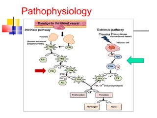
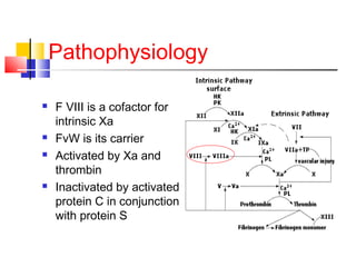
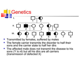
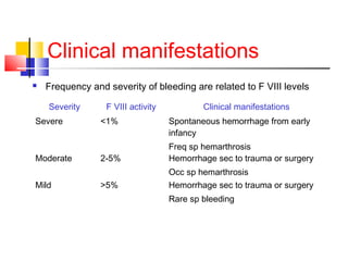
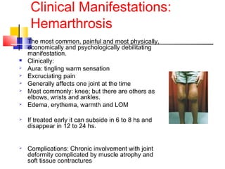
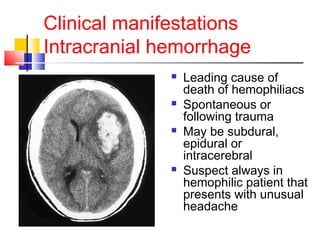
Hemophilia:
The Royal Disease
Dr Ramya
Department of pediatrics
Definition
 Hemophilia- “love of bleeding”
 2 types: A and B
 Hemophilia A: X linked recessive
hereditary disorder that is due to
defective or deficient factor VIII
 Hemophilia B: also X linked inherited
disorder due to deficiency of factor IX
Incidence
 It is the second most common inherited
clotting factor abnormality (after von
Willebrand disease)
 1 in 5000-10000 live male births
 No difference between racial groups
Pathophysiology
Pathophysiology
 F VIII is a cofactor for
intrinsic Xa
 FvW is its carrier
 Activated by Xa and
thrombin
 Inactivated by activated
protein C in conjunction
with protein S
Genetics
 Transmitted by females, suffered by males
 The female carrier transmits the disorder to half their
sons and the carrier state to half her dtrs
 The affected male does not transmit the disease to his
sons (Y is nl) but all his dtrs are all carriers
(transmission of defected X)
Genetics
 In 1/3 of hemophiliac patients, there is
no family history of bleeding.
Clinical manifestations
 Frequency and severity of bleeding are related to F VIII levels
Severity F VIII activity Clinical manifestations
Severe <1% Spontaneous hemorrhage from early
infancy
Freq sp hemarthrosis
Moderate 2-5% Hemorrhage sec to trauma or surgery
Occ sp hemarthrosis
Mild >5% Hemorrhage sec to trauma or surgery
Rare sp bleeding
Clinical Manifestations:
Hemarthrosis
 The most common, painful and most physically,
economically and psychologically debilitating
manifestation.
 Clinically:
 Aura: tingling warm sensation
 Excruciating pain
 Generally affects one joint at the time
 Most commonly: knee; but there are others as
elbows, wrists and ankles.
 Edema, erythema, warmth and LOM
 If treated early it can subside in 6 to 8 hs and
disappear in 12 to 24 hs.
 Complications: Chronic involvement with joint
deformity complicated by muscle atrophy and
soft tissue contractures
Clinical Manifestations:
Hemarthrosis
 Pathophysiology:
 Bleeding probably starts from synovial vessels into
the synovial space.
 Reabsorption of this blood is often incomplete leading
to chronic proliferative synovitis, where the synovium
is more thickened and vascular, creating a “target
joint” with recurrence of bleeding.
 There is destruction of surrounding structures as
well-bone necrosis and cyst formations, osteophytes
 Terminal stage: Chronic Hemophiliac arthropathy:
fibrous or bony ankilosing of the joint.
Clinical Manifestations
Hematomas
 Subcutaneous and muscular
hematomas spread within
fascial spaces
 May compress vital structures:
 Muscle hematomas:
1)calf,2)thigh,3)buttocks,4)fore
arms
 Psoas hematoma- if right
sided may mimic acute
appendicitis
 Retroperitoneal hematoma:
Clinical manifestations
Intracranial hemorrhage
 Leading cause of
death of hemophiliacs
 Spontaneous or
following trauma
 May be subdural,
epidural or
intracerebral
 Suspect always in
hemophilic patient that
presents with unusual
headache
Clinical manifestations
Others
 Gastrointestinal Bleeding:
 Mucous Bleeding:
Epistaxis, gum bleeding.
 Genitourinary Bleeding:
Frequently severe hemophiliac can experience
hematuria and a structural lesion should be ruled out.
Laboratory diagnosis
 Deficit can be quantitative or qualitative
 General Lab: prolonged aPTT, nl PT and BT
 Mixing studies: aPTT corrects with normal
plasma
 Clotting assays: F VIII activity, expressed in
% of normal DecreasedQUANTITATIVE
 Immunoassays: “Cross Reactive Material”
Positive- there is an antigen similar to the F
VIII protein- QUALITATIVE
Differential Diagnosis
 Clinically impossible to differentiate from Hemophilia
B- FIX def- Christmas’ disease
 Type 2N vWD
 Affected patients present with low levels of factor VIII
(usually 5 to 15 percent of normal
 Should be suspected in families in which an
autosomal recessive (rather than X-linked)
inheritance pattern is seen.
Carrier detection and
Antenatal diagnosis
 Family history: if we follow the inheritance
pattern a female is a carrier if she:
 Has an hemophilic father
 Has two hemophilic sons
 Has one hemophilic son and has a family
history
 Has a son but no family history, there is a
67% chance that she is.
Carrier detection and
Antenatal diagnosis
 Coagulation based assays:
Generally heterozygous females have <50% f VIII
levels but if normal it can’t be excluded
 DNA based assays:
Southern blot can detect the inversion in intron 22
If negative for that, there is the need for DNA
sequencing
For prenatal diagnosis: DNA testing on choronic villi
samples obtained by biopsy at week 12
Treatment
General Considerations
 Avoidance of aspirin and
NSAIDs if at all possible
sometimes it is difficult
because of the painful
hemarthrosis
 No IM injections
 Counseling for patient and
family, both genetic and
psychosocial, encouraging
normal socialization
Treatment
Factor replacement
Replacement of F VIII/ IX is the cardinal step to
prevent or reverse acute bleeding episodes
 Dosing: Replacement products can be given on the
basis of body weight or plasma volume ( aprox 5% of
body weight)
 1 U/ml = 100% factor activity
 In a severe hemophiliac, to raise F VIII to 100%
activity or 1 U/ml, we need 50 U/kg
Treatment
Factor replacement
Sources of F VIII
 Plasma
• FFP was used as the only replacement therapy until
1960s.
• Not really much effective since it could only raise f
VIII to 20%, by giving the patient many liters
• Patients used to have to spend most of their time in
the hospital
Treatment
Factor replacement
 Cryoprecipitate
• By mid 1960s
• cold insoluble material obtained from plasma
contained high levels of F VIII and fibrinogen,
achieving a major advance in hemophilia treatment
• 1 unit of FFP prepared by cryoprecipitate contains
50-120 U of VIII
 Plasma Derived f VIII prepared by monoclonal
antibodies.
Treatment
Others
 Antifibrinolyitic Agents

Epsilon aminocaproic acid

Inhibit fibrinolysis by inhibiting plasminogen activator

Adjuvant therapy for dental procedures

Contraindicated in hematuria
 Desmopressin

Transient increase in F VIII levels in pts with mild
hemophilia(2-4 times above baseline)

Mechanism: release from endothelial storage sites

Repeated administration results in a diminished
response- tachyphylaxis

Side effects: hyponatremia, facial flushing and headache
Course and prognosis
 Replacement therapy has its
complications and includes:
 Development of F VIII antibodies
 Liver disease resulting from hepatitis B
and C
 Infection with HIV
So…
WHY IS IT CALLED
THE ROYAL
DISEASE?!!?
History
Why the Royal disease?
 This is because Queen Victoria, Queen of England from 1837 to
1901, was a carrier.
 Most likely a spontaneous mutation since the duke of Kent (her
father) was not affected and her mother did not have any
affected children from the previous marriage.
 Her eighth child, Leopold, had hemophilia and suffered from
frequent hemorrhages. These were reported in the British
Medical Journal in 1868.
 Leopold died of a brain hemorrhage at the age of 31, but not
before he had children. His daughter, Alice, was a carrier and
her son, Viscount Trematon, also died of a brain hemorrhage in
1928.
 The British family descends from Victoria’s first child Edward
who was not affected. Hence this house is disease free.
History
Why the Royal disease?
 Beatrice, Victoria’s youngest child had two
hemophilic sons and a daughter- Victoria
Eugene that was a carrier
 She introduced the hemophilia gene into the
Spanish royal family by marrying king Alfonso
XIII.
 By this time, Queen Victoria’s blood was
recognized as “defective” and the king may
have been warned about Eugene’s carrier
state. However, Royalty was more important
than X chromosomes.
History
Why the Royal disease?
 Alexandra, Queen Victoria's granddaughter, married
Nicholas, the Tsar of Russia in the early 1900's.
 Alexandra, the Tsarina, was a carrier of hemophilia
and her first son, the Tsarevich Alexei, was an
hemophiliac
 The monk Rasputin gained great influence in the
Russian court, partly because he was the only one
able to help the young Tsarevich. He used hypnosis
to relieve Alexei's pain.
 It is speculated that the illness of the heir to the
throne, the strain it placed on the Royal family, and
the influence of the corrupt and alcoholic monk
Rasputin were all factors leading to the Russian
Revolution of 1917.
THANK YOU!

Hemophilia (a) - Pediatrics

  • 1.
    Hemophilia: The Royal Disease DrRamya Department of pediatrics
  • 2.
    Definition  Hemophilia- “loveof bleeding”  2 types: A and B  Hemophilia A: X linked recessive hereditary disorder that is due to defective or deficient factor VIII  Hemophilia B: also X linked inherited disorder due to deficiency of factor IX
  • 3.
    Incidence  It isthe second most common inherited clotting factor abnormality (after von Willebrand disease)  1 in 5000-10000 live male births  No difference between racial groups
  • 4.
  • 5.
    Pathophysiology  F VIIIis a cofactor for intrinsic Xa  FvW is its carrier  Activated by Xa and thrombin  Inactivated by activated protein C in conjunction with protein S
  • 6.
    Genetics  Transmitted byfemales, suffered by males  The female carrier transmits the disorder to half their sons and the carrier state to half her dtrs  The affected male does not transmit the disease to his sons (Y is nl) but all his dtrs are all carriers (transmission of defected X)
  • 7.
    Genetics  In 1/3of hemophiliac patients, there is no family history of bleeding.
  • 8.
    Clinical manifestations  Frequencyand severity of bleeding are related to F VIII levels Severity F VIII activity Clinical manifestations Severe <1% Spontaneous hemorrhage from early infancy Freq sp hemarthrosis Moderate 2-5% Hemorrhage sec to trauma or surgery Occ sp hemarthrosis Mild >5% Hemorrhage sec to trauma or surgery Rare sp bleeding
  • 9.
    Clinical Manifestations: Hemarthrosis  Themost common, painful and most physically, economically and psychologically debilitating manifestation.  Clinically:  Aura: tingling warm sensation  Excruciating pain  Generally affects one joint at the time  Most commonly: knee; but there are others as elbows, wrists and ankles.  Edema, erythema, warmth and LOM  If treated early it can subside in 6 to 8 hs and disappear in 12 to 24 hs.  Complications: Chronic involvement with joint deformity complicated by muscle atrophy and soft tissue contractures
  • 10.
    Clinical Manifestations: Hemarthrosis  Pathophysiology: Bleeding probably starts from synovial vessels into the synovial space.  Reabsorption of this blood is often incomplete leading to chronic proliferative synovitis, where the synovium is more thickened and vascular, creating a “target joint” with recurrence of bleeding.  There is destruction of surrounding structures as well-bone necrosis and cyst formations, osteophytes  Terminal stage: Chronic Hemophiliac arthropathy: fibrous or bony ankilosing of the joint.
  • 11.
    Clinical Manifestations Hematomas  Subcutaneousand muscular hematomas spread within fascial spaces  May compress vital structures:  Muscle hematomas: 1)calf,2)thigh,3)buttocks,4)fore arms  Psoas hematoma- if right sided may mimic acute appendicitis  Retroperitoneal hematoma:
  • 12.
    Clinical manifestations Intracranial hemorrhage Leading cause of death of hemophiliacs  Spontaneous or following trauma  May be subdural, epidural or intracerebral  Suspect always in hemophilic patient that presents with unusual headache
  • 13.
    Clinical manifestations Others  GastrointestinalBleeding:  Mucous Bleeding: Epistaxis, gum bleeding.  Genitourinary Bleeding: Frequently severe hemophiliac can experience hematuria and a structural lesion should be ruled out.
  • 14.
    Laboratory diagnosis  Deficitcan be quantitative or qualitative  General Lab: prolonged aPTT, nl PT and BT  Mixing studies: aPTT corrects with normal plasma  Clotting assays: F VIII activity, expressed in % of normal DecreasedQUANTITATIVE  Immunoassays: “Cross Reactive Material” Positive- there is an antigen similar to the F VIII protein- QUALITATIVE
  • 15.
    Differential Diagnosis  Clinicallyimpossible to differentiate from Hemophilia B- FIX def- Christmas’ disease  Type 2N vWD  Affected patients present with low levels of factor VIII (usually 5 to 15 percent of normal  Should be suspected in families in which an autosomal recessive (rather than X-linked) inheritance pattern is seen.
  • 16.
    Carrier detection and Antenataldiagnosis  Family history: if we follow the inheritance pattern a female is a carrier if she:  Has an hemophilic father  Has two hemophilic sons  Has one hemophilic son and has a family history  Has a son but no family history, there is a 67% chance that she is.
  • 17.
    Carrier detection and Antenataldiagnosis  Coagulation based assays: Generally heterozygous females have <50% f VIII levels but if normal it can’t be excluded  DNA based assays: Southern blot can detect the inversion in intron 22 If negative for that, there is the need for DNA sequencing For prenatal diagnosis: DNA testing on choronic villi samples obtained by biopsy at week 12
  • 18.
    Treatment General Considerations  Avoidanceof aspirin and NSAIDs if at all possible sometimes it is difficult because of the painful hemarthrosis  No IM injections  Counseling for patient and family, both genetic and psychosocial, encouraging normal socialization
  • 19.
    Treatment Factor replacement Replacement ofF VIII/ IX is the cardinal step to prevent or reverse acute bleeding episodes  Dosing: Replacement products can be given on the basis of body weight or plasma volume ( aprox 5% of body weight)  1 U/ml = 100% factor activity  In a severe hemophiliac, to raise F VIII to 100% activity or 1 U/ml, we need 50 U/kg
  • 20.
    Treatment Factor replacement Sources ofF VIII  Plasma • FFP was used as the only replacement therapy until 1960s. • Not really much effective since it could only raise f VIII to 20%, by giving the patient many liters • Patients used to have to spend most of their time in the hospital
  • 21.
    Treatment Factor replacement  Cryoprecipitate •By mid 1960s • cold insoluble material obtained from plasma contained high levels of F VIII and fibrinogen, achieving a major advance in hemophilia treatment • 1 unit of FFP prepared by cryoprecipitate contains 50-120 U of VIII  Plasma Derived f VIII prepared by monoclonal antibodies.
  • 22.
    Treatment Others  Antifibrinolyitic Agents  Epsilonaminocaproic acid  Inhibit fibrinolysis by inhibiting plasminogen activator  Adjuvant therapy for dental procedures  Contraindicated in hematuria  Desmopressin  Transient increase in F VIII levels in pts with mild hemophilia(2-4 times above baseline)  Mechanism: release from endothelial storage sites  Repeated administration results in a diminished response- tachyphylaxis  Side effects: hyponatremia, facial flushing and headache
  • 23.
    Course and prognosis Replacement therapy has its complications and includes:  Development of F VIII antibodies  Liver disease resulting from hepatitis B and C  Infection with HIV
  • 24.
    So… WHY IS ITCALLED THE ROYAL DISEASE?!!?
  • 25.
    History Why the Royaldisease?  This is because Queen Victoria, Queen of England from 1837 to 1901, was a carrier.  Most likely a spontaneous mutation since the duke of Kent (her father) was not affected and her mother did not have any affected children from the previous marriage.  Her eighth child, Leopold, had hemophilia and suffered from frequent hemorrhages. These were reported in the British Medical Journal in 1868.  Leopold died of a brain hemorrhage at the age of 31, but not before he had children. His daughter, Alice, was a carrier and her son, Viscount Trematon, also died of a brain hemorrhage in 1928.  The British family descends from Victoria’s first child Edward who was not affected. Hence this house is disease free.
  • 26.
    History Why the Royaldisease?  Beatrice, Victoria’s youngest child had two hemophilic sons and a daughter- Victoria Eugene that was a carrier  She introduced the hemophilia gene into the Spanish royal family by marrying king Alfonso XIII.  By this time, Queen Victoria’s blood was recognized as “defective” and the king may have been warned about Eugene’s carrier state. However, Royalty was more important than X chromosomes.
  • 27.
    History Why the Royaldisease?  Alexandra, Queen Victoria's granddaughter, married Nicholas, the Tsar of Russia in the early 1900's.  Alexandra, the Tsarina, was a carrier of hemophilia and her first son, the Tsarevich Alexei, was an hemophiliac  The monk Rasputin gained great influence in the Russian court, partly because he was the only one able to help the young Tsarevich. He used hypnosis to relieve Alexei's pain.  It is speculated that the illness of the heir to the throne, the strain it placed on the Royal family, and the influence of the corrupt and alcoholic monk Rasputin were all factors leading to the Russian Revolution of 1917.
  • 28.