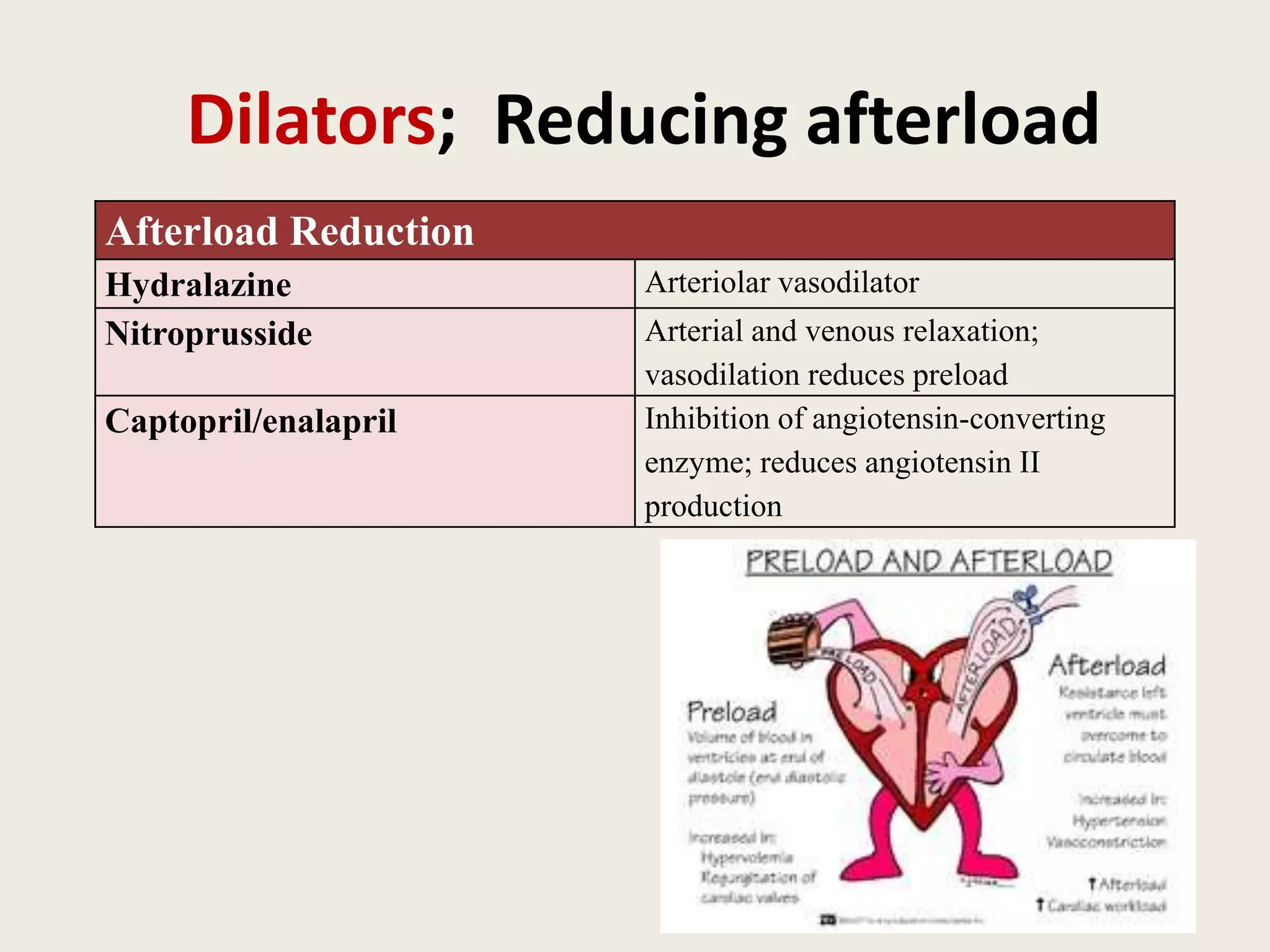
Congestive Heart Failure
Dr.Anup John Thomas
Assistant Professor
Department of Pediatrics,MGMCRI
DEFINITION
• It is a Clinical syndrome in which
– heart is unable to provide the output required to
meet the metabolic demands of the body
– Inability to dispose systemic or pulmonary venous
return adequately
– Combination of above two
Factors Affecting Cardiac Performance
• Cardiac output depends on: stroke volume
and heart rate.
• Stroke volume is dependent on three
important factors:
• preload,
• afterload and
• contractility.
• Preload = volume of blood received by the
heart.
• Basically, preload is stretch.
• Afterload = pressure or resistance the heart has
to overcome to eject blood.
• Afterload is squeeze.
• Preload:
• Preload (volume overload).
• preload (such as in VSD, or valvular insufficiency).
• Afterload:
• Afterload is the resistance (pressure) against which the
heart must pump blood: e.g.; systemic vascular resistance.
• Afterload (such as with aortic stenosis, pulmonary stenosis,
or coarctation of the aorta)
• Contractility
• Contractility (Cardiac Performance Independent of Preload or
Afterload)
• Volume overload is the most common cause of CHF in
children
Compensatory mechanisms in heart failure
(1) Cardiac compensation
– increased HR and cardiac contractility
– Cardiac dilatation (The Frank-Starling mechanism)
– Myocardial hypertrophy
(2) Systemic compensation
– Increase the blood volume
– Redistribution of blood flow
– Increase of erythrocytes
– Increased ability of tissues to utilize oxygen
(3) neurohormonal compensation
– Sympathetic nervous system
– Renin-angiotensin system
– Atrial natriuretic peptide; endothelin
Etiology of Heart Failure
Fetus Premature Neonate
Severe anemia (hemolysis, fetal-maternal
transfusion)
Fluid overload
Supraventricular tachycardia PDA
Ventricular tachycardia VSD
Complete heart block
Full-Term Neonate Infant-Toddler
Asphyxial cardiomyopathy Left-to-right cardiac shunts (VSD)
Left-sided obstructive lesions (coarctation of aorta) Metabolic cardiomyopathy
Transposition of great arteries Acute hypertension (hemolytic-uremic syndrome)
Viral myocarditis Supraventricular tachycardia
Anemia Kawasaki disease
Supraventricular tachycardia
Complete heart block
Child-Adolescent
Rheumatic fever Acute hypertension (glomerulonephritis)
Viral myocarditis Thyrotoxicosis
Endocarditis Cor pulmonale (cystic fibrosis)
Arrhythmias Chronic upper airway obstruction (cor pulmonale)
Cardiomyopathy
Classification of heart failure
(1) According to the course of disease
1) Acute HF
2) Chronic HF
2)According to the cardiac output (CO)
1) Low-output HF: due to volume overload,
pressure overload & contractility problems.
2) High-output HF: Heart Rate is primarily
affected;3A(Anemia, Arrhythmia, AV Fistula)
(3) According to the location of heart failure
1) Left -side heart failure (LHF)
2) Right-side heart failure (RHF)
3) Biventricular failure (whole heart failure)
(4)According to the function impaired
1) systolic failure :Myocarditis, hypertension
2) Diastolic failure: restrictive cardiomyopathy,
cardiac tamponade.
WHEN TO SUSPECT CCF
• Poor wt. gain
• Difficulty in feeding
• Breathes too fast
• Persistent cough and wheezing
• Excessive perspiration, irritability, restlessness
• Puffiness of face
• Pedal edema
• Diaphoresis
APPROACH TO PATIENT
• HISTORY
• PHYSICAL EXAMINATION
• INVESTIGATION
• TREATMENT
Clinical History
NEONATES & INFANTS
• Poor feeding
• Tachypnea worsening during feeding
• Cold sweat on forehead
• Poor weight gain
OLDER CHILDREN
• Fatigue
• Exercise intolerance
• Dyspnea
• Puffy eyes & pedal edema
• Growth failure
CLINICAL MANIFESTATIONS
• The physical examination
findings depend on whether
pulmonary venous congestion,
systemic venous congestion, or
both are present.
• Tachycardia, a gallop rhythm,
and thready pulses may be
present with either cause.
• If left-sided failure is
predominant, tachypnea,
orthopnea, wheezing, and
pulmonary edema are seen.
• If right-sided failure is present,
hepatomegaly, edema, and
distended neck veins are
present.
IMAGING STUDIES
• chest x-ray: cardiomegaly. Pulmonary edema
• ECG: Arrhythmias
• An echocardiogram assesses the heart chamber sizes,
measures myocardial function accurately, and diagnoses
congenital heart defects when present.
• Pulse-oximetry, CBG, hyperoxia test
• CBC, U&E, calcium, creatinine, and LFT
• Blood tests
• Thyroid function
Clinical diagnosis of CHF
E c h o c a r d i o g r a m
Structural diagnosis
(e.g. myopathy, valvular)
Pathophysiological diagnosis
Systolic dysfunction (LVEF < 40%)
Diastolic dysfunction (LVEF > 40%)
Proceed to
treatment guidelines
NYHA CHF classification for infants
• NYHA I - NO SIGN
• NYHA II - RR>50 , WITH OR WITHOUT HEPATOMEGALY
• NYHA III- ALL ABOVE WITH RIB RETRACTION
• NYHA IV- RR>60/min H/R>160/ min,
WITH HEPATOMEGALY,RIB RETRACTION
WITH OR WITHOUT POOR PERFUSION.
Treatment principles
• (1) Correct the underlying causes of HF
• (2) Diet; (low salt and high calories)
• (3) Digitalis; Improve the cardiac contractility
• (4) Diuretics; Reducing preload: frusemide
• (4) Dilators; Reducing afterload; ACE
• Remember 4 D
Treatment of Heart Failure
General Care
Rest Reduces cardiac output
Oxygen Improves oxygenation in presence of
pulmonary edema
Sodium, fluid restrictions Decreases vascular congestion; decreases
preload
Other
Treatment of underlying causes Closure of defects like ASD or VSD
Transplantation Removes diseased heart
Precipitating factors
• Hypertension
• Anemia
• Arrhythmia
• Hyperthyroidism
• Infection
• Fever
Diuretics; Reducing preload
Diuretics
Furosemide Salt excretion by ascending loop of Henle;
reduces preload
Spironolactone Potassium-sparing diuretic
Improve the cardiac contractility
Inotropic Agents
Digitalis Inhibits membrane Na+, K+-ATPase and
increases intracellular Ca2+, improves
cardiac contractility, increases myocardial
oxygen consumption
Dopamine Releases myocardial norepinephrine plus
direct effect on β-receptor, may increase
systemic blood pressure; at low infusion
rates, dilates renal artery, facilitating
diuresis
Dilators; Reducing afterload
Afterload Reduction
Hydralazine Arteriolar vasodilator
Nitroprusside Arterial and venous relaxation;
vasodilation reduces preload
Captopril/enalapril Inhibition of angiotensin-converting
enzyme; reduces angiotensin II
production
ß BLOCKERS
• Effacious in CHF in children due to CHD, Anthracycline
induced cardiomyopathy , dilated cardiomyopathy.
• Improved left ventricular function & exercise
tolerance, decreased need for heart transplant.
• It has been shown to improve clinical symptoms &
neurohormonal markers in infants with CHF due to Lt
to Rt shunts.
• Dose should titrated upwards
• Avoid in decompensated heart failure.
• Carvedilol(initial dose0.08→0.46mg/kg)
Nonpharmacological treatment
modalities
Cardiac resynchronization
therapy:
BiVP
• cardiomyopathy
• LBBB
LV assist device
Surgery:
(depends on the type of defect)
 Blalock Tausig shunt
 Balloon septoplasty
 Mustard Senning
 Jatene’s switch
HEART FAILURE IN SPECIAL CONDITION
• Ductus dependent circulation- PGE2
• Rheumatic carditis -Steroids
• Kawasaki’s disease-IG
• Preterm PDA-Indomethacin
THANK
YOU

Congestive heart failure revised

  • 1.
    Congestive Heart Failure Dr.AnupJohn Thomas Assistant Professor Department of Pediatrics,MGMCRI
  • 2.
    DEFINITION • It isa Clinical syndrome in which – heart is unable to provide the output required to meet the metabolic demands of the body – Inability to dispose systemic or pulmonary venous return adequately – Combination of above two
  • 3.
    Factors Affecting CardiacPerformance • Cardiac output depends on: stroke volume and heart rate. • Stroke volume is dependent on three important factors: • preload, • afterload and • contractility.
  • 4.
    • Preload =volume of blood received by the heart. • Basically, preload is stretch. • Afterload = pressure or resistance the heart has to overcome to eject blood. • Afterload is squeeze.
  • 5.
    • Preload: • Preload(volume overload). • preload (such as in VSD, or valvular insufficiency). • Afterload: • Afterload is the resistance (pressure) against which the heart must pump blood: e.g.; systemic vascular resistance. • Afterload (such as with aortic stenosis, pulmonary stenosis, or coarctation of the aorta) • Contractility • Contractility (Cardiac Performance Independent of Preload or Afterload) • Volume overload is the most common cause of CHF in children
  • 6.
    Compensatory mechanisms inheart failure (1) Cardiac compensation – increased HR and cardiac contractility – Cardiac dilatation (The Frank-Starling mechanism) – Myocardial hypertrophy (2) Systemic compensation – Increase the blood volume – Redistribution of blood flow – Increase of erythrocytes – Increased ability of tissues to utilize oxygen (3) neurohormonal compensation – Sympathetic nervous system – Renin-angiotensin system – Atrial natriuretic peptide; endothelin
  • 7.
    Etiology of HeartFailure Fetus Premature Neonate Severe anemia (hemolysis, fetal-maternal transfusion) Fluid overload Supraventricular tachycardia PDA Ventricular tachycardia VSD Complete heart block Full-Term Neonate Infant-Toddler Asphyxial cardiomyopathy Left-to-right cardiac shunts (VSD) Left-sided obstructive lesions (coarctation of aorta) Metabolic cardiomyopathy Transposition of great arteries Acute hypertension (hemolytic-uremic syndrome) Viral myocarditis Supraventricular tachycardia Anemia Kawasaki disease Supraventricular tachycardia Complete heart block Child-Adolescent Rheumatic fever Acute hypertension (glomerulonephritis) Viral myocarditis Thyrotoxicosis Endocarditis Cor pulmonale (cystic fibrosis) Arrhythmias Chronic upper airway obstruction (cor pulmonale) Cardiomyopathy
  • 8.
    Classification of heartfailure (1) According to the course of disease 1) Acute HF 2) Chronic HF 2)According to the cardiac output (CO) 1) Low-output HF: due to volume overload, pressure overload & contractility problems. 2) High-output HF: Heart Rate is primarily affected;3A(Anemia, Arrhythmia, AV Fistula)
  • 9.
    (3) According tothe location of heart failure 1) Left -side heart failure (LHF) 2) Right-side heart failure (RHF) 3) Biventricular failure (whole heart failure) (4)According to the function impaired 1) systolic failure :Myocarditis, hypertension 2) Diastolic failure: restrictive cardiomyopathy, cardiac tamponade.
  • 10.
    WHEN TO SUSPECTCCF • Poor wt. gain • Difficulty in feeding • Breathes too fast • Persistent cough and wheezing • Excessive perspiration, irritability, restlessness • Puffiness of face • Pedal edema • Diaphoresis
  • 11.
    APPROACH TO PATIENT •HISTORY • PHYSICAL EXAMINATION • INVESTIGATION • TREATMENT
  • 12.
    Clinical History NEONATES &INFANTS • Poor feeding • Tachypnea worsening during feeding • Cold sweat on forehead • Poor weight gain OLDER CHILDREN • Fatigue • Exercise intolerance • Dyspnea • Puffy eyes & pedal edema • Growth failure
  • 13.
    CLINICAL MANIFESTATIONS • Thephysical examination findings depend on whether pulmonary venous congestion, systemic venous congestion, or both are present. • Tachycardia, a gallop rhythm, and thready pulses may be present with either cause. • If left-sided failure is predominant, tachypnea, orthopnea, wheezing, and pulmonary edema are seen. • If right-sided failure is present, hepatomegaly, edema, and distended neck veins are present.
  • 14.
    IMAGING STUDIES • chestx-ray: cardiomegaly. Pulmonary edema • ECG: Arrhythmias • An echocardiogram assesses the heart chamber sizes, measures myocardial function accurately, and diagnoses congenital heart defects when present. • Pulse-oximetry, CBG, hyperoxia test • CBC, U&E, calcium, creatinine, and LFT • Blood tests • Thyroid function
  • 15.
    Clinical diagnosis ofCHF E c h o c a r d i o g r a m Structural diagnosis (e.g. myopathy, valvular) Pathophysiological diagnosis Systolic dysfunction (LVEF < 40%) Diastolic dysfunction (LVEF > 40%) Proceed to treatment guidelines
  • 16.
    NYHA CHF classificationfor infants • NYHA I - NO SIGN • NYHA II - RR>50 , WITH OR WITHOUT HEPATOMEGALY • NYHA III- ALL ABOVE WITH RIB RETRACTION • NYHA IV- RR>60/min H/R>160/ min, WITH HEPATOMEGALY,RIB RETRACTION WITH OR WITHOUT POOR PERFUSION.
  • 17.
    Treatment principles • (1)Correct the underlying causes of HF • (2) Diet; (low salt and high calories) • (3) Digitalis; Improve the cardiac contractility • (4) Diuretics; Reducing preload: frusemide • (4) Dilators; Reducing afterload; ACE • Remember 4 D
  • 18.
    Treatment of HeartFailure General Care Rest Reduces cardiac output Oxygen Improves oxygenation in presence of pulmonary edema Sodium, fluid restrictions Decreases vascular congestion; decreases preload Other Treatment of underlying causes Closure of defects like ASD or VSD Transplantation Removes diseased heart
  • 19.
    Precipitating factors • Hypertension •Anemia • Arrhythmia • Hyperthyroidism • Infection • Fever
  • 20.
    Diuretics; Reducing preload Diuretics FurosemideSalt excretion by ascending loop of Henle; reduces preload Spironolactone Potassium-sparing diuretic
  • 21.
    Improve the cardiaccontractility Inotropic Agents Digitalis Inhibits membrane Na+, K+-ATPase and increases intracellular Ca2+, improves cardiac contractility, increases myocardial oxygen consumption Dopamine Releases myocardial norepinephrine plus direct effect on β-receptor, may increase systemic blood pressure; at low infusion rates, dilates renal artery, facilitating diuresis
  • 22.
    Dilators; Reducing afterload AfterloadReduction Hydralazine Arteriolar vasodilator Nitroprusside Arterial and venous relaxation; vasodilation reduces preload Captopril/enalapril Inhibition of angiotensin-converting enzyme; reduces angiotensin II production
  • 23.
    ß BLOCKERS • Effaciousin CHF in children due to CHD, Anthracycline induced cardiomyopathy , dilated cardiomyopathy. • Improved left ventricular function & exercise tolerance, decreased need for heart transplant. • It has been shown to improve clinical symptoms & neurohormonal markers in infants with CHF due to Lt to Rt shunts. • Dose should titrated upwards • Avoid in decompensated heart failure. • Carvedilol(initial dose0.08→0.46mg/kg)
  • 24.
    Nonpharmacological treatment modalities Cardiac resynchronization therapy: BiVP •cardiomyopathy • LBBB LV assist device Surgery: (depends on the type of defect)  Blalock Tausig shunt  Balloon septoplasty  Mustard Senning  Jatene’s switch
  • 25.
    HEART FAILURE INSPECIAL CONDITION • Ductus dependent circulation- PGE2 • Rheumatic carditis -Steroids • Kawasaki’s disease-IG • Preterm PDA-Indomethacin
  • 26.