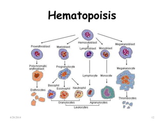
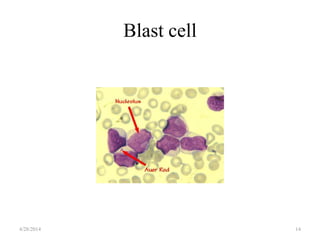
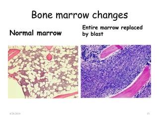
2. Objectives
Definition & Classification of leukemias
The pathophysiology and epidemiology
of Acute lymphoblasticleukemia (ALL)
Review the different drugs, & therapy
strategies in ALL treatment.
Describe some of the newer agents for
the treatment of ALL
4/28/2014 2
3. Definition
• Leukemia is a type of cancer of blood
or bonemarrow
• Characterized by an abnormal increase
of immature white blood cells called
"blasts".
• Leukemia is a broad term covering a
spectrum of diseases.
• Leuka = white, emia = blood
4/28/2014 3
4. The History of Leukemia
1845- Craig and Bennett described a case as
suppuration of the blood
–Virchow discovered this as well, named it “leukemia”
1855- Ernst Neumann discovered that the bone
marrow was the likely origin of leukemia
–
4/28/2014 4
5. The History of Leukemia
1946- Sidney Farber used antifolate agents to
treat leukemia in children
1960s- Addition of vincristine and steroid to
regimens,
– Survival rates increased to over 50%
1970- Beginning of classification, discovery of
cytogenetics and risk based treatment
4/28/2014 5
6. Epidemiology
Most common childhood cancer
– 3,000 new cases each year
Demographics:
–
–
–
Males more commonly than females
Whites more than blacks
More commonly in patients with Down’s
4/28/2014 6
10. • Epidemiology of ALL
peak incidence in 2 to 6 years
more in boys than girls.
median age in adults-35years
• Etiology
less studied
environmental and genetic
factors
4/28/2014 10
13. Blast cell
• Blast cells are immature precursors of either
lymphocytes (lymphoblasts), or granulocytes
(myeloblasts).
• They do not normally appear in peripheral
blood. they can be recognized by their large
size, and primitive nuclei (i.e. the nucleus
contain nucleoli).
• Presence of BS in blood, signify ACUTE
LEUKEMIA.
• Presence of an Auer Rod, is pathognomonic
for Acute Myeloid Leukemia.
4/28/2014 13
17. Classifications of ALL
• FAB CLASSIFICATION
• WHO CLASSIFICATION
• Cyto-genetic CLASSIFICATION
4/28/2014 17
18. FAB CLASSIFICATION OF ALL
CYTOLOGIC
FEATURES
L1 L2 L3
Cell size Small cells
predominate,homo
genous
Large,heterogenou
s in size
Large homogenous
cytoplasm Scanty Variable,often
moderately
abundant
Moderately
abundant
nucleoli Small One or more,often
large
One or
more,prominent
Nuclear size Homogenous Variable,
heterogenous
Stippled,
homogenous
Nuclear shape Regular Irregular clefts regular
Cyt.basophilia variable variable Intensely
basophilic
Cyt.vacuolation variable variable prominent4/28/2014 18
22. Etiology and Pathophysiology
ALL results from mutations of genes
–
–
–
Radiation
Chemicals
Viruses & Other
Malignant immature white blood cells i.e.
–
–
Lymphoblasts crowd out the bone marrow
This includes crowding out of platelets, RBCs,
and mature WBCs
4/28/2014 22
23. CLINICAL FEATURES
Due to infiltration of marrow
• SYMPTOMS
Due to decreased production of
normal marrow elements
4/28/2014 23
26. Clinical features
• Generalized weakness and fatigue
• Anemia
• Frequent or unexplained fever and infection
• Weight loss and/or loss of appetite
• Excessive and unexplained bruising
• Bone pain, joint pain (caused by the spread of
"blast" cells from the marrow cavity)
• Breathlessness
• Enlarged lymph nodes, liver and/or spleen
• Petechiae, which are tiny red spots or lines in
the skin due to low platelet levels
4/28/2014 26
37. Treatment of ALL
Induction 1
cycle chemotherap
y
Dose and schedule
Induction Prednisolon or 1mg/kg p.o days 1-28 days
vincristine 1.5mg/m2 i.v weekly once
x 4 weeks
doxorubicin 30mg/m2 i.v weekly once
x 4 weeks
L-Asparginase 1,00,000 u/m2(total dose)
in divided doses of 10,000
u daily for 10 days
CNS Proph. methotrexate 12mg IT days 1,8,15,22
4/28/2014 37
38. Reassess
• After 4 weeks of phase 1 induction
assess marrow for remission.
• If there is remission taper prednisolone
and after 1 week, start phase2
induction,
• If there is no remission give 2 more
weekly doses of vincristine and doxo and
then assess, if still no remission go for
alternate regimen.
4/28/2014 38
39. Induction 2
Induction2 drugs Dose and
schedule
Cyclophosphamide
Cytosine
arabinoside
650mg/m2 i.v
days 1 and 15
75mg/m2 i.v x 4
days a weeks for
4 week
methotrexate 12mg/m2 IT days
1,8,15,22
Cranial radiation 200 cGy x 9days
4/28/2014 39
40. Reinduction
Re
induction
drug Dose and schedule
vincristine 1.5 mg/m2 i.v weekly one
dose on day 1 and 8
doxorubicin 30mg/m2 i.v. weekly one
dose on day 1 and 8
prednisolone 1mg/kg p.o daily for 14
days
4/28/2014 40
42. Maintenance phase
duration- upto 2 years
maintena
nce
drug Dose and schedule
1st
month
methotrexate 12.5mg i.t on day 1
vincristine 1.4mg/m2 .v day 1
prednisolone 1mg/kg p.o daily day 1-7
6 mercaptopurine 60mg/m2 p.o. daily for
next 3 weeks
methotrexate 15mg/m2 p.o. once a
week for 3 weeks.
2nd
month
6 MCP and
T.Methotxerate
for 4 weeks.4/28/2014 42
43. Follow up
If the patient completes chemotherapy
for 2 years without relapse-stop chemo
and follow up.
No relapse within 5 years-can be declared
as cured.
4/28/2014 43
44. Allogenic stem cell transpantation
• Usually done in second remission.
• Can be done in first remission in high
risk patients
WBC>25000,
philadelphia chromosome positive,
poor initial response to remission
induction.
4/28/2014 44
47. CNS Prophylaxis
CNS involvement at diagnosis <5%
–
–
Without prophylaxis, over 80% of patients in CR
will relapse in the CNS
With prophylaxis, less than 5% have CNS relapse
Intrathecal chemotherapy is now the
mainstay
– Sample intermediate risk regimen: IT MTX alone
or “triple therapy”: IT cytarabine Day 1 of CR
followed by MTX/hydrocortisone/cytarabine
Pui CH, Howard SC. Lancet Oncol 9 (3): 257-68, 2008
4/28/2014 47
48. Prognostic factors in ALL
Determinants Favourable unfavourable
WBC Counts <10,000 >2,00,000
Age 2-10 years <1yr,>10yr
Gender female male
Ethnicity white black
Node,liver,splenomegaly absent massive
Testicular enlargement absent present
CNS involvement absent Csf blast and pleocytosis
FAB Type L1 L2
Cytogenetics T(12;21)(TEL-AML1)
Trsomies 4,10,17
t(9;22)(bcr-abl)
t(4;11)(MLL-AF4)
Ploidy hyperdipoidy hypodiploidy
Time to remission <14days >28days4/28/2014 48