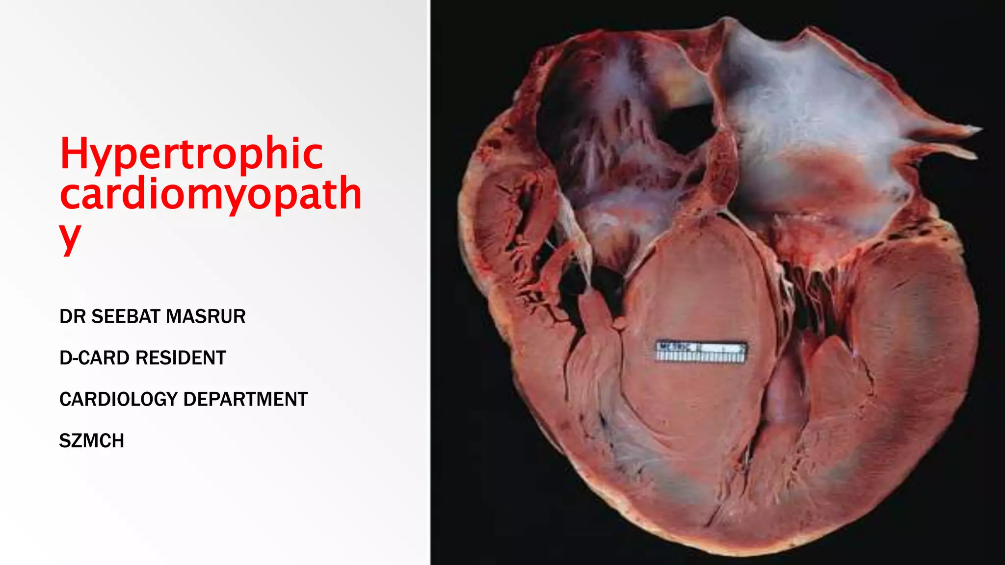
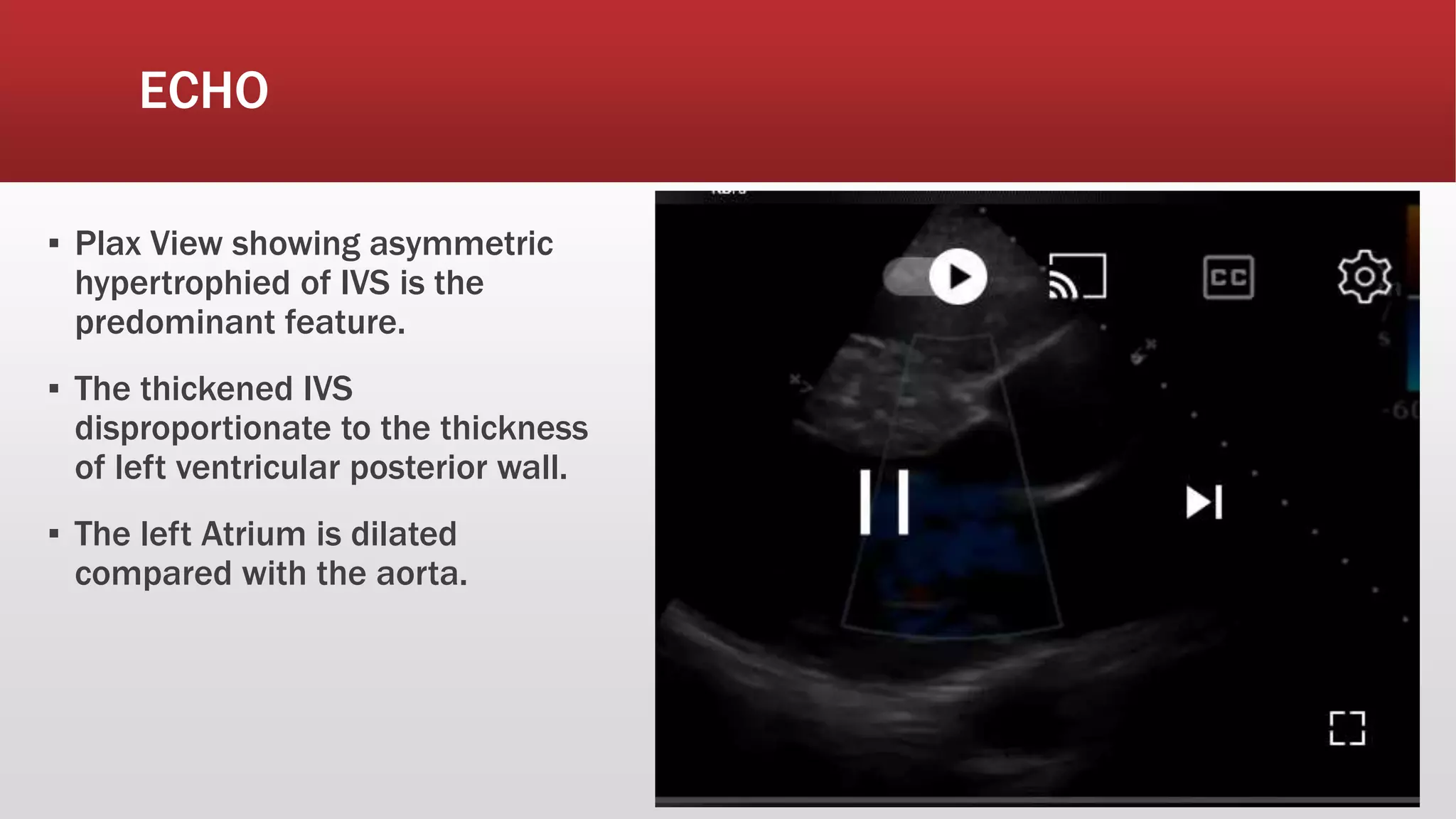
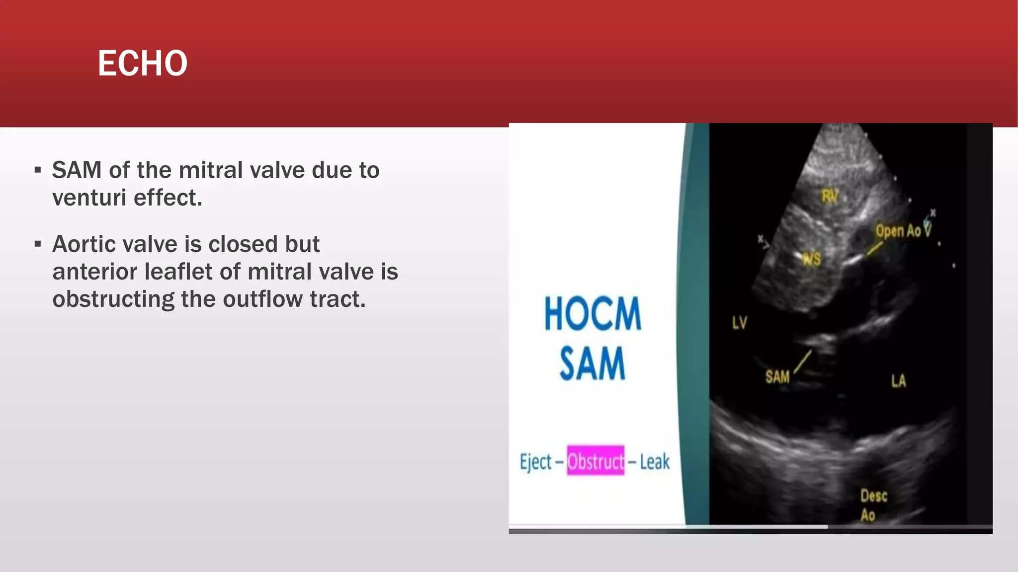
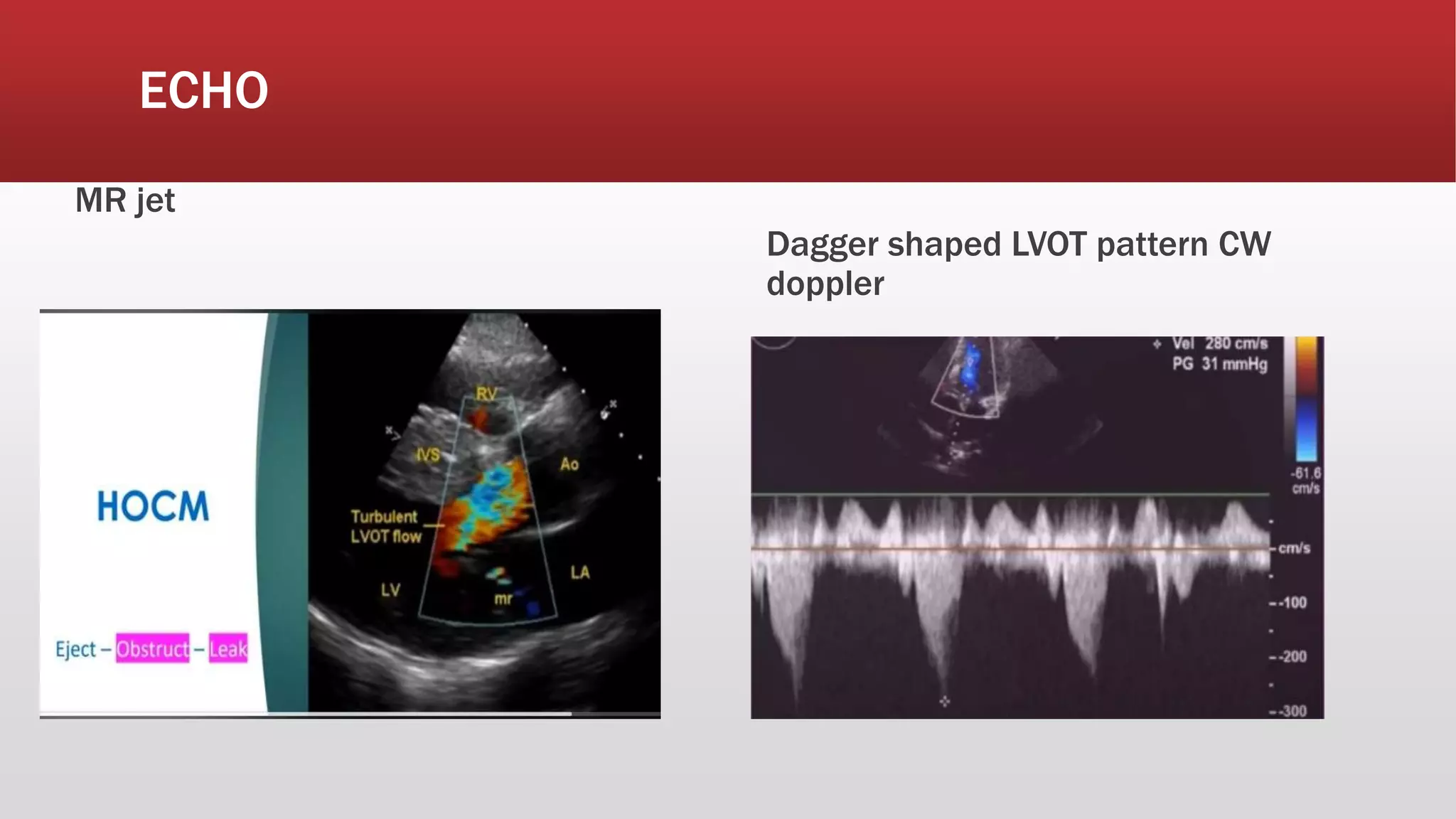
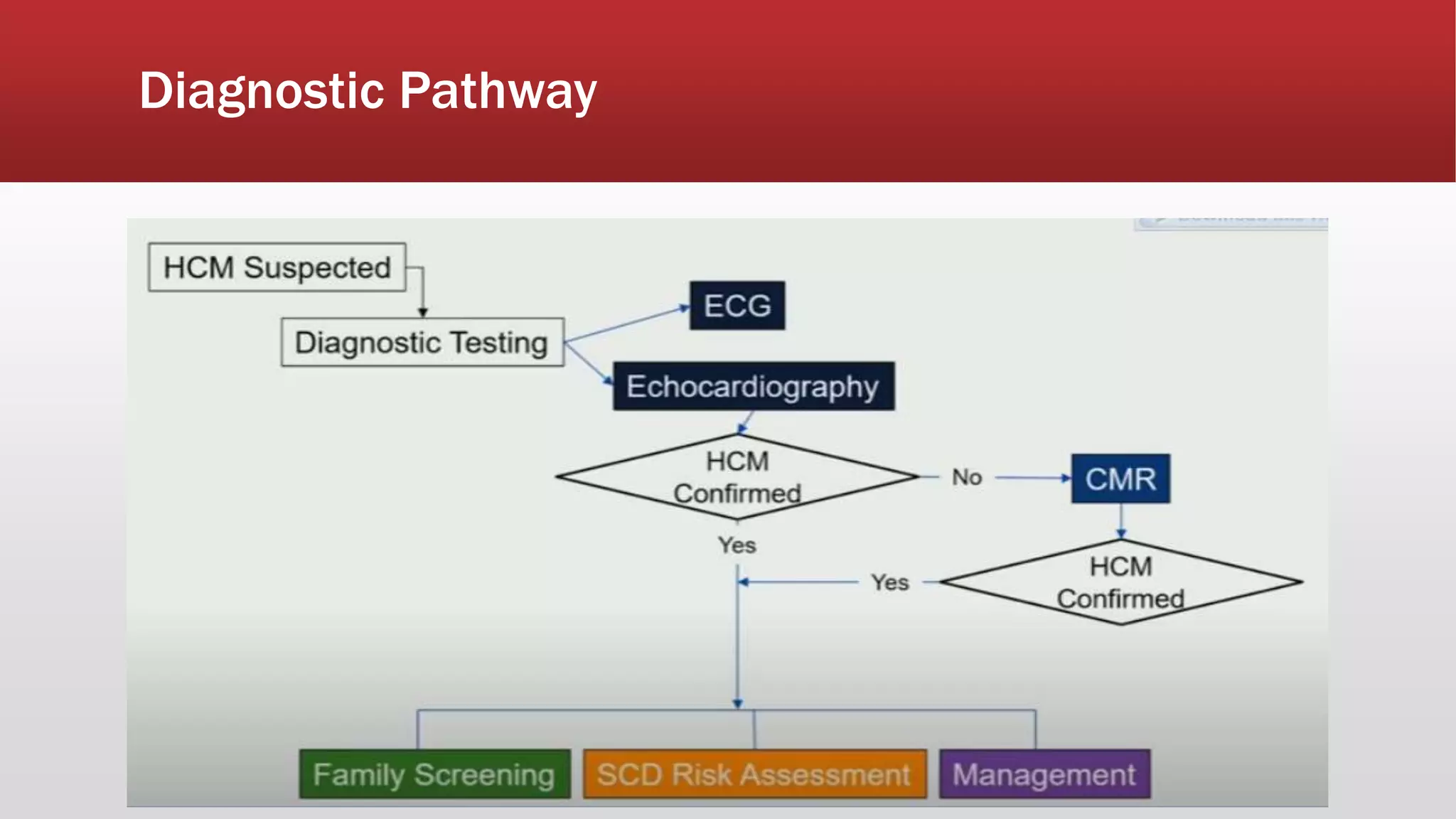
Hypertrophic
cardiomyopath
y
DR SEEBAT MASRUR
D-CARD RESIDENT
CARDIOLOGY DEPARTMENT
SZMCH
Introduction
▪ Autosomal Dominant Disorder
▪ Incidence 1:500 adults (Male predominance)
▪ > 200 familial and sporadic mutation described
▪ 11 genes identified
▪ B-myosin Heavy chain, troponin I, Myosin-binding protein C involved
▪ Associations- Friedrich`s Ataxia, Fabry`s disease, Danon disease, Noonan
& Leopard syndrome
Pathophysiology
▪ Dynamic LV outflow tract obstruction
▪ Diastolic dysfunction
▪ Myocardial ischemia
▪ Mitral regurgitation
▪ Arrhythmias
Pathophysiology
▪ Most Dramatic feature of HOCM is the
dynamic intraventricular pressure
gradient in the left ventricle.
▪ This occur due to contraction of basal
aspect of asymptomatic septum---LVOT
obstruction---blood flow in the midcavity
increased---Anterior leaflet of mitral valve
is sucked in by venturi effect---augments
LVOT obstruction, Mitral regurgitation.
This effect is provoked by
▪ Valsalva maneuver
▪ An extrasystole
▪ Potent vasodilator (Amyl Nitrate)
▪ Inotropic stimulation
▪ Exercise
▪ Large Meal
▪ Alcohol consumption
▪ Fever, Anemia & dehydration
Variants of HCM
▪ Asymmetric Septal Hypertrophy 70%
▪ Basal Septal Hypertrophy 15-20%
▪ Concentric hypertrophy 8-10%
▪ Apical or Lateral wall hypertrophy
<2%
Two Basic Phenotype
▪ Obstructive HCM(HOCM): Two third, at rest or during provocation like
physical exercise, left ventricular outflow is mechanically impeded due to
thickening of the sub-aortic septum interacting with the mitral valve or
thickened muscle itself.
▪ Nonobstructive HCM(NHCM): one third, out flow is always nonobstructive at
all times.
Clinical Feature
Symptoms
▪ Heterogenous, Asymptomatic
▪ Dyspnea
▪ Angina
▪ Syncope
▪ Sudden death
Signs
▪ Jerky carotid pulse
▪ Low systolic BP, Normal diastolic, Narrow pulse
pressure
Arterial pressure Waveform
▪ Spike and Dome pattern
▪ Early spike-rapid ventricular ejection
from hypercontractile LV
▪ Pressure Dip and dome-dynamic
outflow obstruction
Brockenbrough-Braunwald-Morrow sign
After a premature ventricular contraction
(PVC), the left ventricular outflow tract
gradient is close to 100 mm Hg. The pulse
pressure of the ascending aorta (Ao) is
decreased on the beat following the
premature ventricular contraction. This
phenomenon is termed the Brockenbrough
phenomenon
Physical Exam
▪ Apex beat thrusting, may be double apical impulse or even triple
impulse(triple impulse).
▪ A systolic thrill may be palpable at apex.
▪ JVP mostly normal but can show prominent a wave
Auscultation
▪ 1st Heart sound: Normally audible in all 4 areas. Can be loud
▪ 2nd Heart sound: May be split in severe LVOT obstruction.
▪ An S4 is usually present.
▪ Ejection Systolic murmur-at the lower left sternal border and apex.
▪ Pansystolic murmur at the apex due to mitral regurgitations.
Murmur
Dynamic maneuvers
Dynamic
maneuvers
Investigations-ECG
▪ 25% have no symptoms with a normal ECG
▪ LV hypertrophy and ST-T changes, progressive and steeper t-wave inversion(Gaint
negative t wave).
▪ Deep Q wave(Dragger like) in inferior and lateral leads(septal hypertrophy and fibrosis)
▪ LA enlargement(P mitrale)
▪ Left anterior hemiblock
▪ Left bundle branch block
▪ Ventricular ectopic Poor R-wave progression
▪ Pre-excitation and WPW syndrome
▪ VT on ambulatory monitoring
ECG Examples
Classic HCM pattern with
asymmetrical septal hypertrophy:
ECG Examples
Apical HCM
Giant T-wave inversion
CXR
The chest x-ray usually shows mild-to-moderate enlargement of the cardiac
silhouette. The LV contour is rounded, consistent with LV hypertrophy. There is
usually enlargement of the left atrium, and the right-sided chambers are usually
normal.
Echocardiography(Transthoracic ECHO is Diagnostic)
▪ Decreased LV systolic dimensions.
▪ Asymmetrical septal hypertrophy with a ratio of sepum to posterior wall 1.3:1 or greater.
▪ Systolic anterior motion.
▪ Delayed closure of aortic valve.
▪ Midsystolic closure of the aortic valve.
▪ Left atrial enlargement.
▪ Mitral Regurgitation.
▪ Dagger shaped late peaking continuous wave doppler with resting gradient> 30mmHG in
LVOT.
ECHO
▪ Plax View showing asymmetric
hypertrophied of IVS is the
predominant feature.
▪ The thickened IVS
disproportionate to the thickness
of left ventricular posterior wall.
▪ The left Atrium is dilated
compared with the aorta.
ECHO
▪ SAM of the mitral valve due to
venturi effect.
▪ Aortic valve is closed but
anterior leaflet of mitral valve is
obstructing the outflow tract.
ECHO
MR jet
Dagger shaped LVOT pattern CW
doppler
Transesophageal Echocardiography
▪ In most patients, anatomic and hemodynamic information can be obtained by
transthoracic echocardiography alone, but transesophageal echocardiography
may be useful in patients in whom discrete subvalvular stenosis or a primary
abnormality of the mitral valve is suspected.
▪ It is also mandatory for the peri- and intraoperative monitoring of septal
myectomy to confirm of the mechanism of LVOTO, guide the surgical strategy, and
detect postsurgical complications such as ventricular septal defects and residual
LVOTO
Cardiac MRI
Cardiac MRI, with its capabilities in evaluating cardiac morphology and function, has emerged as a
technique particularly well-suited to hypertrophic cardiomyopathy diagnosis and phenotypic
characterization. It is superior to echocardiography in identifying areas of segmental hypertrophy
not reliably visualized or underestimated by echocardiography (i.e. anterolateral and apical
segments). Visualized feat
• Left ventricular systolic dysfunction
• Left ventricular hypertrophy
• with or without right ventricular hypertrophy
• predilection for the basal interventricular septum
• Systolic anterior motion (SAM) of the mitral valve
• mitral regurgitation
• Left ventricular apical aneurysms
• Morphologic variations involving the mitral valvular apparatus (e.g. papillary muscles)
Cardiac MRI
Cardiac MRI also has a role in asymptomatic HCM
mutation carriers by identifying phenotypic markers of
HCM in the absence of left ventricular hypertrophy
including:
• Myocardial crypts
• Elongated mitral valve leaflets
• Late gadolinium enhancement (LGE)
• patchy/streaky intramyocardial patterns at the right
ventricular insertion sites within the hypertrophied
myocardium suggest fibrosis
Nuclear Imaging
▪ Shows reversible and fixed defects in HCM in the absence of epicardial
coronary artery disease. In the most recent guidelines,no specific
recommendation has been made for the use of nuclear imaging for
ischemia assessment.
▪ It is useful in detecting cardiac amyloidosis.
Cardiac Catheterization
▪ Not routinely done
▪ Reserved for situations when invasive modalities of therapy, such as a
pacemaker or surgery are being considered.
▪ Therapeutic catheterization incase of alcohol ablation.
Stress Testing
▪ Exercise stress testing is of limited value for the diagnosis of epicardial coronary
disease in patients with HCM but is helpful in assessing prognosis and the
mechanism of symptoms.
Familial screening for hypertrophic cardiomyopathy
Age Group Recommendation
<12 years Screening optional except in the following situations
1.Presence of symptoms
2.Family history of malignant ventricular tachycardias
3. Competitive athlete
4.Early left ventricular hypertrophy
18-21 years Every 12-18 months
>21 years Every 5 years or more frequent intervals if malignant clinical
course and/or late onset hypertrophic cardiomyopathy
Diagnostic Pathway
Major Risk Factors for Sudden Cardiac Death in HCM
▪ Younger Age.
▪ Nonsustained ventricular tachycardia-Independent predictor, ≥ 3 consecutive beats at ≥ 120 bpm < 30
seconds.
▪ Maximum left ventricular wall thickness-Greatest risk of SCD in patients with a maximum wall thickness of
≥ 30 mm.
▪ Family history of SCD-≥ 1 first-degree relative < 40 years old or any age if relative with an established
diagnosis of HCM.
▪ Syncope
▪ Left atrial enlargement
▪ Left ventricular outflow tract obstruction
▪ Abnormal blood pressure response to exercise-Failure to increase systolic pressure by at least 20 mm Hg
from rest to peak exercise or a fall of > 20 mm Hg from peak pressure
Natural history complicated by
▪ Sudden Cardiac Death 0.5%-1% per year
▪ Atrial fibrillation overall 22% but annual 3%
▪ Heart Failure 5%(Burnt out HOCM)
▪ Infective Endocarditis 4%-5%
Treatment
Nonobstructive:
▪ Betablockers, CCB, disopyramide for
symptomatic relief and prevention of
syncope
▪ Amiodarone may be helpful in
arrhythmia
Treatment
Obstructive:
▪ Medical therapy should be considered the first-line therapy for the relief of symptoms in
patients with obstructive HCM. e.g BB, Non dihydropyridine CCB, Disopyramide. Calcium
channel blockers may improve angina to a greater degree than β-blockers.
▪ Dual Chamber Pacemaker may be used
▪ Outflow tract obstruction can be improved by partial surgical resection( A
myotomy/myomectomy is performed through the aortic root valve-Marrow operation)
▪ Alcohol Septal Ablation: 95-86% absolute alcohol into the septal perforator branch
supplying the LV side of basal or mid-cavity septum. LVOTO gradient ≥ 50 mm Hg,
moderate-tosevere symptoms (New York Heart Association functional class III-IV), or
recurrent exertional syncope despite maximally tolerated drug therapy, other treatment
options such as septal myectomy, septal ablation, or dual-chamber pacing should be
considered
Treatment
Other treatment
▪ ICD (Prevention of Sudden Cardiac Death)
▪ Cardiac Transplantation
▪ Endocarditis prophylaxis
ICD Patient Selection
Drugs to be avoided
▪ Pre-load Reducing agents: Diuretics and Nitroglycerin. Diuretics can be used
cautiously with persistent heart failure.
▪ Afterload Reducing drugs: Dihydropyridine CCB, Nitroglycerin, ACEi/ARBs.
▪ Increased Contractility: Digoxin, Dobutamine, Milrinone.
Stroke Prevention
▪ Anticoagulation is recommended for patients with AF and HCM.
▪ The CHA2 DS2-VASc score does not correlate well with clinical outcome in
patients with HCM and is not recommended to assess thromboembolic risk.
▪ Vitamin K antagonists are recommended.
Advice to be given
▪ Advised against participation in competitive sports and intense physical
activity.
▪ Genetic counselling, as in 50% cases it may be inherited as autosomal
dominant.
▪ First degree family members should be screened by echocardiography.
▪ Avoid dehydration, take plenty of fluid
Pregnancy
▪ All HCM patients who wish to become pregnant should be given prenatal
counseling about the risk of transmission of disease to their offspring.
▪ Patients with HCM usually tolerate pregnancy well if they are not severely
symptomatic.
▪ If patients have been on treatment with β-blockers or calcium blockers,
these drugs should be continued throughout the pregnancy.
▪ Cesarean section should be considered in cases of severe LVOTO.
What`s New
Thank
You
▪ Hurst`s The Heart 14th Ed
▪ ESC 2014 guidelines
▪ AHA 2020 guidelines
▪ Mayo Clinic Lectures
▪ Radiopedia
HCM
▪ Can be asymmetric
▪ Wall thickness >15 mm
▪ LA > 40 mm
▪ LVEDD <45 mm
▪ Diastolic Dysfunction
Athlete heart
▪ Concentric & Regresses
▪ Wall thickness < 15 mm
▪ LA < 40 mm
▪ LVEDD < 45 mm
▪ Diastolic function normal

HCM Hypertrophic cardiomyopathy.pptx

  • 1.
    Hypertrophic cardiomyopath y DR SEEBAT MASRUR D-CARDRESIDENT CARDIOLOGY DEPARTMENT SZMCH
  • 2.
    Introduction ▪ Autosomal DominantDisorder ▪ Incidence 1:500 adults (Male predominance) ▪ > 200 familial and sporadic mutation described ▪ 11 genes identified ▪ B-myosin Heavy chain, troponin I, Myosin-binding protein C involved ▪ Associations- Friedrich`s Ataxia, Fabry`s disease, Danon disease, Noonan & Leopard syndrome
  • 3.
    Pathophysiology ▪ Dynamic LVoutflow tract obstruction ▪ Diastolic dysfunction ▪ Myocardial ischemia ▪ Mitral regurgitation ▪ Arrhythmias
  • 4.
    Pathophysiology ▪ Most Dramaticfeature of HOCM is the dynamic intraventricular pressure gradient in the left ventricle. ▪ This occur due to contraction of basal aspect of asymptomatic septum---LVOT obstruction---blood flow in the midcavity increased---Anterior leaflet of mitral valve is sucked in by venturi effect---augments LVOT obstruction, Mitral regurgitation.
  • 5.
    This effect isprovoked by ▪ Valsalva maneuver ▪ An extrasystole ▪ Potent vasodilator (Amyl Nitrate) ▪ Inotropic stimulation ▪ Exercise ▪ Large Meal ▪ Alcohol consumption ▪ Fever, Anemia & dehydration
  • 6.
    Variants of HCM ▪Asymmetric Septal Hypertrophy 70% ▪ Basal Septal Hypertrophy 15-20% ▪ Concentric hypertrophy 8-10% ▪ Apical or Lateral wall hypertrophy <2%
  • 7.
    Two Basic Phenotype ▪Obstructive HCM(HOCM): Two third, at rest or during provocation like physical exercise, left ventricular outflow is mechanically impeded due to thickening of the sub-aortic septum interacting with the mitral valve or thickened muscle itself. ▪ Nonobstructive HCM(NHCM): one third, out flow is always nonobstructive at all times.
  • 8.
    Clinical Feature Symptoms ▪ Heterogenous,Asymptomatic ▪ Dyspnea ▪ Angina ▪ Syncope ▪ Sudden death Signs ▪ Jerky carotid pulse ▪ Low systolic BP, Normal diastolic, Narrow pulse pressure
  • 9.
    Arterial pressure Waveform ▪Spike and Dome pattern ▪ Early spike-rapid ventricular ejection from hypercontractile LV ▪ Pressure Dip and dome-dynamic outflow obstruction
  • 10.
    Brockenbrough-Braunwald-Morrow sign After apremature ventricular contraction (PVC), the left ventricular outflow tract gradient is close to 100 mm Hg. The pulse pressure of the ascending aorta (Ao) is decreased on the beat following the premature ventricular contraction. This phenomenon is termed the Brockenbrough phenomenon
  • 11.
    Physical Exam ▪ Apexbeat thrusting, may be double apical impulse or even triple impulse(triple impulse). ▪ A systolic thrill may be palpable at apex. ▪ JVP mostly normal but can show prominent a wave
  • 12.
    Auscultation ▪ 1st Heartsound: Normally audible in all 4 areas. Can be loud ▪ 2nd Heart sound: May be split in severe LVOT obstruction. ▪ An S4 is usually present. ▪ Ejection Systolic murmur-at the lower left sternal border and apex. ▪ Pansystolic murmur at the apex due to mitral regurgitations.
  • 13.
  • 14.
  • 15.
  • 16.
    Investigations-ECG ▪ 25% haveno symptoms with a normal ECG ▪ LV hypertrophy and ST-T changes, progressive and steeper t-wave inversion(Gaint negative t wave). ▪ Deep Q wave(Dragger like) in inferior and lateral leads(septal hypertrophy and fibrosis) ▪ LA enlargement(P mitrale) ▪ Left anterior hemiblock ▪ Left bundle branch block ▪ Ventricular ectopic Poor R-wave progression ▪ Pre-excitation and WPW syndrome ▪ VT on ambulatory monitoring
  • 17.
    ECG Examples Classic HCMpattern with asymmetrical septal hypertrophy:
  • 18.
  • 19.
    CXR The chest x-rayusually shows mild-to-moderate enlargement of the cardiac silhouette. The LV contour is rounded, consistent with LV hypertrophy. There is usually enlargement of the left atrium, and the right-sided chambers are usually normal.
  • 20.
    Echocardiography(Transthoracic ECHO isDiagnostic) ▪ Decreased LV systolic dimensions. ▪ Asymmetrical septal hypertrophy with a ratio of sepum to posterior wall 1.3:1 or greater. ▪ Systolic anterior motion. ▪ Delayed closure of aortic valve. ▪ Midsystolic closure of the aortic valve. ▪ Left atrial enlargement. ▪ Mitral Regurgitation. ▪ Dagger shaped late peaking continuous wave doppler with resting gradient> 30mmHG in LVOT.
  • 21.
    ECHO ▪ Plax Viewshowing asymmetric hypertrophied of IVS is the predominant feature. ▪ The thickened IVS disproportionate to the thickness of left ventricular posterior wall. ▪ The left Atrium is dilated compared with the aorta.
  • 22.
    ECHO ▪ SAM ofthe mitral valve due to venturi effect. ▪ Aortic valve is closed but anterior leaflet of mitral valve is obstructing the outflow tract.
  • 23.
    ECHO MR jet Dagger shapedLVOT pattern CW doppler
  • 24.
    Transesophageal Echocardiography ▪ Inmost patients, anatomic and hemodynamic information can be obtained by transthoracic echocardiography alone, but transesophageal echocardiography may be useful in patients in whom discrete subvalvular stenosis or a primary abnormality of the mitral valve is suspected. ▪ It is also mandatory for the peri- and intraoperative monitoring of septal myectomy to confirm of the mechanism of LVOTO, guide the surgical strategy, and detect postsurgical complications such as ventricular septal defects and residual LVOTO
  • 25.
    Cardiac MRI Cardiac MRI,with its capabilities in evaluating cardiac morphology and function, has emerged as a technique particularly well-suited to hypertrophic cardiomyopathy diagnosis and phenotypic characterization. It is superior to echocardiography in identifying areas of segmental hypertrophy not reliably visualized or underestimated by echocardiography (i.e. anterolateral and apical segments). Visualized feat • Left ventricular systolic dysfunction • Left ventricular hypertrophy • with or without right ventricular hypertrophy • predilection for the basal interventricular septum • Systolic anterior motion (SAM) of the mitral valve • mitral regurgitation • Left ventricular apical aneurysms • Morphologic variations involving the mitral valvular apparatus (e.g. papillary muscles)
  • 26.
    Cardiac MRI Cardiac MRIalso has a role in asymptomatic HCM mutation carriers by identifying phenotypic markers of HCM in the absence of left ventricular hypertrophy including: • Myocardial crypts • Elongated mitral valve leaflets • Late gadolinium enhancement (LGE) • patchy/streaky intramyocardial patterns at the right ventricular insertion sites within the hypertrophied myocardium suggest fibrosis
  • 27.
    Nuclear Imaging ▪ Showsreversible and fixed defects in HCM in the absence of epicardial coronary artery disease. In the most recent guidelines,no specific recommendation has been made for the use of nuclear imaging for ischemia assessment. ▪ It is useful in detecting cardiac amyloidosis.
  • 28.
    Cardiac Catheterization ▪ Notroutinely done ▪ Reserved for situations when invasive modalities of therapy, such as a pacemaker or surgery are being considered. ▪ Therapeutic catheterization incase of alcohol ablation.
  • 29.
    Stress Testing ▪ Exercisestress testing is of limited value for the diagnosis of epicardial coronary disease in patients with HCM but is helpful in assessing prognosis and the mechanism of symptoms.
  • 30.
    Familial screening forhypertrophic cardiomyopathy Age Group Recommendation <12 years Screening optional except in the following situations 1.Presence of symptoms 2.Family history of malignant ventricular tachycardias 3. Competitive athlete 4.Early left ventricular hypertrophy 18-21 years Every 12-18 months >21 years Every 5 years or more frequent intervals if malignant clinical course and/or late onset hypertrophic cardiomyopathy
  • 31.
  • 32.
    Major Risk Factorsfor Sudden Cardiac Death in HCM ▪ Younger Age. ▪ Nonsustained ventricular tachycardia-Independent predictor, ≥ 3 consecutive beats at ≥ 120 bpm < 30 seconds. ▪ Maximum left ventricular wall thickness-Greatest risk of SCD in patients with a maximum wall thickness of ≥ 30 mm. ▪ Family history of SCD-≥ 1 first-degree relative < 40 years old or any age if relative with an established diagnosis of HCM. ▪ Syncope ▪ Left atrial enlargement ▪ Left ventricular outflow tract obstruction ▪ Abnormal blood pressure response to exercise-Failure to increase systolic pressure by at least 20 mm Hg from rest to peak exercise or a fall of > 20 mm Hg from peak pressure
  • 33.
    Natural history complicatedby ▪ Sudden Cardiac Death 0.5%-1% per year ▪ Atrial fibrillation overall 22% but annual 3% ▪ Heart Failure 5%(Burnt out HOCM) ▪ Infective Endocarditis 4%-5%
  • 34.
    Treatment Nonobstructive: ▪ Betablockers, CCB,disopyramide for symptomatic relief and prevention of syncope ▪ Amiodarone may be helpful in arrhythmia
  • 35.
    Treatment Obstructive: ▪ Medical therapyshould be considered the first-line therapy for the relief of symptoms in patients with obstructive HCM. e.g BB, Non dihydropyridine CCB, Disopyramide. Calcium channel blockers may improve angina to a greater degree than β-blockers. ▪ Dual Chamber Pacemaker may be used ▪ Outflow tract obstruction can be improved by partial surgical resection( A myotomy/myomectomy is performed through the aortic root valve-Marrow operation) ▪ Alcohol Septal Ablation: 95-86% absolute alcohol into the septal perforator branch supplying the LV side of basal or mid-cavity septum. LVOTO gradient ≥ 50 mm Hg, moderate-tosevere symptoms (New York Heart Association functional class III-IV), or recurrent exertional syncope despite maximally tolerated drug therapy, other treatment options such as septal myectomy, septal ablation, or dual-chamber pacing should be considered
  • 36.
    Treatment Other treatment ▪ ICD(Prevention of Sudden Cardiac Death) ▪ Cardiac Transplantation ▪ Endocarditis prophylaxis
  • 37.
  • 39.
    Drugs to beavoided ▪ Pre-load Reducing agents: Diuretics and Nitroglycerin. Diuretics can be used cautiously with persistent heart failure. ▪ Afterload Reducing drugs: Dihydropyridine CCB, Nitroglycerin, ACEi/ARBs. ▪ Increased Contractility: Digoxin, Dobutamine, Milrinone.
  • 40.
    Stroke Prevention ▪ Anticoagulationis recommended for patients with AF and HCM. ▪ The CHA2 DS2-VASc score does not correlate well with clinical outcome in patients with HCM and is not recommended to assess thromboembolic risk. ▪ Vitamin K antagonists are recommended.
  • 41.
    Advice to begiven ▪ Advised against participation in competitive sports and intense physical activity. ▪ Genetic counselling, as in 50% cases it may be inherited as autosomal dominant. ▪ First degree family members should be screened by echocardiography. ▪ Avoid dehydration, take plenty of fluid
  • 42.
    Pregnancy ▪ All HCMpatients who wish to become pregnant should be given prenatal counseling about the risk of transmission of disease to their offspring. ▪ Patients with HCM usually tolerate pregnancy well if they are not severely symptomatic. ▪ If patients have been on treatment with β-blockers or calcium blockers, these drugs should be continued throughout the pregnancy. ▪ Cesarean section should be considered in cases of severe LVOTO.
  • 43.
  • 44.
    Thank You ▪ Hurst`s TheHeart 14th Ed ▪ ESC 2014 guidelines ▪ AHA 2020 guidelines ▪ Mayo Clinic Lectures ▪ Radiopedia
  • 46.
    HCM ▪ Can beasymmetric ▪ Wall thickness >15 mm ▪ LA > 40 mm ▪ LVEDD <45 mm ▪ Diastolic Dysfunction Athlete heart ▪ Concentric & Regresses ▪ Wall thickness < 15 mm ▪ LA < 40 mm ▪ LVEDD < 45 mm ▪ Diastolic function normal