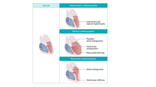
SlideShare a Scribd company logo
CARDIOMYOPATHIES
OUTLINE
• INTRODUCTION
• DEFINITION
• CLASSIFICATION
• DILATED CARDIOMYOPATHY
• HYPERTROPHIC CARDIOMYOPATHY
• RESTRICTIVE CARDIOMYOPATHY
• UNCLASSIFIED CARDIOMYOPATHIES
• CONCLUSION
INTRODUCTION
Cardiomyopathy refers to disease of the heart muscle
Either are confined to the heart or are part of generalized systemic
disorders, often leading to cardiovascular death or progressive heart
failure–related disability
Mostly idiopathic/genetic with clinically relevant disease process
solely/predominantly involving the myocardium- Primary
Known cause due to disease process not limited to the myocardium-
Secondary
DEFINITION
“A myocardial disorder in which the heart muscle is structurally
and functionally abnormal, in the absence of coronary artery
disease, hypertension, valvular disease and congenital heart
disease sufficient to cause the observed myocardial
abnormality”(ESC 2006)
Heterogeneous group of diseases of the myocardium
associated with mechanical and/or electrical dysfunction that
usually (but not invariably) exhibit inappropriate ventricular
hypertrophy or dilatation and are due to a variety of causes that
frequently are genetic(AHA 2008)
Classification
ESC 2007
●Dilated cardiomyopathy (DCM)
●Hypertrophic cardiomyopathy (HCM)
●Restrictive cardiomyopathy (RCM)
●Arrhythmogenic right ventricular cardiomyopathy/dysplasia (ARVC/D
DILATED CARDIOMYOPATHY
(DCM)
INTRODUCTION
Disease of cardiac muscles associated with mechanical and
electrical dysfunction
Characterized by Left or biventricular dilatation with impaired
systolic function
Absence of coronary artery disease, abnormal loading
pressures (e.g., valvular heart disease, hypertension),
or congenital heart disease
EPIDEMIOLOGY
Prevalence: 1:400(between 4-5 million people globally)
Incidence: ∼ 6/100,000 per year (most
common cardiomyopathy)
Sex: M > F (∼ 1.5:1)
Age at presentation: most commonly between 30 and
40 years of age, but can occur at any age
Ethnicity: more common in individuals of African
descent
PRIMARY
 Idiopathic
 Familial(Genetic)
 Substance use (Alcohol, cocaine)
 Cardiotoxic medications (Doxorubicin,
Trastuzumab)
 Infection (infectious myocarditis)
• Coxsackie B, Chagas Dz, HIV
 Infiltrative and autoimmune disorders
• SLE, Sarcoidosis, Vasculitides
 Hemochromatosis
 Pregnancy(PPCM)
 Chronic tachycardia e.g. AFIB
 Radiation
 Endocrinopathies
 Neuromuscular diseases
 Nutritional deficiencies,
e.g., thiamine, selenium, carnitine
SECONDARY
AETIOLOGY
PATHOPHYSIOLOGY
 Mutations mainly involve genes
encoding cytoskeletal, sarcomere or
nuclear envelope proteins(35% of all
DCM)
 Includes TTN(titin), LMNA(LaminA/C),
MYH7(Myosin heavy chain),
MYBC3(Myosin Binding Protein C),
TNNT2(Troponin T), SNC5A(Sodium
Channel Alpha Unit),
PLN(Phospholamban) .etc.
 Mainly Autosomal Dominant with
incomplete penetrance
Genetic
mutation
Secondary
causes
Sarcomere
dysfunction
Impaired
Contractility
Compensatory
Mechanisms
Eccentric
Hypertrophy
DCM
Causative factors decrease myocardial contractility → activation of
compensatory mechanisms (Frank-Starling law) to maintain cardiac
output → ↑ end-diastolic volume (preload) → myocardial
remodeling → eccentric hypertrophy (sarcomeres added in series)
and dilation of the ventricle → reduced myocardial contractility
→ systolic dysfunction and ↓ ejection fraction → heart failure
Decreased LV contractility due to dilation leads to left heart
failure and eventually right heart failure
CLINICAL FEATURES
Gradual development of symptoms of heart failure
• Dyspnea
• Ankle and abdominal swelling
• Fatigue
Social Hx vital(alcohol, cocaine use)
Physical examination
Systolic murmur secondary to MR/TR
S3 gallop
Displacement of the apex beat
Jugular venous distention
Bilateral rales
Peripheral edema
Ascites
DIAGNOSTIC APPROACH
Confirm the diagnosis on echocardiography
Assess for possible precipitants
Assess for complications, e.g., heart failure, arrhythmias.
Evaluate for the underlying etiology
Idiopathic DCM: Refer for genetic counseling and testing to
evaluate for a possible genetic etiology
INVESTIGATIONS
Initial Evaluation
• Echocardiography(Diagnostic requirement)
• ECG, Chest Xray
• FBC
• EUCR
• LFT
• TSH
• BNP or NT-proBNP
ECHOCARDIOGRAPHY
Confirmation of DCM
To screen first-degree relatives of patients with familial DCM
Characteristic findings
• Ventricular dilation with or without atrial dilation
• Normal ventricular wall thickness
• ↓ Left ventricular ejection fraction (LVEF)
• Wall motion abnormalities may be seen in some underlying
etiologies (e.g., muscular dystrophy, acute myocarditis).
Additional studies based on clinical evaluation
• HIV testing
• Ferritin, transferrin saturation
• Urine toxicology screen for suspected substance use
• Inflammatory markers to detect autoimmune disease
or myocarditis
• Specific serologies (e.g., for Lyme disease, Chagas disease)
• Others- Cardiac MRI, Endomyocardial biopsy
TREATMENT
Treat the underlying cause of DCM: e.g. abstinence from alcohol.
Avoid cardiotoxic agents, if possible.
Management of complications(HF, Arrhythmias), if present.
In severe or refractory disease, consider:
• AICD with or without cardiac resynchronization therapy
• Left ventricular assist devices
• Heart transplantation
 Heart failure
• Diuretics, Beta blockers, ACEIs/ARBs/ARNI, SGLT-2i
Mineralocorticoid Receptor Antagonists
 Arrhythmias: Rx according to standard best practice
 Secondary mitral regurgitation: Consider mitral
valve surgery
 Thromboembolic events: Anticoagulation(NOACs,
Warfarin)
Management of complications
Severe or refractory DCM
• Symptomatic with LVEF ≤ 35%: AICD
• Symptomatic with LVEF ≤ 35%, sinus rhythm, and QRS >
150 ms: Cardiac Resynchronization Therapy
Still Refractory
•Left ventricular assist devices(bridge therapy)
•Heart transplant
HYPERTROPIC CARDIOMYOPATHY
(HCM)
INTRODUCTION
• Most common genetic cardiovascular disorder
• Characterized by left ventricular hypertrophy that is not
caused by other cardiac or causative systemic diseases
• Second most common cardiomyopathy
• Two types are distinguished:
• Obstructive HCM; Dynamic Left Ventricular outflow
tract(LVOT) obstruction
• Non-obstructive HCM
EPIDEMIOLOGY
• Affects 1:500 of the population
• Global prevalence between 0.05-2%
• A retrospective study in LUTH showed prevalence of 2%(Mbakwe et al,
2017)
• M>F 3.7:1
• Peak incidence in the third decade of life
• Number one cause of sudden cardiac death in young adults
• Annual mortality is estimated at 1-2 %(Elliot et al)
Characterized by otherwise unexplained left ventricular
hypertrophy
Most commonly single gene defects
Autosomal dominant inheritance with incomplete penetrance
Most commonly caused by mutations of
the sarcomeric protein genes:
• MYH7(Myocyte heavy chain), MYBPC3(Myocyte binding protein C)
genes
• Less commonly due to a mutation in cardiac sarcomeric proteins such
as troponin and tropomyosin
Disorganization of myocyte architecture characterized by
myofibrillar disarray and fibrosis
PATHOPHYSIOLOGY
The pathophysiology of HCM involves 4 interrelated processes
• LVOT obstruction
• Mitral Regurgitation
• Diastolic dysfunction
• Myocardial Ischemia
Mechanisms of obstruction(Dynamic)
• Systolic anterior motion (SAM) of the mitral valve; caused by
either or both:
• Venturi effect: accelerated blood flow through ventricular
outflow tract creates negative pressure that pulls
the mitral valve towards the septum → increased outflow
tract obstruction
• Abnormal Mitral valve location
• Muscular obstruction
• Encroachment of the LVOT by the hypertrophic septum
CLINICAL FEATURES
• Frequently asymptomatic (especially the nonobstructive type)
• Exertional dyspnea
• Angina pectoris
• Dizziness, lightheadedness, syncope
• Palpitations
• Sudden cardiac death (particularly during or after intense physical
activity)
• FHx of SCD or Cardiac disease in young age
• Biphasic pulse(pulsus bisferiens)
• Systolic ejection murmur (crescendo-decrescendo)
• Increases with Valsalva maneuver, standing, inotropic drugs (e.g.,
digitalis)
• Decreases with Hand grip, squatting, or passive leg elevation
• Possible holosystolic murmur
• Sustained apex beat, Double/Triple Apical impulse
• S4
• Paradoxical split of S2
DIAGNOSTIC CRITERIA
• Echocardiography is the best initial and confirmatory test
• Both of following are required to make the diagnosis:
• Left ventricular nondilated hypertrophy (usually ≥ 15 mm in
adults)
• Absence of other cardiac or systemic diseases that could
explain hypertrophy (e.g., long-standing hypertension or aortic
stenosis)
ECHOCARDIOGRAPHY
• Findings in patients with HCM/HOCM
• Wall thickness
• Asymmetrically thickened left ventricular wall, (≥ 15 mm), typically involving the
septum
• LV wall thickness ≥ 30 mm is associated with a high risk of sudden death
• Outflow tract abnormalities
• Systolic anterior motion of the mitral valve(Dynamic LVOT)
• Mitral regurgitation
• ↑ LVOT pressure gradient via Doppler echocardiography
• Other findings
• Left atrial enlargement
• Diastolic dysfunction
• ECG
• LVH with strain pattern
• Deep Q waves, in inferior (II, III, and aVF) and lateral (I, aVL, V4–
6) leads
• Giant inverted T waves in the precordial leads
• Arrhythmias- VF, VTach, AFLUT
• Chest Xray
• Exercise testing
• Ambulatory ECG
• Additional studies: Genetic testing, Coronary
Angiography
TREATMENT
GENERAL APPROACH
• All patients
• Counsel regarding lifestyle changes- Avoidance of dehydration, strenous
exercises
• Risk stratify for sudden cardiac death and consider AICD placement.
• Treat cardiovascular comorbidities.
• Asymptomatic patients: No pharmacological or invasive treatment
is needed
• Symptomatic patients
• Pharmacologic Therapy(1st line).
• Invasive Therapy- ICD, Septal Reduction
• Manage complications (e.g., shock, atrial fibrillation, CHF and ventricular
arrhythmias)
PHARMACOTHERAPY
• Initial therapy
• First-line: Beta blockers (e.g., Propranolol, Atenolol)
• Titrate to goal resting heart rate < 60–65/minute
• Second-line: Non-dihydropyridine CCBs(Verapamil, Diltiazem)
• Additional therapy: Consider if symptoms are persistent
• Obstructive HCM: Disopyramide
• Both types with LVEF > 50%): Oral diuretics, e.g. furosemide(low-dose)
• Mavacamten(cardiac myosin inhibitor); Newly approved
• MEDICATIONS TO AVOID
These are relative contraindications and may not apply to all
patients, e.g., those with acute complications such as heart
failure or AFIB
Medications to be avoided in LVOT obstruction
• High-dose diuretics
• Digoxin
• Spironolactone
• ACE inhibitors and ARBs
• Dihydropyridine CCBs (e.g., nifedipine)
• Vasodilators (e.g., nitrates and PDE-5 inhibitors)
• Positive inotropes (e.g., dopamine, dobutamine, norepinephrine)
Medication to be avoided in nonobstructive HCM
• Digoxin (except in atrial fibrillation with an LVEF ≤ 50%)
AICD:
For prevention of sudden cardiac death in high risk patients.(Prior
history of VFIB,Syncope of unknown cause)
• Septal reduction therapy
• Surgical septal myectomy(Morrow procedure)
• Transcoronary ablation of septal hypertrophy(Alcohol septal ablation)
• Dual chamber pacemaker
• Heart Transplant
INVASIVE THERAPY
COMPLICATIONS
Challenging to manage as many drugs required are relatively
contraindicated in HOCM
• Hypotension
• Heart failure
• Arrhythmias- Always consider Rx with Amiodarone
• Sudden death
RESTRICTIVE CARDIOMYOPATHY
INTRODUCTION
Least common type of cardiomyopathy
Occurs as a result of myocardium distortion due to proliferation
of abnormal tissue or deposition of abnormal compounds
Non-dilated left ventricle, Marked diastolic dysfunction(hallmark)
EPIDEMIOLOGY
The exact prevalence of RCM is unknown
RCM may be idiopathic, familial, or result from various systemic disorders
Familial RCM is often characterized by autosomal dominant
inheritance(mainly mutations in Troponin I & desmin genes)
Endomyocardial Fibrosis(EMF) is the most common cause in sub-Saharan
Africa & the tropics
In SW Nigeria, incidence of EMF noted to be about 0.02% (Patience Aknwusi et al)
Amyloidosis remains the most common cause in Western countries
ETIOLOGY
 Infiltrative Disorders
• Amyloidosis
• Sarcoidosis
• Primary hyperoxaluria
 Storage Disorders
• Haemochromatosis
• Fabry Disease
• Gaucher Disease
• Glycogen storage disorders
 Noninfiltrative Disorders
• Idiopathic
• Scleroderma
• Pseudoxanthoma elasticum
 Endomyocardial Disorders
• Endomyocardial Fibrosis
• Hypereosinophilia(Löffler endocarditis)
• Endocardial Fibroelastosis
• Malignant infiltration(.e.g Carcinoid)
• Iatrogenic(radiation, drugs)
PATHOPHYSIOLOGY
• Infiltration (e.g. abnormal proteins, eosinophils, iron)
or proliferation of connective or fibrotic tissue → ↓
elasticity of myocardium → ↓ ventricular compliance
(severe diastolic dysfunction) → ↓ ventricular filling
in diastole → ↑ left and right-sided filling pressures → ↑
atrial size → ↑ pulmonary and systemic venous congestion
→ late-stage ↓ in LV systolic function
CLINICAL FEATURES
• Dyspnea on exertion
• Othopnea/PND
• Leg swelling
• Palpitations
• Syncope
• Sudden cardiac death
Commonly signs & symptoms of heart failure
 Auscultatory findings
• Prominent S4 gallop
• Possible murmurs(MR/TR)
• Possible crackles
 Features of the underlying disease
• Amyloidosis: carpal tunnel syndrome ,
large tongue
• Hemochromatosis: bronze skin
• Sarcoidosis: erythema nodosum
 Features of right-sided heart failure
• Jugular venous distention
• Peripheral edema
• Hepatomegaly and ascites
• Kussmaul sign
INVESTIGATIONS
INITIAL STUDIES
• ECG; Conduction abnormalities, Large P waves, Low Voltage QRS
• Chest Xray
• Lab Studies
• FBC with differentials
• EUCR
• Troponin & NT-proBNP
• Transthoracic ECHO(TTE)
Low-voltage QRS
complexes
12-lead ECG (paper
speed: 25 mm/s)
- Heart rate approx.
70/min
- Regular sinus
rhythm
- Right axis deviation
(R < S in I, R > S aVF)
- Normal QRS
duration
- Low-voltage QRS
complex (QRS
amplitude < 5 mm in
limb leads, < 10 mm
in precordial leads)
- Poor R wave
progression (R wave
≤ 3 mm in V3)
Non-specific finding seen in the setting of severe ventricular
impairment like pericardial effusion or restrictive cardiomyopathy
ECHO
Characteristic findings of RCM
• Severe diastolic dysfunction
• Normal right and left ventricular volumes
• Preserved EF (may be reduced in late-stage disease)
• Typically normal ventricular wall thickness (may be increased
in amyloidosis, sarcoidosis)
• Left atrial or biatrial enlargement
Findings of underlying disease, e.g.
• Amyloidosis: reflective myocardium (speckling)
• Sarcoidosis: regional wall motion abnormalities that do not match coronary
blood flow distribution
Additional Diagnostic Studies
Assessment for associated disease states
• Ambulatory ECG monitoring: Abnormal ECG findings, Hx of Sarcoidosis
• Cardiac catheterization
• Evaluation of secondary end-organ damage e.g. urine analysis and renal
ultrasound in renal impairment
Screening studies for the suspected underlying cause
• Elevated ferritin and transferrin saturation(Hemochromatosis)
• Angiotensin-converting enzyme levels(Sarcoidosis)
• Serum free light chains, serum and urine electrophoresis(Amyloidosis)
• Enzyme and genetic testing(Storage disorders)
Advanced studies; Cardiac MRI, Endomyocardial Biopsy
TREATMENT
Most Challenging form of cardiomyopathy to manage due to limited
treatment options
Approach
• Start symptomatic treatment of heart failure, including education on
lifestyle modification.
• Screen for and treat associated diseases(arrhythmias, thromboembolism)
• Treat the underlying cause, when possible.
• Patients with severe or refractory symptoms:
• Consider heart transplant
• Palliative care; If not suitable for heart transplant
Heart failure management in RCM
Medication Treatment mechanism
Additional considerations
specific to RCM
Diuretics
•First-line treatment for heart
failure secondary to RCM
•Slowly decrease fluid
overload with gentle diuresis
and sodium restriction
•Avoid rapid and/or high
volume diuresis: may
cause hypotension
Beta blockers and calcium
channel blockers
•Reduce sympathetic tone
•Decrease arrhythmias
•Increase diastolic filling time
•Introduce slowly:
Profound hypotension may
occur
ACEIs/ARBs
•Reduce afterload
•Reduce
ventricular hypertrophy
•Use with caution in RCM:
may cause hypotension
Management of associated arrhythmias
• Treat according to best standard practice; Cardioversion, pharmacotherapy
• Avoid Digoxin
• Consider the following, depending on ECG and Holter monitor findings:
• Pacemaker placement
• Automated Implantable Cardioverter Defibrillator
Disease-specific treatment of RCM
Etiology Treatment
Infiltrative
Amyloidosis: lIght chain
•Chemotherapy
•Autologous stem cell transplant
•Monoclonal antibodies against plasma cells
Amyloidosis: transthyretin amyloidosis (ATTR)
•Liver transplant
•Transthyretin stabilizers
•RNA antisense therapies (e.g., inotersen,
patisiran)
•Monoclonal antibodies
•Doxycycline with tauroursodeoxycholic acid
Sarcoidosis
•Immunosuppressive therapy
•Pacemaker with or without AICD
Storage disorder
Hereditary hemochromatosis
•Frequent phlebotomy
•Iron chelation therapy
Iron overload •Iron chelation therapy
Endomyocardial disorder
Hypereosinophilic syndrome
•Corticosteroids
•IFN-α, Hydroxyurea
Radiotherapy
•Symptomatic treatment
•Possible heart transplant
Noninfiltrative Systemic sclerosis
•Symptomatic treatment
•Early treatment with dihydropyridine CCB
Restrictive cardiomyopathy versus constrictive pericarditis
RCM Constrictive pericarditis
Clinical presentation
•Signs and symptoms of CHF
•Positive Kussmaul sign
Auscultation
•Mitral or tricuspid
regurgitation
•S4 gallop
•Pericardial knock
X-ray chest •Atrial enlargement •Pericardial calcification
Echocardiography
•Decreased respiratory
variation in Doppler flow
•Ventricular septum shift
with respiration
•Thickened pericardium may
be seen.
CT or MRI chest •Normal pericardium
•Calcified or
thickened pericardium
Cardiac catheterization
•LVEDP > RVEDP
•Right
ventricular systolic pressure
(RVSP) ≥ 55 mm Hg
•LVEDP similar to RVEDP
•Normal RVSP
Differential diagnosis of major cardiomyopathies
Types Dilated cardiomyopathy Hypertrophic cardiomyopathy
Restrictive
cardiomyopathy
Etiology
•Mostly idiopathic
•Genetic predisposition (TTN
gene mutation)
•Other causes; Infections, Toxins
•Inherited (AD) mutation of:
• Myosin binding protein C
• β-Myosin heavy chain
•Mostly idiopathic
•Amyloidosis, EMF
Pathophysiology
•Eccentric hypertrophy of the left
ventricle → ↓ ventricular
contractility → ↓ LVEF
•Concentric hypertrophy of the left
ventricle
• Dynamic LVOT obstrution
•Proliferation of
connective tissue
→↓ Elasticity of
cardiac tissue
•Severe diastolic
dysfnction
Distinctive clinical features •S3 gallop
•PanSystolic murmur
• Syncope
•Arrhythmias
•S4 gallop
•Systolic ejection murmur
•Sudden death
•Predominant Right-
sided signs
Echocardiography
LV cavity size •Significantly increased •Decreased •Decreased
EF •Significantly decreased •Normal •Normal or increased
Wall thickness •Normal or decreased •Significantly increased •Usually increased
Additional findings
•Left or biventricular dilation (with or
without atrial dilation)
•Wall motion abnormalities
•Systolic dysfunction
•Normal diastolic function
•Outflow tract obstruction (SAM,
interventricular septum hypertrophy)
•Reduced diastolic filling
•Dilated atria,
nondilated ventricles
•Reduced diastolic fil
ling
Other characteristics •Most common cardiomyopathy
•Second most common cardiomyopathy
•Most common cause of sudden heart
failurt in athletes and teenagers
•Poor prognosis
without heart
transplant
Arrhythmogenic Right Ventricular
Cardiomyopathy
Fibro-fatty replacement of cardiac myocytes in the ventricular
wall
Most common in young adults (mean age at diagnosis: ∼ 30
years)
Prevalence: 1:1,000–2,000
Mutations of various genes(e.g., plakoglobin
(JUP), desmoplakin (DSP), plakophilin-2 (PKP2), desmoglein-
2 (DSG2)
Autosomal recessive or autosomal dominant inheritance
PATHOPHYSIOLOGY
• Right ventricular myocardial
cell death
→fatty/fibrotic tissue replace
ment) → thinning of the right
ventricular wall → dilation of
the ventricle → ventricular
arrhythmia and dysfunction
• The left ventricle can also be
affected, but consequences
are usually less severe.
 Clinical features
• Predominantly right-sided symptoms
• Increased propensity for arrhythmias & Sudden Cardiac death
Diagnositc Approach
ARVC is diagnosed based on the AHA criteria which include the
following features:
• Dysfunction and structural abnormalities of RV (can be revealed
by echocardiography, MRI, or RV angiography)
• Histological characteristics (require myocardial biopsy)
• ECG findings Arrhythmias, Abnormal repolarization,
Depolarization/conduction abnormalities
• Family history (confirmation of ARVC in a relative either by criteria,
pathological examination in surgery or autopsy, or by genetic testing)
INVESTIGATIONS
ECG
• Repolarization disturbances in the right precordial leads (V1-3)
• Possibly epsilon wave (at the end of a widened QRS complex)
• Highly specific for ARVC but only occurs in ∼ ⅓ of patients
• Increased QRS duration
• Ventricular tachycardia or Ventricular extrasystoles
Echocardiography and cardiac MRI
• RV enlargement
• RV wall motion abnormalities
• ↓ RV EF
• Localized RV aneurysms
Endomyocardial biopsy
Genetic testing
MANAGEMENT
Avoid intense physical exertion.
Antiarrhythmic treatment
• Pharmacologic: beta blockers (e.g., sotalol), amiodarone, CCBs
• Invasive
• AICD implantation (in high-risk patients, e.g., patients with left ventricular involvement)
• Radiofrequency ablation (only as ancillary treatment)
Heart transplant (in severe cases that are refractory to all other
treatments)
Screening and genetic counseling for first-degree relatives
UNCLASSIFIED CARDIOMYOPATHIES
 Definition: rare inherited cardiomyopathy which is associated with structural
abnormalities of the left ventricular myocardium (prominent trabeculations and deep
intertrabecular recesses)
 Clinical findings
• Signs of heart failure and arrhythmia
• Thromboembolic phenomena(stroke, mesenteric ischemia)
 Diagnostics:
•ECHO and/or cardiac MRI: LV wall thickening, prominent trabecular meshwork,
detection of abnormal flow (within the deep intertrabecular recesses)
 Treatment: no causal treatment available
• Avoid intense physical exertion
• Symptomatic treatment of complications (e.g., heart failure)
• Prevention of thromboembolism
• AICD
• Heart transplant
• Family and genetic counseling
Left ventricular noncompaction
Arrhythmia-Induced Cardiomyopathy
Definition: recurring or persistent atrial or ventricular arrhythmias causing
structural cardiac changes and left ventricular dysfunction (potentially
reversible)
Etiology
• Supraventricular tachyarrhythmias (i.e., atrial fibrillation, atrial flutter)
• Ventricular tachyarrhythmia
• Atrial or ventricular ectopy (with or without tachycardia)
Clinical features
• Signs of underlying arrhythmia (e.g., palpitations, syncope)
• Signs of left heart failure (e.g., dyspnea, chest pain, pulmonary edema)
Diagnostics
• ECG
• Echocardiography &/or Cardiac MRI
• To exclude other causes (e.g., coronary heart disease via coronary
angiography)
Treatment
• Beta blockers: management of CHF, rate control in tachyarrhythmias
• Antiarrhythmics (e.g., amiodarone)
• Catheter ablation: rhythm control in tachyarrhythmias, ectopic foci
Takotsubo Cardiomyopathy
Definition: acute, stress-induced, reversible dysfunction of the left
ventricle that can mimic acute coronary syndrome
Classification:
• Primary form: Symptoms have led the patient to seek medical attention
• Secondary form : The patient is already seriously ill with another condition, meaning
that the presentation may be more insidious.
90% of affected individuals are postmenopausal women.
More common in patients with preexisting mental illness
Triggers: Intense emotional stress- usually negative (i.e., “broken heart
syndrome”)
Less common: strong, positive emotions (i.e., “happy heart syndrome”)
Severe illness, Drugs
Pathophysiology: Emotional/physical stress → activation of the
sympathetic nervous system → massive catecholamine discharge →
cardiotoxicity, multivessel spasms, and dysfunction → myocardial stunning
Clinical features: shows overlap with ACS
• Retrosternal chest pain, dyspnea
• Syncope
• Signs of heart failure &/or cardiogenic shock(hypotension, pulmonary
edema
Investigations – ECG
Cardiac biomarkers(↑ Troponin T/I, ↑ BNP)
Transthoracic ECHO
Diagnostic Criteria
ECG; ST elevations, absent reciprocal depressions, T-wave inversions
prolonged QTc
• ST elevation in aVR, in combination with ST elevations in V1–
V3(100% specific)
ECHO:
• ↓ LVEF, Global LV dyskinesis involving the apex (most common)
• Regional wall motion abnormalities
• Apical left ventricular ballooning
• More rarely, midventricular ballooning (10–20% of cases) or basal
ballooning (< 5% of cases)
• LVOT obstruction (up to 25% of cases)
Additional studies: Coronary Angiography, Cardiac MRI
TREATMENT
Hemodynamically Unstable;
• No LVOT obstruction
• Inotropic +/- Vasopressor support
• LVOT obstruction
• IV Fluids
• Beta blockers; (Esmolol, metoprolol)
• Vasopressor support(if in shock); Phenylephrine, Vasopressin
• Avoid Inotropes, Vasodilators, Diuretics
Hemodynamically stable: ACEIs, BB
Additional Considerations
Empiric Rx for ACS(until ruled out)
Venous thromboembolism prophylaxis
Prevention of Arrhythmias
Identify & treat underlying cause(.e.g SSRIs for depression)
Prognosis
Recovery: within 1–2 weeks in most cases
Recurrence rate: 2–4% per year
In-hospital mortality: up to 5%
CONCLUSION
• Cardiomyopathies remain a significant cause of morbidity &
mortality despite advances in healthcare
• Knowledge of possible reversible causes(especially with DCM)
can be extremely life-saving
• Disease impact should necessitate further research &
epidemiological studies in Sub-Saharan Africa
• Hopefully, with further genetic understanding if the disease,
specific treatment modalities with significant benefits would be
identified
REFERENCES
• Longo D, Fauci A, Kasper D, Hauser S, Jameson J, Loscalzo J. Harrisons’ Principles of Internal Medicine 21st Edition, 2021, McGraw-Hill Medical; 2021
• .Schultheiss HP, Fairweather D, Caforio ALP, et al. Dilated cardiomyopathy. Nat Rev Dis Primers. 2019; 5(1). doi: 10.1038/s41572-019-0084-1
• Brieler J, Breeden MA, Tucker J. Cardiomyopathy: An Overview.. Am Fam Physician. 2017; 96(10): p.640-646. pmid: 29431384
• Reichart D, Magnussen C, Zeller T, Blankenberg S. Dilated cardiomyopathy: from epidemiologic to genetic phenotypes. J Intern Med. 2019; 286(4):
p.362-372. doi: 10.1111/joim.12944
• .Nishimura RA, Seggewiss H, Schaff HV. Hypertrophic Obstructive Cardiomyopathy. Circ Res. 2017; 121(7): p.771-
783. doi: 10.1161/circresaha.116.309348
• Diagnosis of Hypertrophic Cardiomyopathy: What Every Cardiologist Needs to Know. https://www.acc.org/latest-in-
cardiology/articles/2020/02/25/06/34/diagnosis-of-hypertrophic-cardiomyopathy. Updated: February 27, 2020.
• Pereira NL, Grogan M, Dec GW. Spectrum of Restrictive and Infiltrative Cardiomyopathies. J Am Coll Cardiol. 2018; 71(10): p.1130-
1148. doi: 10.1016/j.jacc.2018.01.016
• Muchtar E, Blauwet LA, Gertz MA. Restrictive Cardiomyopathy. Circ Res. 2017; 121(7): p.819-837. doi: 10.1161/circresaha.117.310982
• AMBOSS
• MEDSCAPE

More Related Content

Similar to Cardiomyopathies.pptx

hypertrophic cardiomyopathy-a case report
hypertrophic cardiomyopathy-a case reporthypertrophic cardiomyopathy-a case report
hypertrophic cardiomyopathy-a case report
Debarshee Chakraborty
 
12. diseases of the myocardium
12. diseases of the myocardium12. diseases of the myocardium
12. diseases of the myocardium
Ahmad Hamadi
 
Cardiomyopathy 2.1 -Feisal.pptx
Cardiomyopathy 2.1 -Feisal.pptxCardiomyopathy 2.1 -Feisal.pptx
Cardiomyopathy 2.1 -Feisal.pptx
HanaaMohamedSheikhOm
 
2.7. Cardiomyopathies_ lecture.ppt
2.7. Cardiomyopathies_ lecture.ppt2.7. Cardiomyopathies_ lecture.ppt
2.7. Cardiomyopathies_ lecture.ppt
AmareDejene
 
Cardiomyopathies
CardiomyopathiesCardiomyopathies
Cardiomyopathies
MilDoc
 
Restrictive cardiomyopathy
Restrictive cardiomyopathyRestrictive cardiomyopathy
Restrictive cardiomyopathy
Johnson Velukkaran
 
Management of Acute Myocardial Infarction.pptx
Management of Acute Myocardial Infarction.pptxManagement of Acute Myocardial Infarction.pptx
Management of Acute Myocardial Infarction.pptx
Dr. Adamu Ibrahim
 
3. ARRHYTHMIAS.pptx
3. ARRHYTHMIAS.pptx3. ARRHYTHMIAS.pptx
3. ARRHYTHMIAS.pptx
mariaidrees3
 
Restrictive cardiomyopathy
Restrictive cardiomyopathyRestrictive cardiomyopathy
Restrictive cardiomyopathy
Nizam Uddin
 
Basic science and forensic pathology aspects of cardiac conduction system dis...
Basic science and forensic pathology aspects of cardiac conduction system dis...Basic science and forensic pathology aspects of cardiac conduction system dis...
Basic science and forensic pathology aspects of cardiac conduction system dis...
Luchengam
 
Myocardial disease.pdf
Myocardial disease.pdfMyocardial disease.pdf
Myocardial disease.pdf
DrAliAlsaady1
 
A diverse spectrum of cardiomyopathies including atypical variants ijar nove...
A diverse spectrum of cardiomyopathies including atypical variants  ijar nove...A diverse spectrum of cardiomyopathies including atypical variants  ijar nove...
A diverse spectrum of cardiomyopathies including atypical variants ijar nove...
Sachin Adukia
 
Pathology of blood vessels
Pathology of blood vesselsPathology of blood vessels
Pathology of blood vessels
martin osodo
 
Cardiomyopathiesclassification,oetiology and treatment
Cardiomyopathiesclassification,oetiology and treatmentCardiomyopathiesclassification,oetiology and treatment
Cardiomyopathiesclassification,oetiology and treatment
Pijush Kanti Mandal
 
Cardiomyopathy
CardiomyopathyCardiomyopathy
Cardiomyopathy
Ramachandra Barik
 
Cardiovascular manifestation in systemic disease
Cardiovascular manifestation in systemic diseaseCardiovascular manifestation in systemic disease
Cardiovascular manifestation in systemic disease
Anil Khatri
 
4. CARDIOMYOPATHIES.pptx
4. CARDIOMYOPATHIES.pptx4. CARDIOMYOPATHIES.pptx
4. CARDIOMYOPATHIES.pptx
mariaidrees3
 
2. Congestive heart failure by Dawit.pptx
2. Congestive heart failure by Dawit.pptx2. Congestive heart failure by Dawit.pptx
2. Congestive heart failure by Dawit.pptx
RebiraWorkineh
 
Cardiomyopathy Presentation
Cardiomyopathy PresentationCardiomyopathy Presentation
Cardiomyopathy Presentation
Paul Pasco
 
Cardiomyopathy
CardiomyopathyCardiomyopathy
Cardiomyopathy
sunil JMI
 

Similar to Cardiomyopathies.pptx (20)

hypertrophic cardiomyopathy-a case report
hypertrophic cardiomyopathy-a case reporthypertrophic cardiomyopathy-a case report
hypertrophic cardiomyopathy-a case report
 
12. diseases of the myocardium
12. diseases of the myocardium12. diseases of the myocardium
12. diseases of the myocardium
 
Cardiomyopathy 2.1 -Feisal.pptx
Cardiomyopathy 2.1 -Feisal.pptxCardiomyopathy 2.1 -Feisal.pptx
Cardiomyopathy 2.1 -Feisal.pptx
 
2.7. Cardiomyopathies_ lecture.ppt
2.7. Cardiomyopathies_ lecture.ppt2.7. Cardiomyopathies_ lecture.ppt
2.7. Cardiomyopathies_ lecture.ppt
 
Cardiomyopathies
CardiomyopathiesCardiomyopathies
Cardiomyopathies
 
Restrictive cardiomyopathy
Restrictive cardiomyopathyRestrictive cardiomyopathy
Restrictive cardiomyopathy
 
Management of Acute Myocardial Infarction.pptx
Management of Acute Myocardial Infarction.pptxManagement of Acute Myocardial Infarction.pptx
Management of Acute Myocardial Infarction.pptx
 
3. ARRHYTHMIAS.pptx
3. ARRHYTHMIAS.pptx3. ARRHYTHMIAS.pptx
3. ARRHYTHMIAS.pptx
 
Restrictive cardiomyopathy
Restrictive cardiomyopathyRestrictive cardiomyopathy
Restrictive cardiomyopathy
 
Basic science and forensic pathology aspects of cardiac conduction system dis...
Basic science and forensic pathology aspects of cardiac conduction system dis...Basic science and forensic pathology aspects of cardiac conduction system dis...
Basic science and forensic pathology aspects of cardiac conduction system dis...
 
Myocardial disease.pdf
Myocardial disease.pdfMyocardial disease.pdf
Myocardial disease.pdf
 
A diverse spectrum of cardiomyopathies including atypical variants ijar nove...
A diverse spectrum of cardiomyopathies including atypical variants  ijar nove...A diverse spectrum of cardiomyopathies including atypical variants  ijar nove...
A diverse spectrum of cardiomyopathies including atypical variants ijar nove...
 
Pathology of blood vessels
Pathology of blood vesselsPathology of blood vessels
Pathology of blood vessels
 
Cardiomyopathiesclassification,oetiology and treatment
Cardiomyopathiesclassification,oetiology and treatmentCardiomyopathiesclassification,oetiology and treatment
Cardiomyopathiesclassification,oetiology and treatment
 
Cardiomyopathy
CardiomyopathyCardiomyopathy
Cardiomyopathy
 
Cardiovascular manifestation in systemic disease
Cardiovascular manifestation in systemic diseaseCardiovascular manifestation in systemic disease
Cardiovascular manifestation in systemic disease
 
4. CARDIOMYOPATHIES.pptx
4. CARDIOMYOPATHIES.pptx4. CARDIOMYOPATHIES.pptx
4. CARDIOMYOPATHIES.pptx
 
2. Congestive heart failure by Dawit.pptx
2. Congestive heart failure by Dawit.pptx2. Congestive heart failure by Dawit.pptx
2. Congestive heart failure by Dawit.pptx
 
Cardiomyopathy Presentation
Cardiomyopathy PresentationCardiomyopathy Presentation
Cardiomyopathy Presentation
 
Cardiomyopathy
CardiomyopathyCardiomyopathy
Cardiomyopathy
 

More from AsmauBelko

SupraventricularTachycardia.pptx
SupraventricularTachycardia.pptxSupraventricularTachycardia.pptx
SupraventricularTachycardia.pptx
AsmauBelko
 
ACROMEGALY.pptx
ACROMEGALY.pptxACROMEGALY.pptx
ACROMEGALY.pptx
AsmauBelko
 
Ultimate SERONEGATIVE.pptx
Ultimate  SERONEGATIVE.pptxUltimate  SERONEGATIVE.pptx
Ultimate SERONEGATIVE.pptx
AsmauBelko
 
ATRIAL FIBULLATION.pptx
ATRIAL  FIBULLATION.pptxATRIAL  FIBULLATION.pptx
ATRIAL FIBULLATION.pptx
AsmauBelko
 
multipleendocrineneoplasiamensyndromes-180316184612.pdf
multipleendocrineneoplasiamensyndromes-180316184612.pdfmultipleendocrineneoplasiamensyndromes-180316184612.pdf
multipleendocrineneoplasiamensyndromes-180316184612.pdf
AsmauBelko
 
Disorders of Calcium Metabolism.pdf
Disorders of Calcium Metabolism.pdfDisorders of Calcium Metabolism.pdf
Disorders of Calcium Metabolism.pdf
AsmauBelko
 

More from AsmauBelko (6)

SupraventricularTachycardia.pptx
SupraventricularTachycardia.pptxSupraventricularTachycardia.pptx
SupraventricularTachycardia.pptx
 
ACROMEGALY.pptx
ACROMEGALY.pptxACROMEGALY.pptx
ACROMEGALY.pptx
 
Ultimate SERONEGATIVE.pptx
Ultimate  SERONEGATIVE.pptxUltimate  SERONEGATIVE.pptx
Ultimate SERONEGATIVE.pptx
 
ATRIAL FIBULLATION.pptx
ATRIAL  FIBULLATION.pptxATRIAL  FIBULLATION.pptx
ATRIAL FIBULLATION.pptx
 
multipleendocrineneoplasiamensyndromes-180316184612.pdf
multipleendocrineneoplasiamensyndromes-180316184612.pdfmultipleendocrineneoplasiamensyndromes-180316184612.pdf
multipleendocrineneoplasiamensyndromes-180316184612.pdf
 
Disorders of Calcium Metabolism.pdf
Disorders of Calcium Metabolism.pdfDisorders of Calcium Metabolism.pdf
Disorders of Calcium Metabolism.pdf
 

Recently uploaded

K CỔ TỬ CUNG.pdf tự ghi chép, chữ hơi xấu
K CỔ TỬ CUNG.pdf tự ghi chép, chữ hơi xấuK CỔ TỬ CUNG.pdf tự ghi chép, chữ hơi xấu
K CỔ TỬ CUNG.pdf tự ghi chép, chữ hơi xấu
HongBiThi1
 
Cell Therapy Expansion and Challenges in Autoimmune Disease
Cell Therapy Expansion and Challenges in Autoimmune DiseaseCell Therapy Expansion and Challenges in Autoimmune Disease
Cell Therapy Expansion and Challenges in Autoimmune Disease
Health Advances
 
A Classical Text Review on Basavarajeeyam
A Classical Text Review on BasavarajeeyamA Classical Text Review on Basavarajeeyam
A Classical Text Review on Basavarajeeyam
Dr. Jyothirmai Paindla
 
Osteoporosis - Definition , Evaluation and Management .pdf
Osteoporosis - Definition , Evaluation and Management .pdfOsteoporosis - Definition , Evaluation and Management .pdf
Osteoporosis - Definition , Evaluation and Management .pdf
Jim Jacob Roy
 
Role of Mukta Pishti in the Management of Hyperthyroidism
Role of Mukta Pishti in the Management of HyperthyroidismRole of Mukta Pishti in the Management of Hyperthyroidism
Role of Mukta Pishti in the Management of Hyperthyroidism
Dr. Jyothirmai Paindla
 
CHEMOTHERAPY_RDP_CHAPTER 3_ANTIFUNGAL AGENT.pdf
CHEMOTHERAPY_RDP_CHAPTER 3_ANTIFUNGAL AGENT.pdfCHEMOTHERAPY_RDP_CHAPTER 3_ANTIFUNGAL AGENT.pdf
CHEMOTHERAPY_RDP_CHAPTER 3_ANTIFUNGAL AGENT.pdf
rishi2789
 
Diabetic nephropathy diagnosis treatment
Diabetic nephropathy diagnosis treatmentDiabetic nephropathy diagnosis treatment
Diabetic nephropathy diagnosis treatment
arahmanzai5
 
Top Effective Soaps for Fungal Skin Infections in India
Top Effective Soaps for Fungal Skin Infections in IndiaTop Effective Soaps for Fungal Skin Infections in India
Top Effective Soaps for Fungal Skin Infections in India
SwisschemDerma
 
OCT Training Course for clinical practice Part 1
OCT Training Course for clinical practice Part 1OCT Training Course for clinical practice Part 1
OCT Training Course for clinical practice Part 1
KafrELShiekh University
 
Journal Article Review on Rasamanikya
Journal Article Review on RasamanikyaJournal Article Review on Rasamanikya
Journal Article Review on Rasamanikya
Dr. Jyothirmai Paindla
 
The Electrocardiogram - Physiologic Principles
The Electrocardiogram - Physiologic PrinciplesThe Electrocardiogram - Physiologic Principles
The Electrocardiogram - Physiologic Principles
MedicoseAcademics
 
Artificial Intelligence Symposium (THAIS)
Artificial Intelligence Symposium (THAIS)Artificial Intelligence Symposium (THAIS)
Artificial Intelligence Symposium (THAIS)
Josep Vidal-Alaball
 
8 Surprising Reasons To Meditate 40 Minutes A Day That Can Change Your Life.pptx
8 Surprising Reasons To Meditate 40 Minutes A Day That Can Change Your Life.pptx8 Surprising Reasons To Meditate 40 Minutes A Day That Can Change Your Life.pptx
8 Surprising Reasons To Meditate 40 Minutes A Day That Can Change Your Life.pptx
Holistified Wellness
 
Adhd Medication Shortage Uk - trinexpharmacy.com
Adhd Medication Shortage Uk - trinexpharmacy.comAdhd Medication Shortage Uk - trinexpharmacy.com
Adhd Medication Shortage Uk - trinexpharmacy.com
reignlana06
 
Ketone bodies and metabolism-biochemistry
Ketone bodies and metabolism-biochemistryKetone bodies and metabolism-biochemistry
Ketone bodies and metabolism-biochemistry
Dhayanithi C
 
Top-Vitamin-Supplement-Brands-in-India List
Top-Vitamin-Supplement-Brands-in-India ListTop-Vitamin-Supplement-Brands-in-India List
Top-Vitamin-Supplement-Brands-in-India List
SwisschemDerma
 
Vestibulocochlear Nerve by Dr. Rabia Inam Gandapore.pptx
Vestibulocochlear Nerve by Dr. Rabia Inam Gandapore.pptxVestibulocochlear Nerve by Dr. Rabia Inam Gandapore.pptx
Vestibulocochlear Nerve by Dr. Rabia Inam Gandapore.pptx
Dr. Rabia Inam Gandapore
 
Clinic ^%[+27633867063*Abortion Pills For Sale In Tembisa Central
Clinic ^%[+27633867063*Abortion Pills For Sale In Tembisa CentralClinic ^%[+27633867063*Abortion Pills For Sale In Tembisa Central
Clinic ^%[+27633867063*Abortion Pills For Sale In Tembisa Central
19various
 
TEST BANK For Basic and Clinical Pharmacology, 14th Edition by Bertram G. Kat...
TEST BANK For Basic and Clinical Pharmacology, 14th Edition by Bertram G. Kat...TEST BANK For Basic and Clinical Pharmacology, 14th Edition by Bertram G. Kat...
TEST BANK For Basic and Clinical Pharmacology, 14th Edition by Bertram G. Kat...
rightmanforbloodline
 
Histololgy of Female Reproductive System.pptx
Histololgy of Female Reproductive System.pptxHistololgy of Female Reproductive System.pptx
Histololgy of Female Reproductive System.pptx
AyeshaZaid1
 

Recently uploaded (20)

K CỔ TỬ CUNG.pdf tự ghi chép, chữ hơi xấu
K CỔ TỬ CUNG.pdf tự ghi chép, chữ hơi xấuK CỔ TỬ CUNG.pdf tự ghi chép, chữ hơi xấu
K CỔ TỬ CUNG.pdf tự ghi chép, chữ hơi xấu
 
Cell Therapy Expansion and Challenges in Autoimmune Disease
Cell Therapy Expansion and Challenges in Autoimmune DiseaseCell Therapy Expansion and Challenges in Autoimmune Disease
Cell Therapy Expansion and Challenges in Autoimmune Disease
 
A Classical Text Review on Basavarajeeyam
A Classical Text Review on BasavarajeeyamA Classical Text Review on Basavarajeeyam
A Classical Text Review on Basavarajeeyam
 
Osteoporosis - Definition , Evaluation and Management .pdf
Osteoporosis - Definition , Evaluation and Management .pdfOsteoporosis - Definition , Evaluation and Management .pdf
Osteoporosis - Definition , Evaluation and Management .pdf
 
Role of Mukta Pishti in the Management of Hyperthyroidism
Role of Mukta Pishti in the Management of HyperthyroidismRole of Mukta Pishti in the Management of Hyperthyroidism
Role of Mukta Pishti in the Management of Hyperthyroidism
 
CHEMOTHERAPY_RDP_CHAPTER 3_ANTIFUNGAL AGENT.pdf
CHEMOTHERAPY_RDP_CHAPTER 3_ANTIFUNGAL AGENT.pdfCHEMOTHERAPY_RDP_CHAPTER 3_ANTIFUNGAL AGENT.pdf
CHEMOTHERAPY_RDP_CHAPTER 3_ANTIFUNGAL AGENT.pdf
 
Diabetic nephropathy diagnosis treatment
Diabetic nephropathy diagnosis treatmentDiabetic nephropathy diagnosis treatment
Diabetic nephropathy diagnosis treatment
 
Top Effective Soaps for Fungal Skin Infections in India
Top Effective Soaps for Fungal Skin Infections in IndiaTop Effective Soaps for Fungal Skin Infections in India
Top Effective Soaps for Fungal Skin Infections in India
 
OCT Training Course for clinical practice Part 1
OCT Training Course for clinical practice Part 1OCT Training Course for clinical practice Part 1
OCT Training Course for clinical practice Part 1
 
Journal Article Review on Rasamanikya
Journal Article Review on RasamanikyaJournal Article Review on Rasamanikya
Journal Article Review on Rasamanikya
 
The Electrocardiogram - Physiologic Principles
The Electrocardiogram - Physiologic PrinciplesThe Electrocardiogram - Physiologic Principles
The Electrocardiogram - Physiologic Principles
 
Artificial Intelligence Symposium (THAIS)
Artificial Intelligence Symposium (THAIS)Artificial Intelligence Symposium (THAIS)
Artificial Intelligence Symposium (THAIS)
 
8 Surprising Reasons To Meditate 40 Minutes A Day That Can Change Your Life.pptx
8 Surprising Reasons To Meditate 40 Minutes A Day That Can Change Your Life.pptx8 Surprising Reasons To Meditate 40 Minutes A Day That Can Change Your Life.pptx
8 Surprising Reasons To Meditate 40 Minutes A Day That Can Change Your Life.pptx
 
Adhd Medication Shortage Uk - trinexpharmacy.com
Adhd Medication Shortage Uk - trinexpharmacy.comAdhd Medication Shortage Uk - trinexpharmacy.com
Adhd Medication Shortage Uk - trinexpharmacy.com
 
Ketone bodies and metabolism-biochemistry
Ketone bodies and metabolism-biochemistryKetone bodies and metabolism-biochemistry
Ketone bodies and metabolism-biochemistry
 
Top-Vitamin-Supplement-Brands-in-India List
Top-Vitamin-Supplement-Brands-in-India ListTop-Vitamin-Supplement-Brands-in-India List
Top-Vitamin-Supplement-Brands-in-India List
 
Vestibulocochlear Nerve by Dr. Rabia Inam Gandapore.pptx
Vestibulocochlear Nerve by Dr. Rabia Inam Gandapore.pptxVestibulocochlear Nerve by Dr. Rabia Inam Gandapore.pptx
Vestibulocochlear Nerve by Dr. Rabia Inam Gandapore.pptx
 
Clinic ^%[+27633867063*Abortion Pills For Sale In Tembisa Central
Clinic ^%[+27633867063*Abortion Pills For Sale In Tembisa CentralClinic ^%[+27633867063*Abortion Pills For Sale In Tembisa Central
Clinic ^%[+27633867063*Abortion Pills For Sale In Tembisa Central
 
TEST BANK For Basic and Clinical Pharmacology, 14th Edition by Bertram G. Kat...
TEST BANK For Basic and Clinical Pharmacology, 14th Edition by Bertram G. Kat...TEST BANK For Basic and Clinical Pharmacology, 14th Edition by Bertram G. Kat...
TEST BANK For Basic and Clinical Pharmacology, 14th Edition by Bertram G. Kat...
 
Histololgy of Female Reproductive System.pptx
Histololgy of Female Reproductive System.pptxHistololgy of Female Reproductive System.pptx
Histololgy of Female Reproductive System.pptx
 

Cardiomyopathies.pptx

  • 2. OUTLINE • INTRODUCTION • DEFINITION • CLASSIFICATION • DILATED CARDIOMYOPATHY • HYPERTROPHIC CARDIOMYOPATHY • RESTRICTIVE CARDIOMYOPATHY • UNCLASSIFIED CARDIOMYOPATHIES • CONCLUSION
  • 3. INTRODUCTION Cardiomyopathy refers to disease of the heart muscle Either are confined to the heart or are part of generalized systemic disorders, often leading to cardiovascular death or progressive heart failure–related disability Mostly idiopathic/genetic with clinically relevant disease process solely/predominantly involving the myocardium- Primary Known cause due to disease process not limited to the myocardium- Secondary
  • 4. DEFINITION “A myocardial disorder in which the heart muscle is structurally and functionally abnormal, in the absence of coronary artery disease, hypertension, valvular disease and congenital heart disease sufficient to cause the observed myocardial abnormality”(ESC 2006) Heterogeneous group of diseases of the myocardium associated with mechanical and/or electrical dysfunction that usually (but not invariably) exhibit inappropriate ventricular hypertrophy or dilatation and are due to a variety of causes that frequently are genetic(AHA 2008)
  • 5. Classification ESC 2007 ●Dilated cardiomyopathy (DCM) ●Hypertrophic cardiomyopathy (HCM) ●Restrictive cardiomyopathy (RCM) ●Arrhythmogenic right ventricular cardiomyopathy/dysplasia (ARVC/D
  • 6.
  • 8. INTRODUCTION Disease of cardiac muscles associated with mechanical and electrical dysfunction Characterized by Left or biventricular dilatation with impaired systolic function Absence of coronary artery disease, abnormal loading pressures (e.g., valvular heart disease, hypertension), or congenital heart disease
  • 9. EPIDEMIOLOGY Prevalence: 1:400(between 4-5 million people globally) Incidence: ∼ 6/100,000 per year (most common cardiomyopathy) Sex: M > F (∼ 1.5:1) Age at presentation: most commonly between 30 and 40 years of age, but can occur at any age Ethnicity: more common in individuals of African descent
  • 10. PRIMARY  Idiopathic  Familial(Genetic)  Substance use (Alcohol, cocaine)  Cardiotoxic medications (Doxorubicin, Trastuzumab)  Infection (infectious myocarditis) • Coxsackie B, Chagas Dz, HIV  Infiltrative and autoimmune disorders • SLE, Sarcoidosis, Vasculitides  Hemochromatosis  Pregnancy(PPCM)  Chronic tachycardia e.g. AFIB  Radiation  Endocrinopathies  Neuromuscular diseases  Nutritional deficiencies, e.g., thiamine, selenium, carnitine SECONDARY AETIOLOGY
  • 12.  Mutations mainly involve genes encoding cytoskeletal, sarcomere or nuclear envelope proteins(35% of all DCM)  Includes TTN(titin), LMNA(LaminA/C), MYH7(Myosin heavy chain), MYBC3(Myosin Binding Protein C), TNNT2(Troponin T), SNC5A(Sodium Channel Alpha Unit), PLN(Phospholamban) .etc.  Mainly Autosomal Dominant with incomplete penetrance
  • 14. Causative factors decrease myocardial contractility → activation of compensatory mechanisms (Frank-Starling law) to maintain cardiac output → ↑ end-diastolic volume (preload) → myocardial remodeling → eccentric hypertrophy (sarcomeres added in series) and dilation of the ventricle → reduced myocardial contractility → systolic dysfunction and ↓ ejection fraction → heart failure Decreased LV contractility due to dilation leads to left heart failure and eventually right heart failure
  • 15. CLINICAL FEATURES Gradual development of symptoms of heart failure • Dyspnea • Ankle and abdominal swelling • Fatigue Social Hx vital(alcohol, cocaine use) Physical examination Systolic murmur secondary to MR/TR S3 gallop Displacement of the apex beat Jugular venous distention Bilateral rales Peripheral edema Ascites
  • 16. DIAGNOSTIC APPROACH Confirm the diagnosis on echocardiography Assess for possible precipitants Assess for complications, e.g., heart failure, arrhythmias. Evaluate for the underlying etiology Idiopathic DCM: Refer for genetic counseling and testing to evaluate for a possible genetic etiology
  • 17. INVESTIGATIONS Initial Evaluation • Echocardiography(Diagnostic requirement) • ECG, Chest Xray • FBC • EUCR • LFT • TSH • BNP or NT-proBNP
  • 18. ECHOCARDIOGRAPHY Confirmation of DCM To screen first-degree relatives of patients with familial DCM Characteristic findings • Ventricular dilation with or without atrial dilation • Normal ventricular wall thickness • ↓ Left ventricular ejection fraction (LVEF) • Wall motion abnormalities may be seen in some underlying etiologies (e.g., muscular dystrophy, acute myocarditis).
  • 19.
  • 20. Additional studies based on clinical evaluation • HIV testing • Ferritin, transferrin saturation • Urine toxicology screen for suspected substance use • Inflammatory markers to detect autoimmune disease or myocarditis • Specific serologies (e.g., for Lyme disease, Chagas disease) • Others- Cardiac MRI, Endomyocardial biopsy
  • 21. TREATMENT Treat the underlying cause of DCM: e.g. abstinence from alcohol. Avoid cardiotoxic agents, if possible. Management of complications(HF, Arrhythmias), if present. In severe or refractory disease, consider: • AICD with or without cardiac resynchronization therapy • Left ventricular assist devices • Heart transplantation
  • 22.  Heart failure • Diuretics, Beta blockers, ACEIs/ARBs/ARNI, SGLT-2i Mineralocorticoid Receptor Antagonists  Arrhythmias: Rx according to standard best practice  Secondary mitral regurgitation: Consider mitral valve surgery  Thromboembolic events: Anticoagulation(NOACs, Warfarin) Management of complications
  • 23. Severe or refractory DCM • Symptomatic with LVEF ≤ 35%: AICD • Symptomatic with LVEF ≤ 35%, sinus rhythm, and QRS > 150 ms: Cardiac Resynchronization Therapy Still Refractory •Left ventricular assist devices(bridge therapy) •Heart transplant
  • 25. INTRODUCTION • Most common genetic cardiovascular disorder • Characterized by left ventricular hypertrophy that is not caused by other cardiac or causative systemic diseases
  • 26. • Second most common cardiomyopathy • Two types are distinguished: • Obstructive HCM; Dynamic Left Ventricular outflow tract(LVOT) obstruction • Non-obstructive HCM
  • 27. EPIDEMIOLOGY • Affects 1:500 of the population • Global prevalence between 0.05-2% • A retrospective study in LUTH showed prevalence of 2%(Mbakwe et al, 2017) • M>F 3.7:1 • Peak incidence in the third decade of life • Number one cause of sudden cardiac death in young adults • Annual mortality is estimated at 1-2 %(Elliot et al)
  • 28. Characterized by otherwise unexplained left ventricular hypertrophy Most commonly single gene defects Autosomal dominant inheritance with incomplete penetrance Most commonly caused by mutations of the sarcomeric protein genes: • MYH7(Myocyte heavy chain), MYBPC3(Myocyte binding protein C) genes • Less commonly due to a mutation in cardiac sarcomeric proteins such as troponin and tropomyosin Disorganization of myocyte architecture characterized by myofibrillar disarray and fibrosis
  • 29. PATHOPHYSIOLOGY The pathophysiology of HCM involves 4 interrelated processes • LVOT obstruction • Mitral Regurgitation • Diastolic dysfunction • Myocardial Ischemia
  • 30.
  • 31.
  • 32.
  • 33. Mechanisms of obstruction(Dynamic) • Systolic anterior motion (SAM) of the mitral valve; caused by either or both: • Venturi effect: accelerated blood flow through ventricular outflow tract creates negative pressure that pulls the mitral valve towards the septum → increased outflow tract obstruction • Abnormal Mitral valve location • Muscular obstruction • Encroachment of the LVOT by the hypertrophic septum
  • 34. CLINICAL FEATURES • Frequently asymptomatic (especially the nonobstructive type) • Exertional dyspnea • Angina pectoris • Dizziness, lightheadedness, syncope • Palpitations • Sudden cardiac death (particularly during or after intense physical activity) • FHx of SCD or Cardiac disease in young age
  • 35. • Biphasic pulse(pulsus bisferiens) • Systolic ejection murmur (crescendo-decrescendo) • Increases with Valsalva maneuver, standing, inotropic drugs (e.g., digitalis) • Decreases with Hand grip, squatting, or passive leg elevation • Possible holosystolic murmur • Sustained apex beat, Double/Triple Apical impulse • S4 • Paradoxical split of S2
  • 36. DIAGNOSTIC CRITERIA • Echocardiography is the best initial and confirmatory test • Both of following are required to make the diagnosis: • Left ventricular nondilated hypertrophy (usually ≥ 15 mm in adults) • Absence of other cardiac or systemic diseases that could explain hypertrophy (e.g., long-standing hypertension or aortic stenosis)
  • 37.
  • 38. ECHOCARDIOGRAPHY • Findings in patients with HCM/HOCM • Wall thickness • Asymmetrically thickened left ventricular wall, (≥ 15 mm), typically involving the septum • LV wall thickness ≥ 30 mm is associated with a high risk of sudden death • Outflow tract abnormalities • Systolic anterior motion of the mitral valve(Dynamic LVOT) • Mitral regurgitation • ↑ LVOT pressure gradient via Doppler echocardiography • Other findings • Left atrial enlargement • Diastolic dysfunction
  • 39. • ECG • LVH with strain pattern • Deep Q waves, in inferior (II, III, and aVF) and lateral (I, aVL, V4– 6) leads • Giant inverted T waves in the precordial leads • Arrhythmias- VF, VTach, AFLUT • Chest Xray • Exercise testing • Ambulatory ECG • Additional studies: Genetic testing, Coronary Angiography
  • 40. TREATMENT GENERAL APPROACH • All patients • Counsel regarding lifestyle changes- Avoidance of dehydration, strenous exercises • Risk stratify for sudden cardiac death and consider AICD placement. • Treat cardiovascular comorbidities. • Asymptomatic patients: No pharmacological or invasive treatment is needed • Symptomatic patients • Pharmacologic Therapy(1st line). • Invasive Therapy- ICD, Septal Reduction • Manage complications (e.g., shock, atrial fibrillation, CHF and ventricular arrhythmias)
  • 41. PHARMACOTHERAPY • Initial therapy • First-line: Beta blockers (e.g., Propranolol, Atenolol) • Titrate to goal resting heart rate < 60–65/minute • Second-line: Non-dihydropyridine CCBs(Verapamil, Diltiazem) • Additional therapy: Consider if symptoms are persistent • Obstructive HCM: Disopyramide • Both types with LVEF > 50%): Oral diuretics, e.g. furosemide(low-dose) • Mavacamten(cardiac myosin inhibitor); Newly approved
  • 42. • MEDICATIONS TO AVOID These are relative contraindications and may not apply to all patients, e.g., those with acute complications such as heart failure or AFIB Medications to be avoided in LVOT obstruction • High-dose diuretics • Digoxin • Spironolactone • ACE inhibitors and ARBs • Dihydropyridine CCBs (e.g., nifedipine) • Vasodilators (e.g., nitrates and PDE-5 inhibitors) • Positive inotropes (e.g., dopamine, dobutamine, norepinephrine) Medication to be avoided in nonobstructive HCM • Digoxin (except in atrial fibrillation with an LVEF ≤ 50%)
  • 43. AICD: For prevention of sudden cardiac death in high risk patients.(Prior history of VFIB,Syncope of unknown cause) • Septal reduction therapy • Surgical septal myectomy(Morrow procedure) • Transcoronary ablation of septal hypertrophy(Alcohol septal ablation) • Dual chamber pacemaker • Heart Transplant INVASIVE THERAPY
  • 44. COMPLICATIONS Challenging to manage as many drugs required are relatively contraindicated in HOCM • Hypotension • Heart failure • Arrhythmias- Always consider Rx with Amiodarone • Sudden death
  • 46. INTRODUCTION Least common type of cardiomyopathy Occurs as a result of myocardium distortion due to proliferation of abnormal tissue or deposition of abnormal compounds Non-dilated left ventricle, Marked diastolic dysfunction(hallmark)
  • 47. EPIDEMIOLOGY The exact prevalence of RCM is unknown RCM may be idiopathic, familial, or result from various systemic disorders Familial RCM is often characterized by autosomal dominant inheritance(mainly mutations in Troponin I & desmin genes) Endomyocardial Fibrosis(EMF) is the most common cause in sub-Saharan Africa & the tropics In SW Nigeria, incidence of EMF noted to be about 0.02% (Patience Aknwusi et al) Amyloidosis remains the most common cause in Western countries
  • 48. ETIOLOGY  Infiltrative Disorders • Amyloidosis • Sarcoidosis • Primary hyperoxaluria  Storage Disorders • Haemochromatosis • Fabry Disease • Gaucher Disease • Glycogen storage disorders  Noninfiltrative Disorders • Idiopathic • Scleroderma • Pseudoxanthoma elasticum  Endomyocardial Disorders • Endomyocardial Fibrosis • Hypereosinophilia(Löffler endocarditis) • Endocardial Fibroelastosis • Malignant infiltration(.e.g Carcinoid) • Iatrogenic(radiation, drugs)
  • 49. PATHOPHYSIOLOGY • Infiltration (e.g. abnormal proteins, eosinophils, iron) or proliferation of connective or fibrotic tissue → ↓ elasticity of myocardium → ↓ ventricular compliance (severe diastolic dysfunction) → ↓ ventricular filling in diastole → ↑ left and right-sided filling pressures → ↑ atrial size → ↑ pulmonary and systemic venous congestion → late-stage ↓ in LV systolic function
  • 50. CLINICAL FEATURES • Dyspnea on exertion • Othopnea/PND • Leg swelling • Palpitations • Syncope • Sudden cardiac death Commonly signs & symptoms of heart failure  Auscultatory findings • Prominent S4 gallop • Possible murmurs(MR/TR) • Possible crackles  Features of the underlying disease • Amyloidosis: carpal tunnel syndrome , large tongue • Hemochromatosis: bronze skin • Sarcoidosis: erythema nodosum  Features of right-sided heart failure • Jugular venous distention • Peripheral edema • Hepatomegaly and ascites • Kussmaul sign
  • 51. INVESTIGATIONS INITIAL STUDIES • ECG; Conduction abnormalities, Large P waves, Low Voltage QRS • Chest Xray • Lab Studies • FBC with differentials • EUCR • Troponin & NT-proBNP • Transthoracic ECHO(TTE)
  • 52. Low-voltage QRS complexes 12-lead ECG (paper speed: 25 mm/s) - Heart rate approx. 70/min - Regular sinus rhythm - Right axis deviation (R < S in I, R > S aVF) - Normal QRS duration - Low-voltage QRS complex (QRS amplitude < 5 mm in limb leads, < 10 mm in precordial leads) - Poor R wave progression (R wave ≤ 3 mm in V3) Non-specific finding seen in the setting of severe ventricular impairment like pericardial effusion or restrictive cardiomyopathy
  • 53. ECHO Characteristic findings of RCM • Severe diastolic dysfunction • Normal right and left ventricular volumes • Preserved EF (may be reduced in late-stage disease) • Typically normal ventricular wall thickness (may be increased in amyloidosis, sarcoidosis) • Left atrial or biatrial enlargement Findings of underlying disease, e.g. • Amyloidosis: reflective myocardium (speckling) • Sarcoidosis: regional wall motion abnormalities that do not match coronary blood flow distribution
  • 54.
  • 55. Additional Diagnostic Studies Assessment for associated disease states • Ambulatory ECG monitoring: Abnormal ECG findings, Hx of Sarcoidosis • Cardiac catheterization • Evaluation of secondary end-organ damage e.g. urine analysis and renal ultrasound in renal impairment Screening studies for the suspected underlying cause • Elevated ferritin and transferrin saturation(Hemochromatosis) • Angiotensin-converting enzyme levels(Sarcoidosis) • Serum free light chains, serum and urine electrophoresis(Amyloidosis) • Enzyme and genetic testing(Storage disorders) Advanced studies; Cardiac MRI, Endomyocardial Biopsy
  • 56. TREATMENT Most Challenging form of cardiomyopathy to manage due to limited treatment options Approach • Start symptomatic treatment of heart failure, including education on lifestyle modification. • Screen for and treat associated diseases(arrhythmias, thromboembolism) • Treat the underlying cause, when possible. • Patients with severe or refractory symptoms: • Consider heart transplant • Palliative care; If not suitable for heart transplant
  • 57. Heart failure management in RCM Medication Treatment mechanism Additional considerations specific to RCM Diuretics •First-line treatment for heart failure secondary to RCM •Slowly decrease fluid overload with gentle diuresis and sodium restriction •Avoid rapid and/or high volume diuresis: may cause hypotension Beta blockers and calcium channel blockers •Reduce sympathetic tone •Decrease arrhythmias •Increase diastolic filling time •Introduce slowly: Profound hypotension may occur ACEIs/ARBs •Reduce afterload •Reduce ventricular hypertrophy •Use with caution in RCM: may cause hypotension
  • 58. Management of associated arrhythmias • Treat according to best standard practice; Cardioversion, pharmacotherapy • Avoid Digoxin • Consider the following, depending on ECG and Holter monitor findings: • Pacemaker placement • Automated Implantable Cardioverter Defibrillator
  • 59. Disease-specific treatment of RCM Etiology Treatment Infiltrative Amyloidosis: lIght chain •Chemotherapy •Autologous stem cell transplant •Monoclonal antibodies against plasma cells Amyloidosis: transthyretin amyloidosis (ATTR) •Liver transplant •Transthyretin stabilizers •RNA antisense therapies (e.g., inotersen, patisiran) •Monoclonal antibodies •Doxycycline with tauroursodeoxycholic acid Sarcoidosis •Immunosuppressive therapy •Pacemaker with or without AICD Storage disorder Hereditary hemochromatosis •Frequent phlebotomy •Iron chelation therapy Iron overload •Iron chelation therapy Endomyocardial disorder Hypereosinophilic syndrome •Corticosteroids •IFN-α, Hydroxyurea Radiotherapy •Symptomatic treatment •Possible heart transplant Noninfiltrative Systemic sclerosis •Symptomatic treatment •Early treatment with dihydropyridine CCB
  • 60. Restrictive cardiomyopathy versus constrictive pericarditis RCM Constrictive pericarditis Clinical presentation •Signs and symptoms of CHF •Positive Kussmaul sign Auscultation •Mitral or tricuspid regurgitation •S4 gallop •Pericardial knock X-ray chest •Atrial enlargement •Pericardial calcification Echocardiography •Decreased respiratory variation in Doppler flow •Ventricular septum shift with respiration •Thickened pericardium may be seen. CT or MRI chest •Normal pericardium •Calcified or thickened pericardium Cardiac catheterization •LVEDP > RVEDP •Right ventricular systolic pressure (RVSP) ≥ 55 mm Hg •LVEDP similar to RVEDP •Normal RVSP
  • 61. Differential diagnosis of major cardiomyopathies Types Dilated cardiomyopathy Hypertrophic cardiomyopathy Restrictive cardiomyopathy Etiology •Mostly idiopathic •Genetic predisposition (TTN gene mutation) •Other causes; Infections, Toxins •Inherited (AD) mutation of: • Myosin binding protein C • β-Myosin heavy chain •Mostly idiopathic •Amyloidosis, EMF Pathophysiology •Eccentric hypertrophy of the left ventricle → ↓ ventricular contractility → ↓ LVEF •Concentric hypertrophy of the left ventricle • Dynamic LVOT obstrution •Proliferation of connective tissue →↓ Elasticity of cardiac tissue •Severe diastolic dysfnction Distinctive clinical features •S3 gallop •PanSystolic murmur • Syncope •Arrhythmias •S4 gallop •Systolic ejection murmur •Sudden death •Predominant Right- sided signs Echocardiography LV cavity size •Significantly increased •Decreased •Decreased EF •Significantly decreased •Normal •Normal or increased Wall thickness •Normal or decreased •Significantly increased •Usually increased Additional findings •Left or biventricular dilation (with or without atrial dilation) •Wall motion abnormalities •Systolic dysfunction •Normal diastolic function •Outflow tract obstruction (SAM, interventricular septum hypertrophy) •Reduced diastolic filling •Dilated atria, nondilated ventricles •Reduced diastolic fil ling Other characteristics •Most common cardiomyopathy •Second most common cardiomyopathy •Most common cause of sudden heart failurt in athletes and teenagers •Poor prognosis without heart transplant
  • 62. Arrhythmogenic Right Ventricular Cardiomyopathy Fibro-fatty replacement of cardiac myocytes in the ventricular wall Most common in young adults (mean age at diagnosis: ∼ 30 years) Prevalence: 1:1,000–2,000 Mutations of various genes(e.g., plakoglobin (JUP), desmoplakin (DSP), plakophilin-2 (PKP2), desmoglein- 2 (DSG2) Autosomal recessive or autosomal dominant inheritance
  • 63. PATHOPHYSIOLOGY • Right ventricular myocardial cell death →fatty/fibrotic tissue replace ment) → thinning of the right ventricular wall → dilation of the ventricle → ventricular arrhythmia and dysfunction • The left ventricle can also be affected, but consequences are usually less severe.  Clinical features • Predominantly right-sided symptoms • Increased propensity for arrhythmias & Sudden Cardiac death
  • 64. Diagnositc Approach ARVC is diagnosed based on the AHA criteria which include the following features: • Dysfunction and structural abnormalities of RV (can be revealed by echocardiography, MRI, or RV angiography) • Histological characteristics (require myocardial biopsy) • ECG findings Arrhythmias, Abnormal repolarization, Depolarization/conduction abnormalities • Family history (confirmation of ARVC in a relative either by criteria, pathological examination in surgery or autopsy, or by genetic testing)
  • 65. INVESTIGATIONS ECG • Repolarization disturbances in the right precordial leads (V1-3) • Possibly epsilon wave (at the end of a widened QRS complex) • Highly specific for ARVC but only occurs in ∼ ⅓ of patients • Increased QRS duration • Ventricular tachycardia or Ventricular extrasystoles Echocardiography and cardiac MRI • RV enlargement • RV wall motion abnormalities • ↓ RV EF • Localized RV aneurysms Endomyocardial biopsy Genetic testing
  • 66. MANAGEMENT Avoid intense physical exertion. Antiarrhythmic treatment • Pharmacologic: beta blockers (e.g., sotalol), amiodarone, CCBs • Invasive • AICD implantation (in high-risk patients, e.g., patients with left ventricular involvement) • Radiofrequency ablation (only as ancillary treatment) Heart transplant (in severe cases that are refractory to all other treatments) Screening and genetic counseling for first-degree relatives
  • 68.  Definition: rare inherited cardiomyopathy which is associated with structural abnormalities of the left ventricular myocardium (prominent trabeculations and deep intertrabecular recesses)  Clinical findings • Signs of heart failure and arrhythmia • Thromboembolic phenomena(stroke, mesenteric ischemia)  Diagnostics: •ECHO and/or cardiac MRI: LV wall thickening, prominent trabecular meshwork, detection of abnormal flow (within the deep intertrabecular recesses)  Treatment: no causal treatment available • Avoid intense physical exertion • Symptomatic treatment of complications (e.g., heart failure) • Prevention of thromboembolism • AICD • Heart transplant • Family and genetic counseling Left ventricular noncompaction
  • 69. Arrhythmia-Induced Cardiomyopathy Definition: recurring or persistent atrial or ventricular arrhythmias causing structural cardiac changes and left ventricular dysfunction (potentially reversible) Etiology • Supraventricular tachyarrhythmias (i.e., atrial fibrillation, atrial flutter) • Ventricular tachyarrhythmia • Atrial or ventricular ectopy (with or without tachycardia) Clinical features • Signs of underlying arrhythmia (e.g., palpitations, syncope) • Signs of left heart failure (e.g., dyspnea, chest pain, pulmonary edema)
  • 70. Diagnostics • ECG • Echocardiography &/or Cardiac MRI • To exclude other causes (e.g., coronary heart disease via coronary angiography) Treatment • Beta blockers: management of CHF, rate control in tachyarrhythmias • Antiarrhythmics (e.g., amiodarone) • Catheter ablation: rhythm control in tachyarrhythmias, ectopic foci
  • 71. Takotsubo Cardiomyopathy Definition: acute, stress-induced, reversible dysfunction of the left ventricle that can mimic acute coronary syndrome Classification: • Primary form: Symptoms have led the patient to seek medical attention • Secondary form : The patient is already seriously ill with another condition, meaning that the presentation may be more insidious. 90% of affected individuals are postmenopausal women. More common in patients with preexisting mental illness Triggers: Intense emotional stress- usually negative (i.e., “broken heart syndrome”) Less common: strong, positive emotions (i.e., “happy heart syndrome”) Severe illness, Drugs
  • 72. Pathophysiology: Emotional/physical stress → activation of the sympathetic nervous system → massive catecholamine discharge → cardiotoxicity, multivessel spasms, and dysfunction → myocardial stunning Clinical features: shows overlap with ACS • Retrosternal chest pain, dyspnea • Syncope • Signs of heart failure &/or cardiogenic shock(hypotension, pulmonary edema Investigations – ECG Cardiac biomarkers(↑ Troponin T/I, ↑ BNP) Transthoracic ECHO
  • 74. ECG; ST elevations, absent reciprocal depressions, T-wave inversions prolonged QTc • ST elevation in aVR, in combination with ST elevations in V1– V3(100% specific) ECHO: • ↓ LVEF, Global LV dyskinesis involving the apex (most common) • Regional wall motion abnormalities • Apical left ventricular ballooning • More rarely, midventricular ballooning (10–20% of cases) or basal ballooning (< 5% of cases) • LVOT obstruction (up to 25% of cases) Additional studies: Coronary Angiography, Cardiac MRI
  • 75. TREATMENT Hemodynamically Unstable; • No LVOT obstruction • Inotropic +/- Vasopressor support • LVOT obstruction • IV Fluids • Beta blockers; (Esmolol, metoprolol) • Vasopressor support(if in shock); Phenylephrine, Vasopressin • Avoid Inotropes, Vasodilators, Diuretics Hemodynamically stable: ACEIs, BB
  • 76. Additional Considerations Empiric Rx for ACS(until ruled out) Venous thromboembolism prophylaxis Prevention of Arrhythmias Identify & treat underlying cause(.e.g SSRIs for depression) Prognosis Recovery: within 1–2 weeks in most cases Recurrence rate: 2–4% per year In-hospital mortality: up to 5%
  • 77. CONCLUSION • Cardiomyopathies remain a significant cause of morbidity & mortality despite advances in healthcare • Knowledge of possible reversible causes(especially with DCM) can be extremely life-saving • Disease impact should necessitate further research & epidemiological studies in Sub-Saharan Africa • Hopefully, with further genetic understanding if the disease, specific treatment modalities with significant benefits would be identified
  • 78. REFERENCES • Longo D, Fauci A, Kasper D, Hauser S, Jameson J, Loscalzo J. Harrisons’ Principles of Internal Medicine 21st Edition, 2021, McGraw-Hill Medical; 2021 • .Schultheiss HP, Fairweather D, Caforio ALP, et al. Dilated cardiomyopathy. Nat Rev Dis Primers. 2019; 5(1). doi: 10.1038/s41572-019-0084-1 • Brieler J, Breeden MA, Tucker J. Cardiomyopathy: An Overview.. Am Fam Physician. 2017; 96(10): p.640-646. pmid: 29431384 • Reichart D, Magnussen C, Zeller T, Blankenberg S. Dilated cardiomyopathy: from epidemiologic to genetic phenotypes. J Intern Med. 2019; 286(4): p.362-372. doi: 10.1111/joim.12944 • .Nishimura RA, Seggewiss H, Schaff HV. Hypertrophic Obstructive Cardiomyopathy. Circ Res. 2017; 121(7): p.771- 783. doi: 10.1161/circresaha.116.309348 • Diagnosis of Hypertrophic Cardiomyopathy: What Every Cardiologist Needs to Know. https://www.acc.org/latest-in- cardiology/articles/2020/02/25/06/34/diagnosis-of-hypertrophic-cardiomyopathy. Updated: February 27, 2020. • Pereira NL, Grogan M, Dec GW. Spectrum of Restrictive and Infiltrative Cardiomyopathies. J Am Coll Cardiol. 2018; 71(10): p.1130- 1148. doi: 10.1016/j.jacc.2018.01.016 • Muchtar E, Blauwet LA, Gertz MA. Restrictive Cardiomyopathy. Circ Res. 2017; 121(7): p.819-837. doi: 10.1161/circresaha.117.310982 • AMBOSS • MEDSCAPE

Editor's Notes

  1. They were classified according to anatomy and physiology into the following types, each of which has multiple different causes: ●Dilated cardiomyopathy (DCM) ●Hypertrophic cardiomyopathy (HCM) ●Restrictive cardiomyopathy (RCM) ●Arrhythmogenic right ventricular cardiomyopathy/dysplasia (ARVC/D) ●Unclassified cardiomyopathies Etiologies include a host of genetic, inflammatory, metabolic, toxic, and other diseases