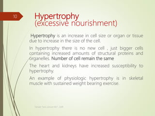
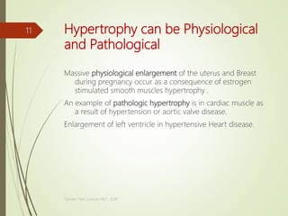
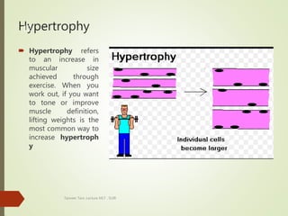
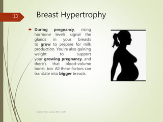
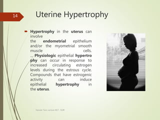
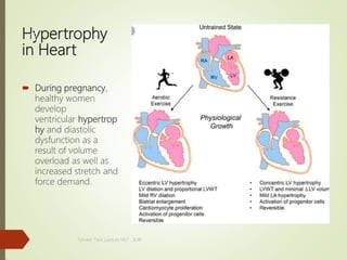
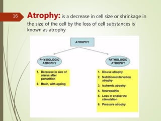
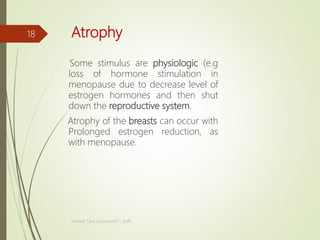
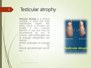
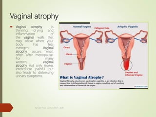
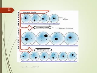
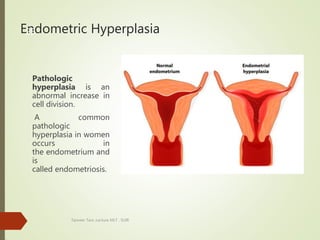
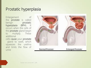
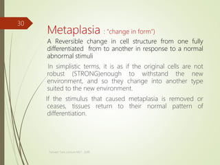
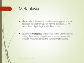
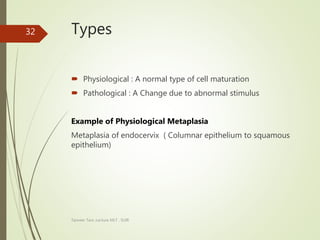
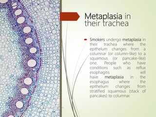
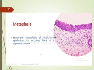
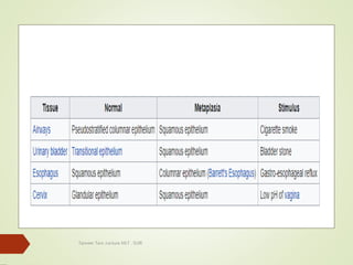
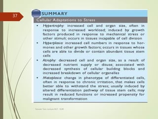
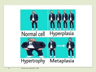
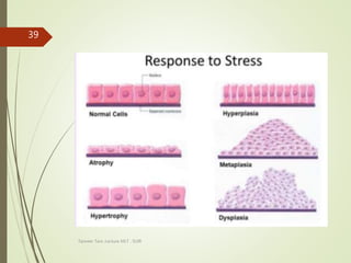
Tanveer Tara ,Lecture MLT , SUIR
1
Cellular adaptation
Adaptation
The process of change by which an organism or species
becomes better suited to its environment.
Cellular adaptation
In cell biology and pathophysiology, cellular
adaptation refers to changes made by a cell in response to
adverse environmental changes. The adaptation may be
physiologic(al) (normal) or pathologic(al) (abnormal). Five
minor types of adaptation include atrophy, hypertrophy,
hyperplasia, dysplasia, and metaplasia
Tanveer Tara ,Lecture MLT , SUIR
2
Cell
Tanveer Tara ,Lecture MLT , SUIR
3
CELL Biology
Cell has three components.
Cytoplasm ,Cell membrane , nucleus
In cytoplasm there are some organelles which have
some various function these are
Mitochondria
Power house of the cell.
Ribosomes
Synthesize protein in the cell
Lysosome
Intracellular digestion and removed unwanted substances.
Tanveer Tara ,Lecture MLT , SUIR
4
CELL Biology
Endoplasmic reticulum
Detoxifies the damaging substance to the cell.
Cell Membrane.
It maintain fluid ,ionic balance and provide pathway for
controlled transport of various substances.
Nucleus.
Control center of the cell and composed of DNA.
Tanveer Tara ,Lecture MLT , SUIR
5
Tanveer Tara ,Lecture MLT , SUIR
6
Adaption
Tanveer Tara ,Lecture MLT , SUIR
7
Cellular adaptation
In cell biology and Pathology cellular adaption refers to
change made by a cell in response to adverse
Environmental changes.
Adaptation is basically an adjustment of the cell of the
cell with in limits to the environment in which altered
steady state is achieved to preserve the viability of the
cell despite continued stress.
Stress may physiological or pathologic stimuli,
Tanveer Tara ,Lecture MLT , SUIR
8
Examples of Cellular adaptation
Hypertrophy
Atrophy
Hyperplasia
Metaplasia
Tanveer Tara ,Lecture MLT , SUIR
9
Plasia means cellular multiplication
Hypertrophy
(excessive nourishment)
Hypertrophy is an increase in cell size or organ or tissue
due to increase in the size of the cell.
In hypertrophy there is no new cell , just bigger cells
containing increased amounts of structural proteins and
organelles. Number of cell remain the same
The heart and kidneys have increased susceptibility to
hypertrophy.
An example of physiologic hypertrophy is in skeletal
muscle with sustained weight bearing exercise.
Tanveer Tara ,Lecture MLT , SUIR
10
Hypertrophy can be Physiological
and Pathological
Massive physiological enlargement of the uterus and Breast
during pregnancy occur as a consequence of estrogen
stimulated smooth muscles hypertrophy .
An example of pathologic hypertrophy is in cardiac muscle as
a result of hypertension or aortic valve disease.
Enlargement of left ventricle in hypertensive Heart disease.
Tanveer Tara ,Lecture MLT , SUIR
11
Hypertrophy
 Hypertrophy refers
to an increase in
muscular size
achieved through
exercise. When you
work out, if you want
to tone or improve
muscle definition,
lifting weights is the
most common way to
increase hypertroph
y
Tanveer Tara ,Lecture MLT , SUIR
12
Breast Hypertrophy
 During pregnancy, rising
hormone levels signal the
glands in your breasts
to grow to prepare for milk
production. You're also gaining
weight to support
your growing pregnancy, and
there's that blood-volume
boost, too. All these factors can
translate into bigger breasts
Tanveer Tara ,Lecture MLT , SUIR
13
Uterine Hypertrophy
 Hypertrophy in the uterus can
involve
the endometrial epithelium
and/or the myometrial smooth
muscle cells.
... Physiologic epithelial hypertro
phy can occur in response to
increased circulating estrogen
levels during the estrous cycle.
Compounds that have estrogenic
activity can induce
epithelial hypertrophy in
the uterus.
Tanveer Tara ,Lecture MLT , SUIR
14
Hypertrophy
in Heart
 During pregnancy,
healthy women
develop
ventricular hypertrop
hy and diastolic
dysfunction as a
result of volume
overload as well as
increased stretch and
force demand.
.
Tanveer Tara ,Lecture MLT , SUIR
15
Tanveer Tara ,Lecture MLT , SUIR
16 Atrophy: is a decrease in cell size or shrinkage in
the size of the cell by the loss of cell substances is
known as atrophy
Causes of atrophy
Decreased workload( e.g immobilization of a
limb to permit healing of a fracture)
Loss of innervation
Diminished blood supply
Inadequate nutrition
Loss of endocrine stimulation
Aging(loss of teeth, hair, thinning of skin that
creates wrinkles, weakening of muscles, loss of
weight in organs and sluggish mental activity)
Tanveer Tara ,Lecture MLT , SUIR
17
Atrophy
Some stimulus are physiologic (e.g
loss of hormone stimulation in
menopause due to decrease level of
estrogen hormones and then shut
down the reproductive system.
Atrophy of the breasts can occur with
Prolonged estrogen reduction, as
with menopause.
Tanveer Tara ,Lecture MLT , SUIR
18
Testicular atrophy
Testicular atrophy is a medical
condition in which the male
reproductive organs (the
testes, which in humans are
located in the scrotum)
diminish in size and may be
accompanied by loss of
function. with prolonged use
of enough exogenous sex
steroid
(either androgen or estroge
n) to
reduce gonadotropin secret
ion.
Tanveer Tara ,Lecture MLT , SUIR
19
Vaginal atrophy
 Vaginal atrophy is
thinning, drying and
inflammation of
the vaginal walls that
may occur when your
body has less
estrogen. Vaginal
atrophy occurs most
often after menopause.
For many
women, vaginal
atrophy not only makes
intercourse painful but
also leads to distressing
urinary symptoms.
Tanveer Tara ,Lecture MLT , SUIR
20
Penis Atrophy
 As men age, fatty deposits build up
in the arteries causing reduced
blood flow to the penis. This
results in the muscle cells in the
erectile tubes inside
the penis becoming weaker. ... This
accumulation of scar tissue affects
the spongy erectile tissues of
the penis, causes penis shrinkage,
and limits erection size.
Tanveer Tara ,Lecture MLT , SUIR
21
Mechanism of atrophy
Atrophy mechanism consist of a combination of
decrease protein synthesis and increased protein
degradation in cell.
Protein synthesis decrease b/c of reduced metabolic
activity.
In many situations , atrophy is also accompanied by
increase autophagy,.
Autophagy (self eating) is the process in which the
starved cell eats its own components in an attempt to
survive.
Tanveer Tara ,Lecture MLT , SUIR
22
Tanveer Tara ,Lecture MLT , SUIR
23
Hyperplasia
Hyperplasia is an increase in the number of cells which
result increase the size of an organ.
It is the result of increased cell mitosis, or division .
Hyperplasia can be physiological or pathological.
.
Tanveer Tara ,Lecture MLT , SUIR
24
Endometric Hyperplasia
Pathologic
hyperplasia is an
abnormal increase in
cell division.
A common
pathologic
hyperplasia in women
occurs in
the endometrium and
is
called endometriosis.
Tanveer Tara ,Lecture MLT , SUIR
25
Prostatic hyperplasia
Enlargement of
the prostate is called
benign prostatic
hyperplasia (BPH). It
occurs when the cells of
the prostate gland begin
to multiply. These
additional
cells cause your prostate
gland to swell, which
squeezes the urethra
and limits the flow of
urine
Tanveer Tara ,Lecture MLT , SUIR
26
vaginal hyperplasia,
 vaginal hyperplasia, a
proliferation of
the vaginal mucosa, usually
originating from the floor of
the vagina anterior to the
urethral orifice, occurs during
proestrus and estrus as a
result of estrogenic
stimulation. Occasionally,
the prolapse continues
throughout pregnancy or
recurs at parturition.
Tanveer Tara ,Lecture MLT , SUIR
27
Sebaceous hyperplasia
 In this condition, small
yellowish growths
develop on the skin,
usually on the face. This
condition is neither
contagious nor
dangerous.
For example, the
estrogen-dependent
uterine cells undergo
hyperplasia and
hypertrophy following
pregnancy.
Tanveer Tara ,Lecture MLT , SUIR
28
Penile shaft
 Sebaceous hyperplasia of the
scrotum and penile shaft.
... Sebaceous hyperplasia (SH)
is a proliferative abnormality
of sebaceous glands, most
often seen in elderly
individuals. It consists of single
or multiple, asymptomatic,
small yellow papules with a
central depression.
https://www.ncbi.nlm.nih.gov/pubmed/22
346273
Tanveer Tara ,Lecture MLT , SUIR
29
Metaplasia : "change in form")
A Reversible change in cell structure from one fully
differentiated from to another in response to a normal
abnormal stimuli
In simplistic terms, it is as if the original cells are not
robust (STRONG)enough to withstand the new
environment, and so they change into another type
suited to the new environment.
If the stimulus that caused metaplasia is removed or
ceases, tissues return to their normal pattern of
differentiation.
Tanveer Tara ,Lecture MLT , SUIR
30
Metaplasia
 Metaplasia is the conversion from one type of normal
adult cell to another type of normal adult cell. ... An
example of physiologic metaplasia is the
 squamous metaplasia that occurs in the uterine cervix
during the menstrual cycle as the squamocolumnar
junction migrates across the transformation zone
Tanveer Tara ,Lecture MLT , SUIR
31
Types
 Physiological : A normal type of cell maturation
 Pathological : A Change due to abnormal stimulus
Example of Physiological Metaplasia
Metaplasia of endocervix ( Columnar epithelium to squamous
epithelium)
Tanveer Tara ,Lecture MLT , SUIR
32
These transformed cells may become dysplastic or cancerous
if the stimulus (e.g., cigarette smoking) is not Stop.
If stress persists, metaplasia can progress to dysplasia and
eventually carcinoma;
Tanveer Tara ,Lecture MLT , SUIR
33
Metaplasia in
their trachea
 Smokers undergo metaplasia in
their trachea where the
epithelum changes from a
columnar (or column-like) to a
squamous (or pancake-like)
one. People who have
conditions such as reflux
esophagitis will
have metaplasia in the
esophagus where the
epithelium changes from
stratified squamous (stack of
pancakes) to columnar.
Tanveer Tara ,Lecture MLT , SUIR
34
Tanveer Tara ,Lecture MLT , SUIR
35
Tanveer Tara ,Lecture MLT , SUIR
36
Tanveer Tara ,Lecture MLT , SUIR
37
38 Tanveer Tara ,Lecture MLT , SUIR
Tanveer Tara ,Lecture MLT , SUIR
39
Tanveer Tara ,Lecture MLT , SUIR
40
Tanveer Tara ,Lecture MLT , SUIR
41
Tanveer Tara ,Lecture MLT , SUIR
42
Tanveer Tara ,Lecture MLT , SUIR
43
APLASIA vs Hypolasia
is that hypoplasia is
underdevelopment or incomplete
development of a tissue or organ,
especially when caused by an
inadequate or below-normal number
of cells while aplasia is (pathology) a
condition marked by the incomplete
development, or absence, of an organ
or tissue.
Tanveer Tara ,Lecture MLT , SUIR
44
THANK YOU
Tanveer Tara ,Lecture MLT , SUIR
45

cell adaptation cell injury

  • 1.
  • 2.
    Cellular adaptation Adaptation The processof change by which an organism or species becomes better suited to its environment. Cellular adaptation In cell biology and pathophysiology, cellular adaptation refers to changes made by a cell in response to adverse environmental changes. The adaptation may be physiologic(al) (normal) or pathologic(al) (abnormal). Five minor types of adaptation include atrophy, hypertrophy, hyperplasia, dysplasia, and metaplasia Tanveer Tara ,Lecture MLT , SUIR 2
  • 3.
  • 4.
    CELL Biology Cell hasthree components. Cytoplasm ,Cell membrane , nucleus In cytoplasm there are some organelles which have some various function these are Mitochondria Power house of the cell. Ribosomes Synthesize protein in the cell Lysosome Intracellular digestion and removed unwanted substances. Tanveer Tara ,Lecture MLT , SUIR 4
  • 5.
    CELL Biology Endoplasmic reticulum Detoxifiesthe damaging substance to the cell. Cell Membrane. It maintain fluid ,ionic balance and provide pathway for controlled transport of various substances. Nucleus. Control center of the cell and composed of DNA. Tanveer Tara ,Lecture MLT , SUIR 5
  • 6.
  • 7.
  • 8.
    Cellular adaptation In cellbiology and Pathology cellular adaption refers to change made by a cell in response to adverse Environmental changes. Adaptation is basically an adjustment of the cell of the cell with in limits to the environment in which altered steady state is achieved to preserve the viability of the cell despite continued stress. Stress may physiological or pathologic stimuli, Tanveer Tara ,Lecture MLT , SUIR 8
  • 9.
    Examples of Cellularadaptation Hypertrophy Atrophy Hyperplasia Metaplasia Tanveer Tara ,Lecture MLT , SUIR 9 Plasia means cellular multiplication
  • 10.
    Hypertrophy (excessive nourishment) Hypertrophy isan increase in cell size or organ or tissue due to increase in the size of the cell. In hypertrophy there is no new cell , just bigger cells containing increased amounts of structural proteins and organelles. Number of cell remain the same The heart and kidneys have increased susceptibility to hypertrophy. An example of physiologic hypertrophy is in skeletal muscle with sustained weight bearing exercise. Tanveer Tara ,Lecture MLT , SUIR 10
  • 11.
    Hypertrophy can bePhysiological and Pathological Massive physiological enlargement of the uterus and Breast during pregnancy occur as a consequence of estrogen stimulated smooth muscles hypertrophy . An example of pathologic hypertrophy is in cardiac muscle as a result of hypertension or aortic valve disease. Enlargement of left ventricle in hypertensive Heart disease. Tanveer Tara ,Lecture MLT , SUIR 11
  • 12.
    Hypertrophy  Hypertrophy refers toan increase in muscular size achieved through exercise. When you work out, if you want to tone or improve muscle definition, lifting weights is the most common way to increase hypertroph y Tanveer Tara ,Lecture MLT , SUIR 12
  • 13.
    Breast Hypertrophy  Duringpregnancy, rising hormone levels signal the glands in your breasts to grow to prepare for milk production. You're also gaining weight to support your growing pregnancy, and there's that blood-volume boost, too. All these factors can translate into bigger breasts Tanveer Tara ,Lecture MLT , SUIR 13
  • 14.
    Uterine Hypertrophy  Hypertrophyin the uterus can involve the endometrial epithelium and/or the myometrial smooth muscle cells. ... Physiologic epithelial hypertro phy can occur in response to increased circulating estrogen levels during the estrous cycle. Compounds that have estrogenic activity can induce epithelial hypertrophy in the uterus. Tanveer Tara ,Lecture MLT , SUIR 14
  • 15.
    Hypertrophy in Heart  Duringpregnancy, healthy women develop ventricular hypertrop hy and diastolic dysfunction as a result of volume overload as well as increased stretch and force demand. . Tanveer Tara ,Lecture MLT , SUIR 15
  • 16.
    Tanveer Tara ,LectureMLT , SUIR 16 Atrophy: is a decrease in cell size or shrinkage in the size of the cell by the loss of cell substances is known as atrophy
  • 17.
    Causes of atrophy Decreasedworkload( e.g immobilization of a limb to permit healing of a fracture) Loss of innervation Diminished blood supply Inadequate nutrition Loss of endocrine stimulation Aging(loss of teeth, hair, thinning of skin that creates wrinkles, weakening of muscles, loss of weight in organs and sluggish mental activity) Tanveer Tara ,Lecture MLT , SUIR 17
  • 18.
    Atrophy Some stimulus arephysiologic (e.g loss of hormone stimulation in menopause due to decrease level of estrogen hormones and then shut down the reproductive system. Atrophy of the breasts can occur with Prolonged estrogen reduction, as with menopause. Tanveer Tara ,Lecture MLT , SUIR 18
  • 19.
    Testicular atrophy Testicular atrophyis a medical condition in which the male reproductive organs (the testes, which in humans are located in the scrotum) diminish in size and may be accompanied by loss of function. with prolonged use of enough exogenous sex steroid (either androgen or estroge n) to reduce gonadotropin secret ion. Tanveer Tara ,Lecture MLT , SUIR 19
  • 20.
    Vaginal atrophy  Vaginalatrophy is thinning, drying and inflammation of the vaginal walls that may occur when your body has less estrogen. Vaginal atrophy occurs most often after menopause. For many women, vaginal atrophy not only makes intercourse painful but also leads to distressing urinary symptoms. Tanveer Tara ,Lecture MLT , SUIR 20
  • 21.
    Penis Atrophy  Asmen age, fatty deposits build up in the arteries causing reduced blood flow to the penis. This results in the muscle cells in the erectile tubes inside the penis becoming weaker. ... This accumulation of scar tissue affects the spongy erectile tissues of the penis, causes penis shrinkage, and limits erection size. Tanveer Tara ,Lecture MLT , SUIR 21
  • 22.
    Mechanism of atrophy Atrophymechanism consist of a combination of decrease protein synthesis and increased protein degradation in cell. Protein synthesis decrease b/c of reduced metabolic activity. In many situations , atrophy is also accompanied by increase autophagy,. Autophagy (self eating) is the process in which the starved cell eats its own components in an attempt to survive. Tanveer Tara ,Lecture MLT , SUIR 22
  • 23.
    Tanveer Tara ,LectureMLT , SUIR 23
  • 24.
    Hyperplasia Hyperplasia is anincrease in the number of cells which result increase the size of an organ. It is the result of increased cell mitosis, or division . Hyperplasia can be physiological or pathological. . Tanveer Tara ,Lecture MLT , SUIR 24
  • 25.
    Endometric Hyperplasia Pathologic hyperplasia isan abnormal increase in cell division. A common pathologic hyperplasia in women occurs in the endometrium and is called endometriosis. Tanveer Tara ,Lecture MLT , SUIR 25
  • 26.
    Prostatic hyperplasia Enlargement of theprostate is called benign prostatic hyperplasia (BPH). It occurs when the cells of the prostate gland begin to multiply. These additional cells cause your prostate gland to swell, which squeezes the urethra and limits the flow of urine Tanveer Tara ,Lecture MLT , SUIR 26
  • 27.
    vaginal hyperplasia,  vaginalhyperplasia, a proliferation of the vaginal mucosa, usually originating from the floor of the vagina anterior to the urethral orifice, occurs during proestrus and estrus as a result of estrogenic stimulation. Occasionally, the prolapse continues throughout pregnancy or recurs at parturition. Tanveer Tara ,Lecture MLT , SUIR 27
  • 28.
    Sebaceous hyperplasia  Inthis condition, small yellowish growths develop on the skin, usually on the face. This condition is neither contagious nor dangerous. For example, the estrogen-dependent uterine cells undergo hyperplasia and hypertrophy following pregnancy. Tanveer Tara ,Lecture MLT , SUIR 28
  • 29.
    Penile shaft  Sebaceoushyperplasia of the scrotum and penile shaft. ... Sebaceous hyperplasia (SH) is a proliferative abnormality of sebaceous glands, most often seen in elderly individuals. It consists of single or multiple, asymptomatic, small yellow papules with a central depression. https://www.ncbi.nlm.nih.gov/pubmed/22 346273 Tanveer Tara ,Lecture MLT , SUIR 29
  • 30.
    Metaplasia : "changein form") A Reversible change in cell structure from one fully differentiated from to another in response to a normal abnormal stimuli In simplistic terms, it is as if the original cells are not robust (STRONG)enough to withstand the new environment, and so they change into another type suited to the new environment. If the stimulus that caused metaplasia is removed or ceases, tissues return to their normal pattern of differentiation. Tanveer Tara ,Lecture MLT , SUIR 30
  • 31.
    Metaplasia  Metaplasia isthe conversion from one type of normal adult cell to another type of normal adult cell. ... An example of physiologic metaplasia is the  squamous metaplasia that occurs in the uterine cervix during the menstrual cycle as the squamocolumnar junction migrates across the transformation zone Tanveer Tara ,Lecture MLT , SUIR 31
  • 32.
    Types  Physiological :A normal type of cell maturation  Pathological : A Change due to abnormal stimulus Example of Physiological Metaplasia Metaplasia of endocervix ( Columnar epithelium to squamous epithelium) Tanveer Tara ,Lecture MLT , SUIR 32
  • 33.
    These transformed cellsmay become dysplastic or cancerous if the stimulus (e.g., cigarette smoking) is not Stop. If stress persists, metaplasia can progress to dysplasia and eventually carcinoma; Tanveer Tara ,Lecture MLT , SUIR 33
  • 34.
    Metaplasia in their trachea Smokers undergo metaplasia in their trachea where the epithelum changes from a columnar (or column-like) to a squamous (or pancake-like) one. People who have conditions such as reflux esophagitis will have metaplasia in the esophagus where the epithelium changes from stratified squamous (stack of pancakes) to columnar. Tanveer Tara ,Lecture MLT , SUIR 34
  • 35.
    Tanveer Tara ,LectureMLT , SUIR 35
  • 36.
    Tanveer Tara ,LectureMLT , SUIR 36
  • 37.
    Tanveer Tara ,LectureMLT , SUIR 37
  • 38.
    38 Tanveer Tara,Lecture MLT , SUIR
  • 39.
    Tanveer Tara ,LectureMLT , SUIR 39
  • 40.
    Tanveer Tara ,LectureMLT , SUIR 40
  • 41.
    Tanveer Tara ,LectureMLT , SUIR 41
  • 42.
    Tanveer Tara ,LectureMLT , SUIR 42
  • 43.
    Tanveer Tara ,LectureMLT , SUIR 43
  • 44.
    APLASIA vs Hypolasia isthat hypoplasia is underdevelopment or incomplete development of a tissue or organ, especially when caused by an inadequate or below-normal number of cells while aplasia is (pathology) a condition marked by the incomplete development, or absence, of an organ or tissue. Tanveer Tara ,Lecture MLT , SUIR 44
  • 45.
    THANK YOU Tanveer Tara,Lecture MLT , SUIR 45

Editor's Notes

  • #12 Aortic valve disease is a condition in which the valve between the main pumping chamber of your heart (left ventricle) and the main artery to your body (aorta) doesn't work properly. Aortic valve disease may be a condition present at birth (congenital heart disease), or it may result from other causes.
  • #17 Parturition: Giving Birth , defecation : Releasing of stool , micturition : Urine (Brain mass: Shrinkage in the frontal lobe and hippocampus - areas involved in higher cognitive function and encoding new memories - starting around the age of 60 or 70 years. ... Myelin is thought to shrink with age, and as a result, slow processing and reduce cognitive function
  • #18 When ultimately these motor units lose their innervation and there are no healthy axons left to connect with them, all their fibers shrink together (group atrophy). ... Denervation atrophy is caused by peripheral neuropathies and motor neuron diseases.
  • #26 Endometrial hyperplasia is a condition in which the endometrium (the lining of the uterus) becomes abnormally thick. Although endometrial hyperplasia is not cancer, it can lead to uterine cancer in some women. Endometrial hyperplasia is usually caused by an excess of estrogen without progesterone (female hormones).
  • #27 The mean weight of the normal prostate in adult males is about 11 grams, usually ranging between 7 and 16 grams.t
  • #29 sebaceous glandThe endometrium is the innermost lining layer of the uterus, and functions to prevent adhesions between the opposed walls of the myometrium, thereby maintaining the patency of the uterine cavity. During the menstrual cycle or estrous cycle, the endometrium grows to a thick, blood vessel-rich, glandular tissue layer. noun a small gland in the skin which secretes a lubricating oily matter (sebum) into the hair follicles to lubricate the skin and hair.