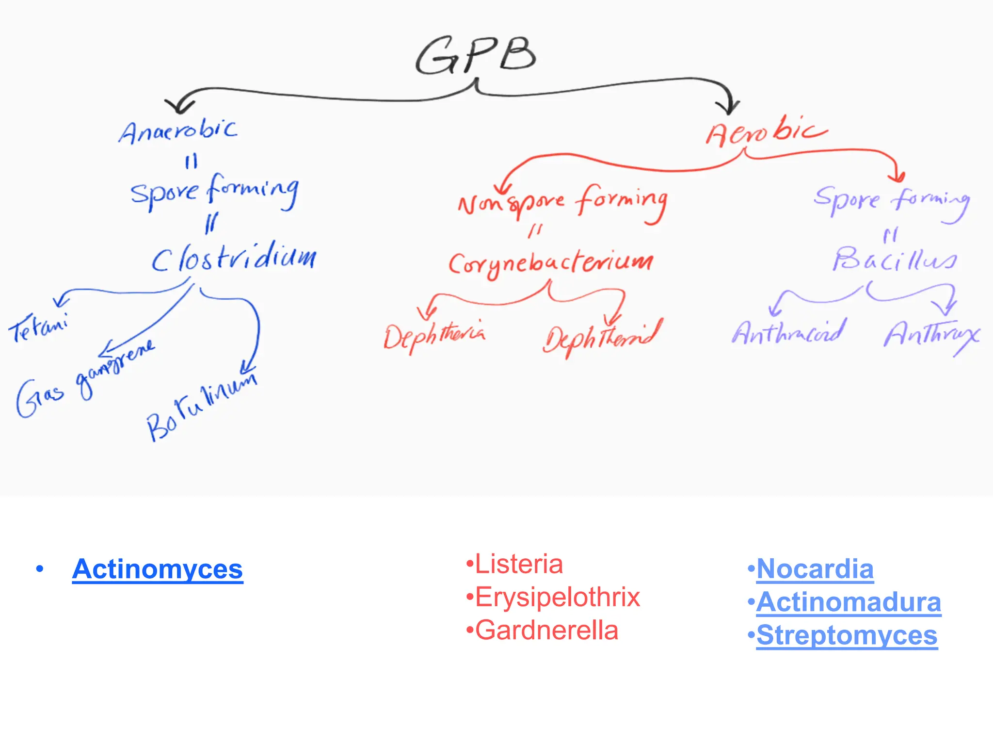
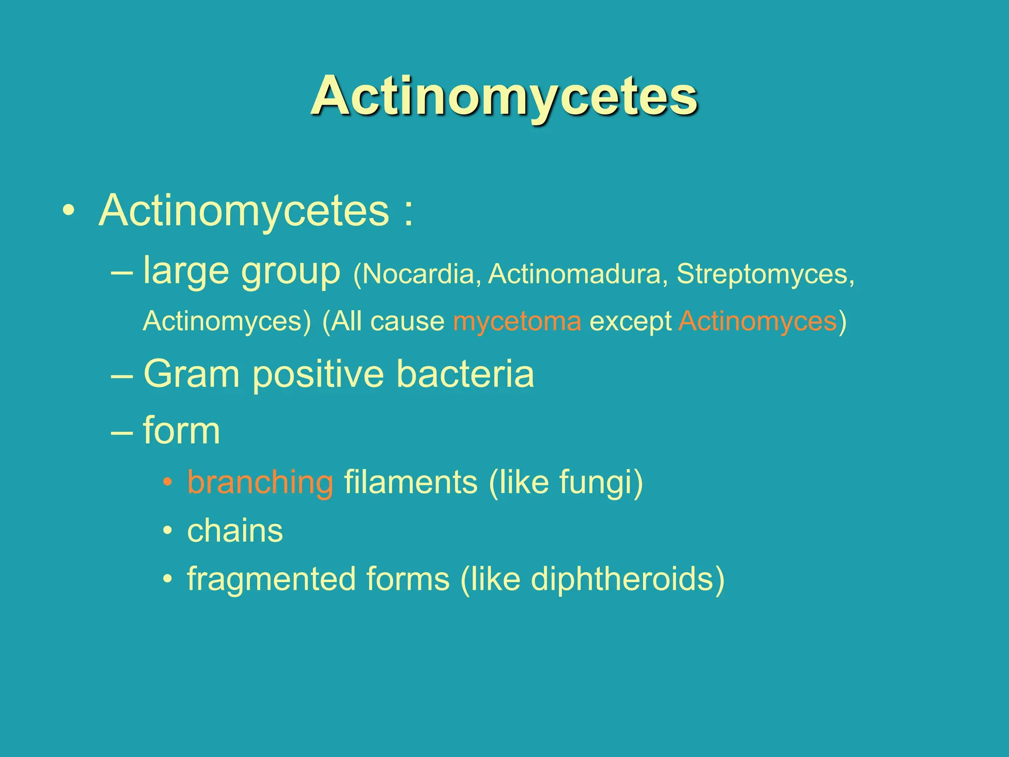
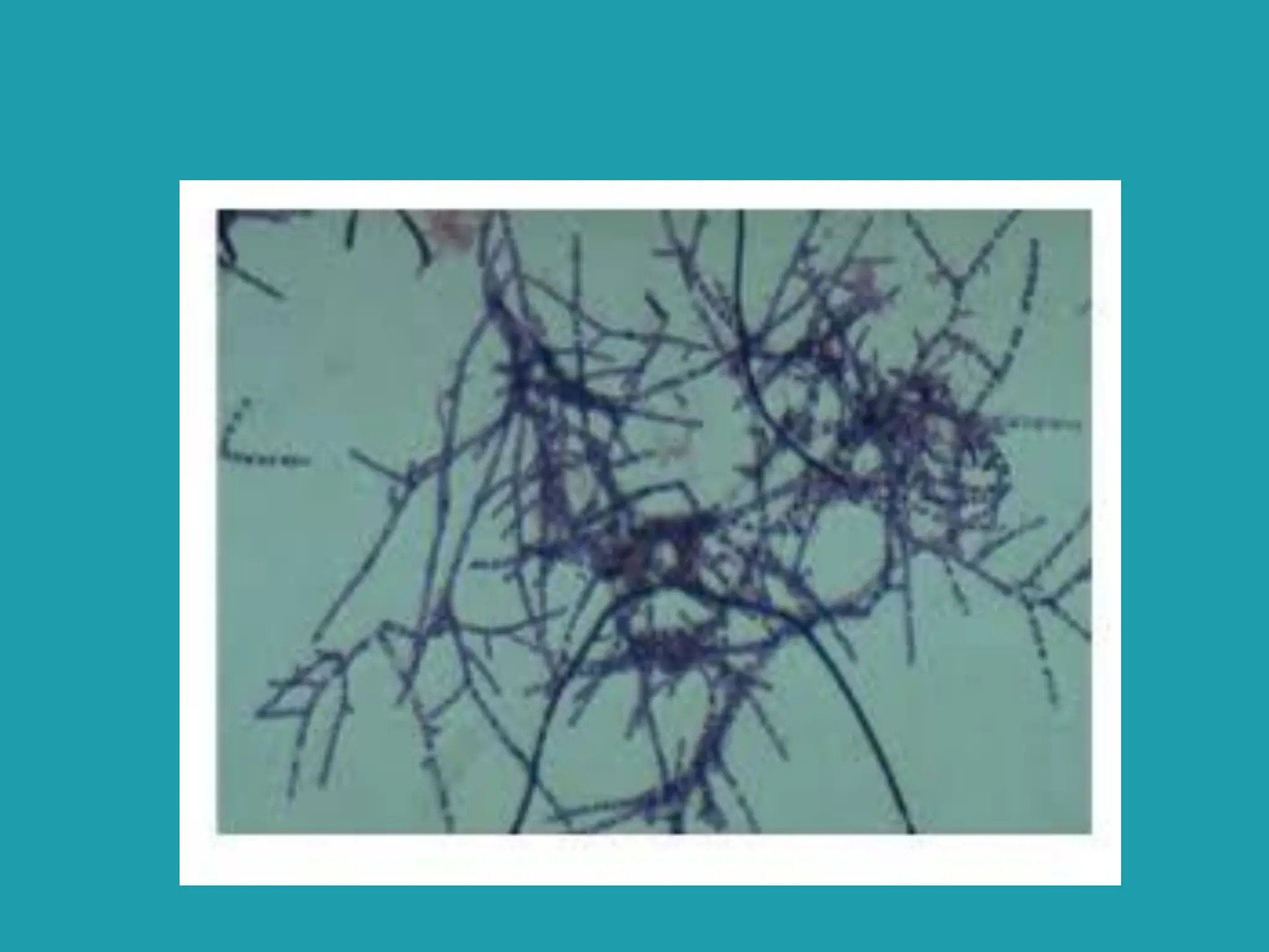
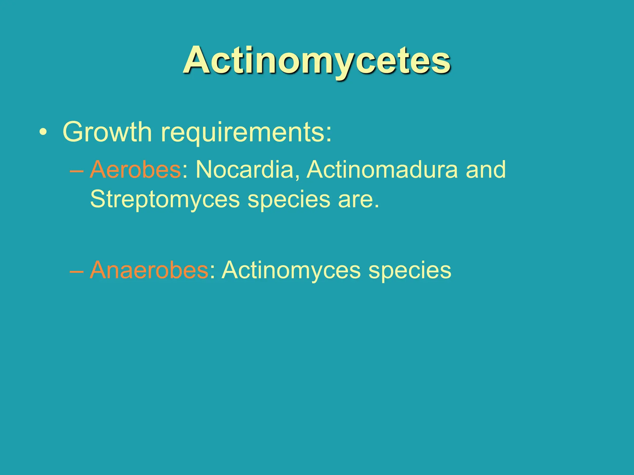
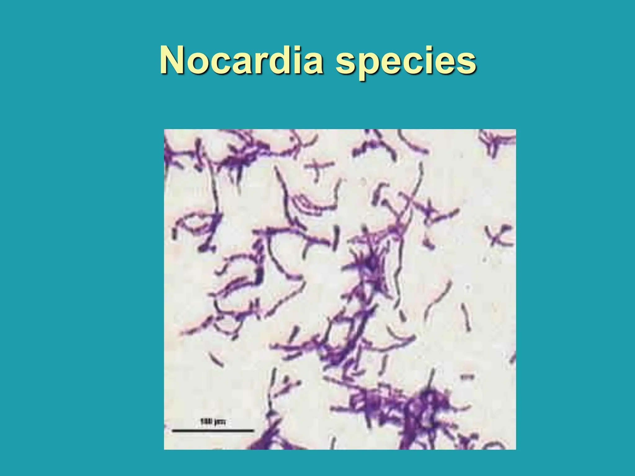
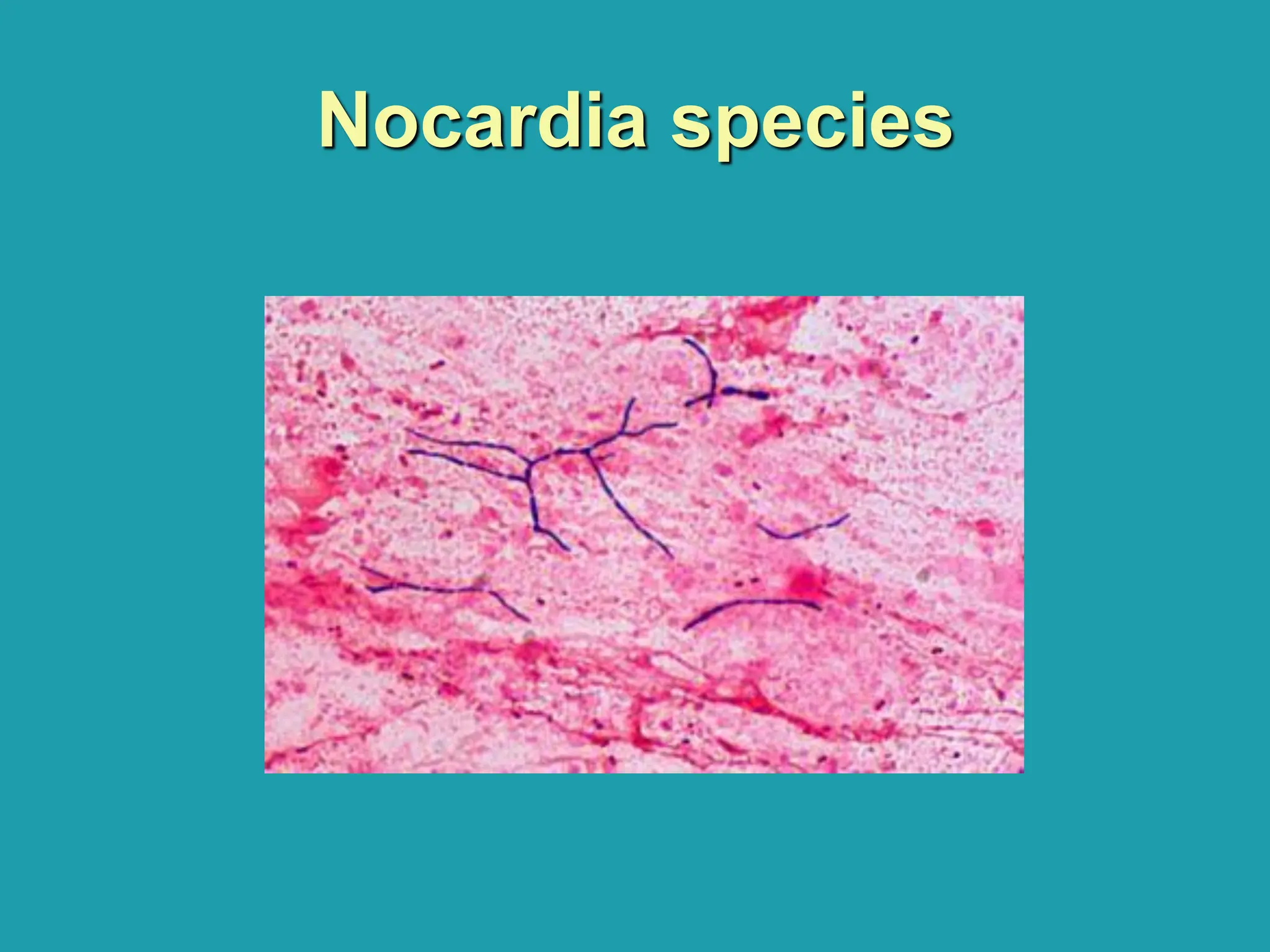
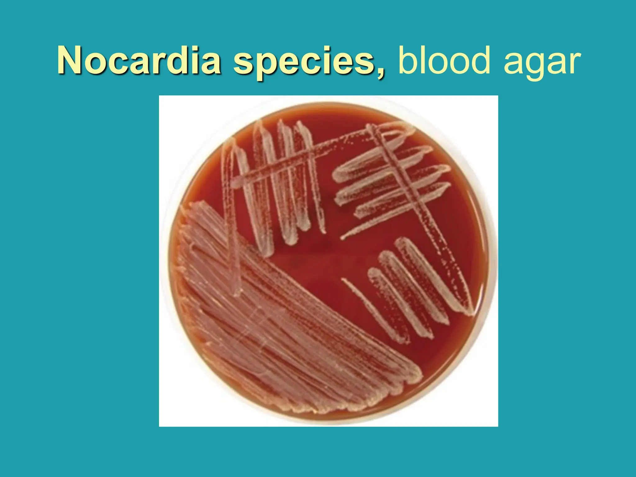
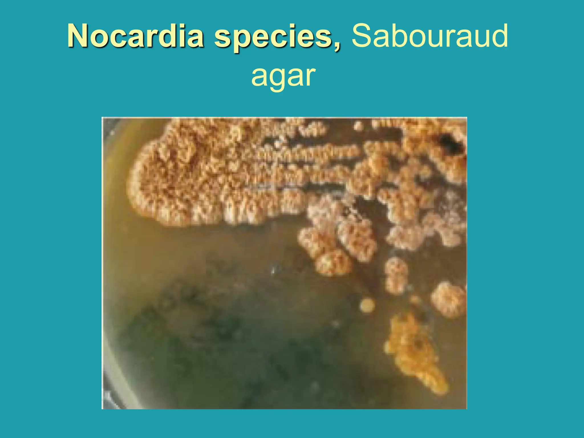
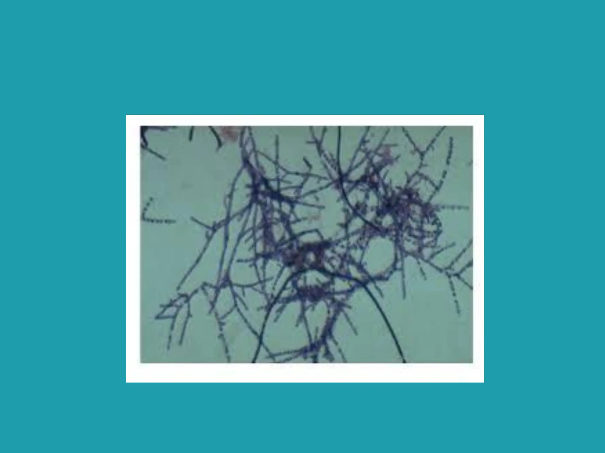
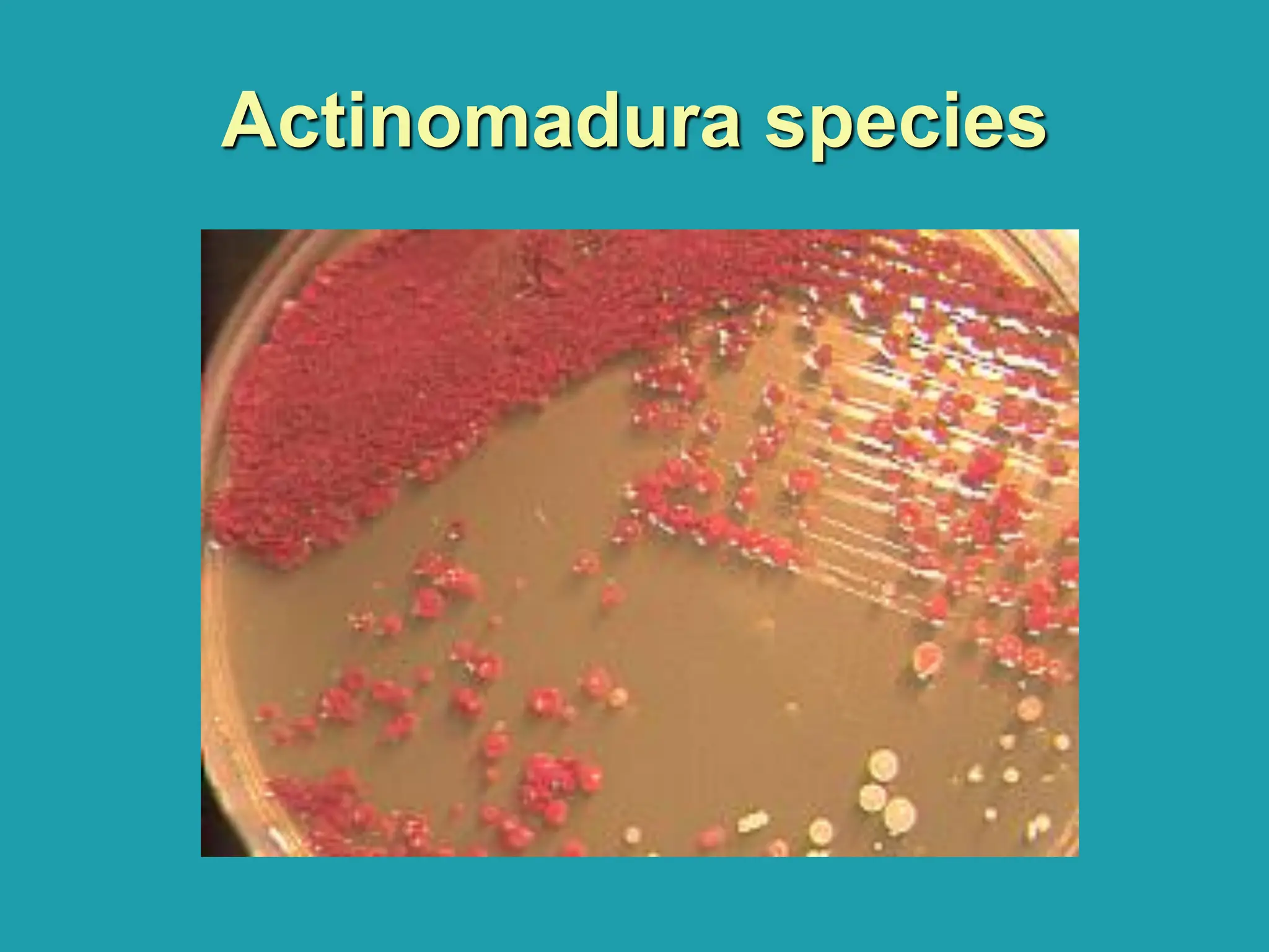
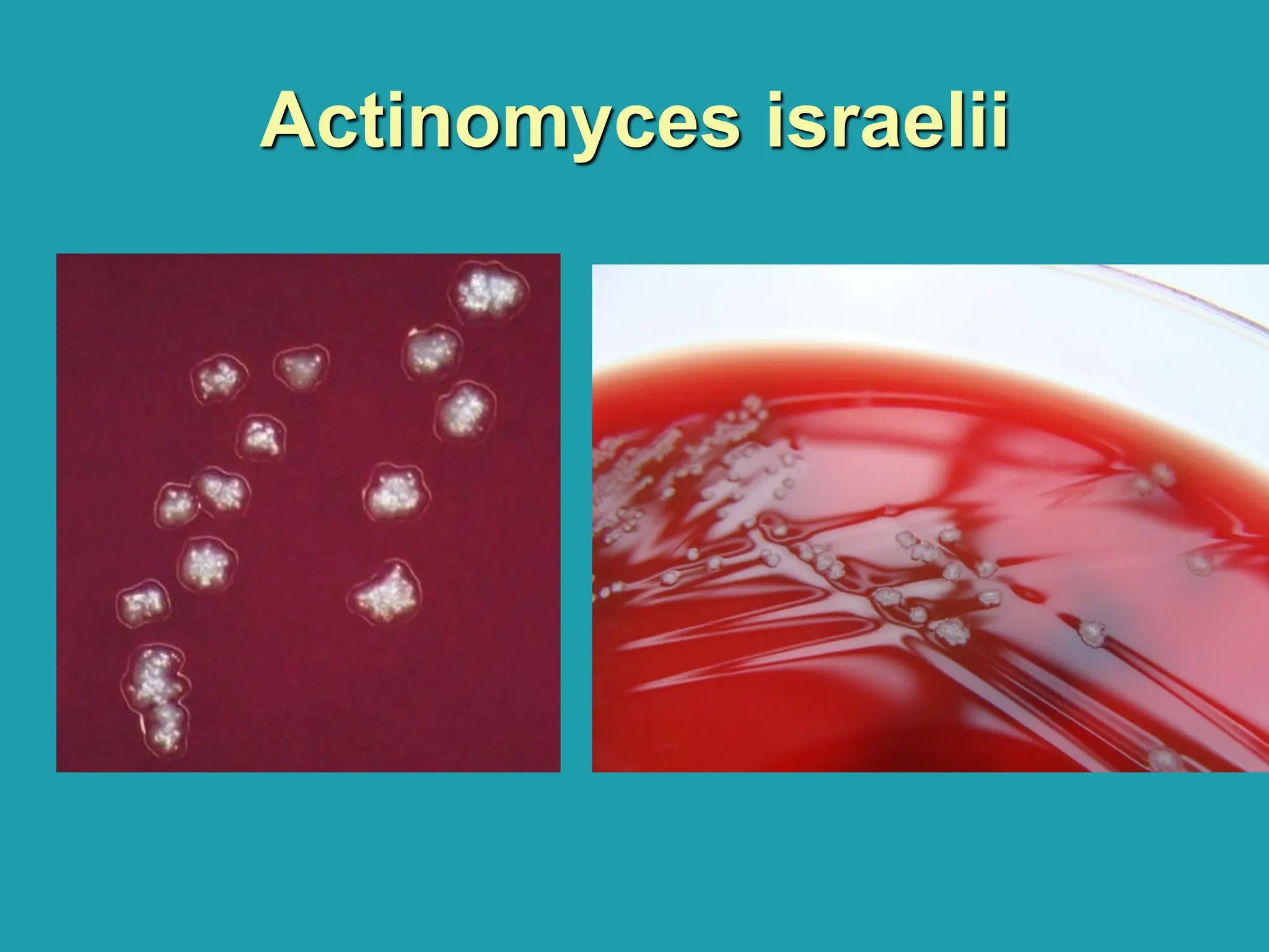
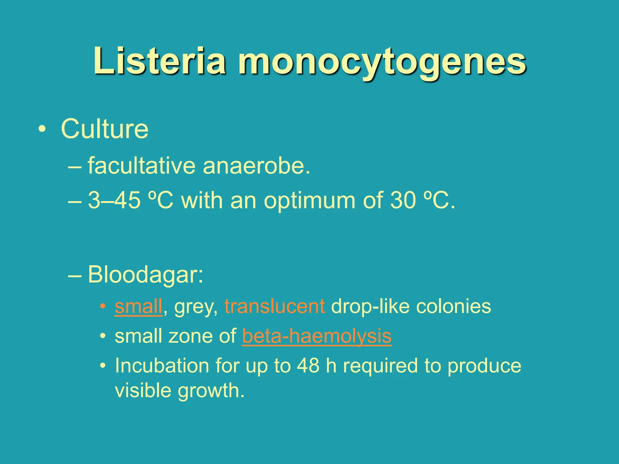
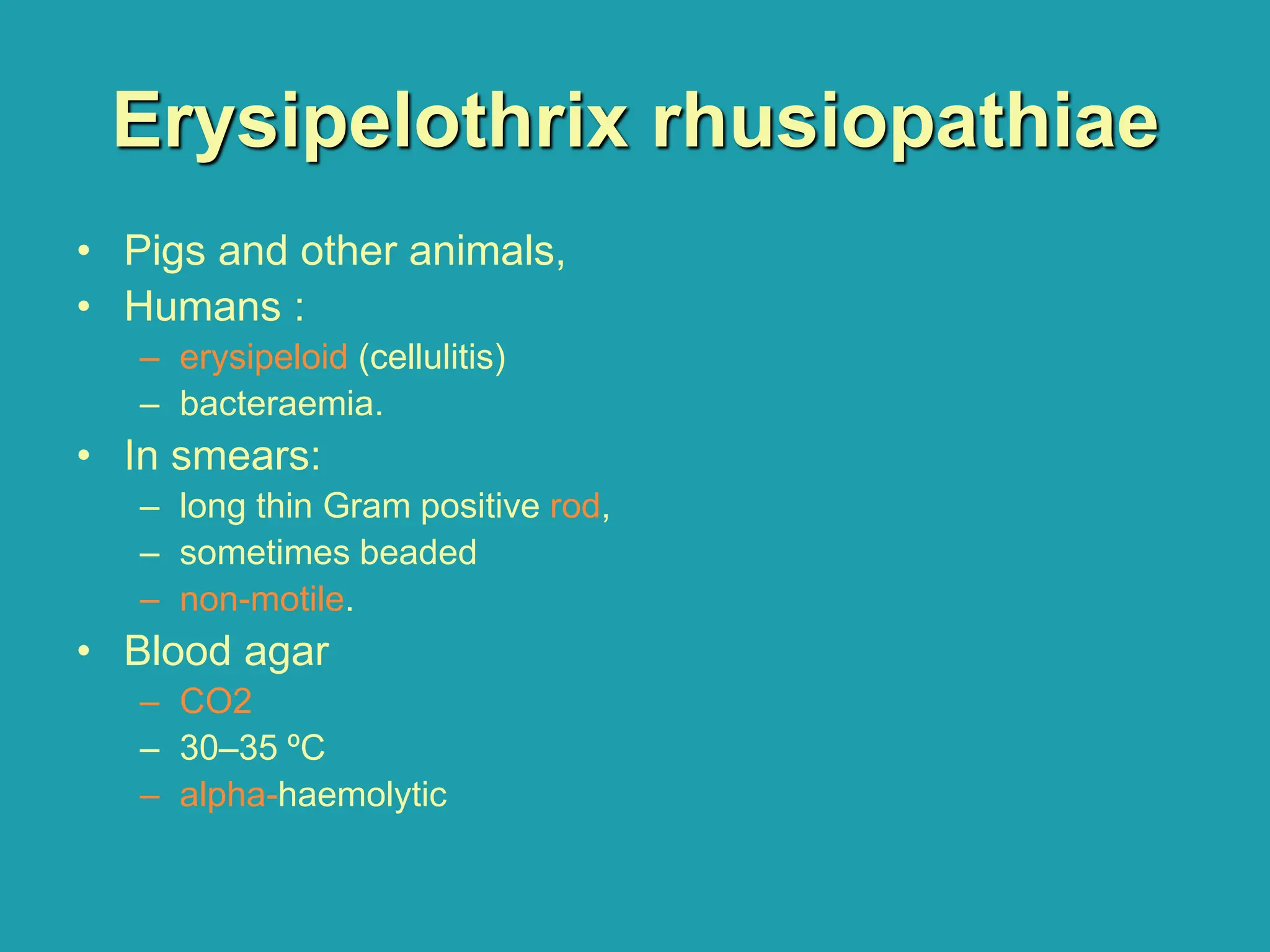
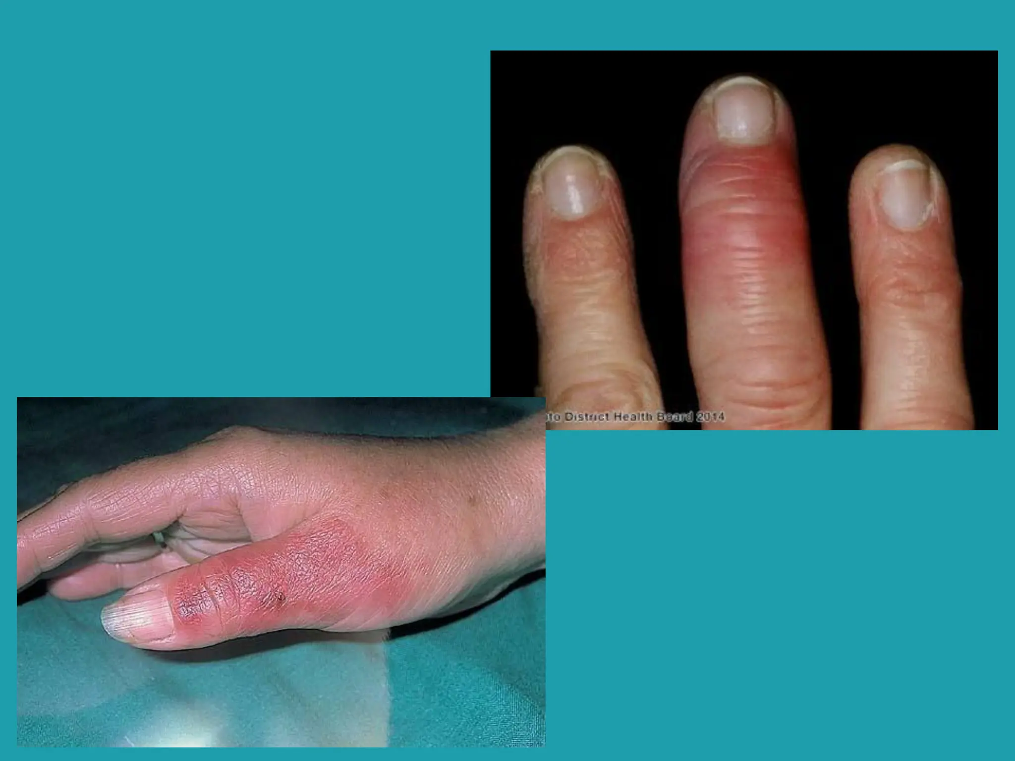
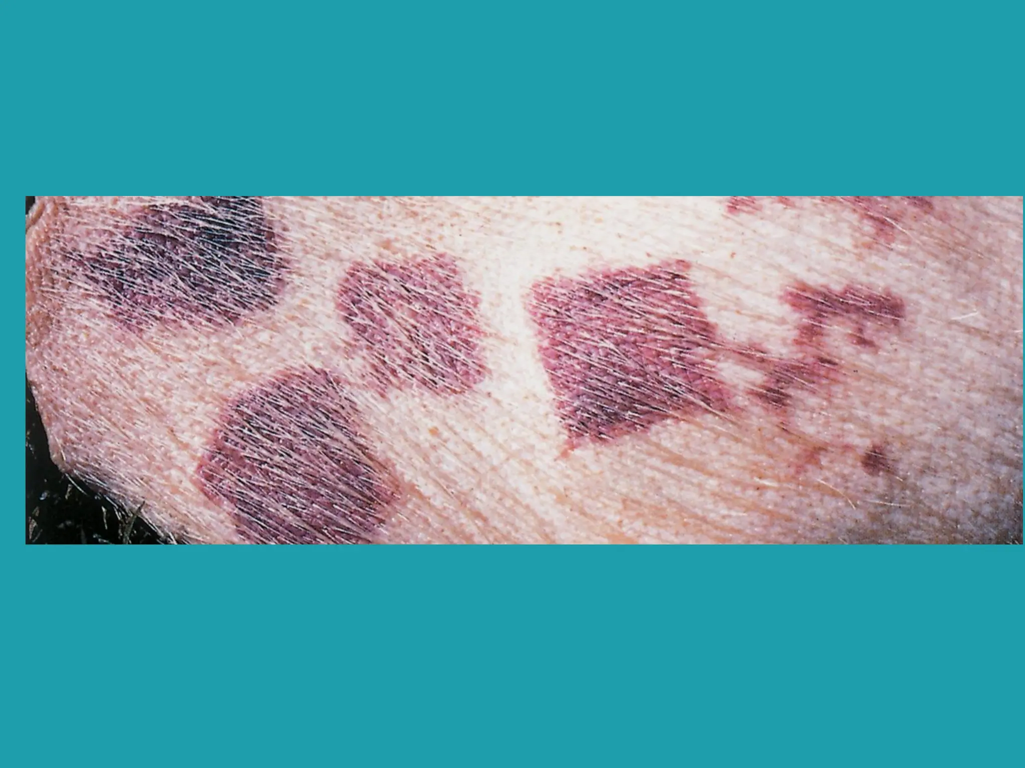
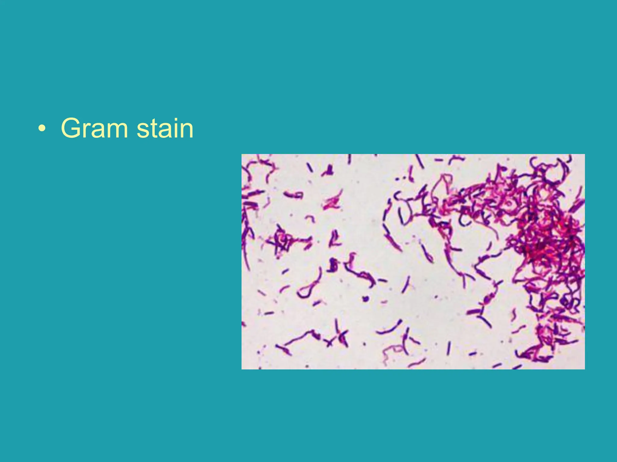
Other GPB
Dr. Mohammed Elkady
Clinical Pathology Consultant,
M.B.B.CH, EFCpath
• Actinomyces •Listeria
•Erysipelothrix
•Gardnerella
•Nocardia
•Actinomadura
•Streptomyces
Actinomycetes
• Actinomycetes :
– large group (Nocardia, Actinomadura, Streptomyces,
Actinomyces) (All cause mycetoma except Actinomyces)
– Gram positive bacteria
– form
• branching filaments (like fungi)
• chains
• fragmented forms (like diphtheroids)
Actinomycetes
• Growth requirements:
– Aerobes: Nocardia, Actinomadura and
Streptomyces species are.
– Anaerobes: Actinomyces species
Nocardia species
• Pathogenicity
– Actinomycetoma:
• Chronic granulomatous disease
• Affecting subcutaneous and deep tissues:
– pulmonary and lympho-cutaneous infection.
• specimen =mucopus, sputum, and
infected tissue
Mycetoma
• Granulomatous
disease caused by
actinomycetes and
fungi
Mycetoma, sampling
Nocardia species
• Morphology
– non-motile
– branching threads.
– Gram positive but often stain unevenly.
– weak acid fast.
• In mycetoma: small granules are
discharged in the pus.
Nocardia species
Nocardia species
Nocardia species
Nocardia species
• Culture
– Obligate aerobic.
– 45 ºC (help to isolate Nocardia from
specimens that contain commensals) as well
as at 35–37 ºC and room temperature.
– Can be cultured on Sabouraud agar at 35–37
ºC. for 3–14 days,
– Orange to pink, waxy folded colonies
• Catalase positive and urease positive
Nocardia species, blood agar
Nocardia species, Sabouraud
agar
Actinomadura species
• Pathogenicity
– mycetoma
• Specimens: Pus from draining sinuses
Actinomadura species
• Morphology
– non-motile and
– branching threads.
– NOT acid fast.
Actinomadura species
• Culture
– Grow slowly
– Require an enriched medium.
– Leathery appearance
– Red pigmentation
• Urease negative
Actinomadura species
Streptomyces
• Pathogenicity
– mycetoma
Streptomyces
• Morphology
Like other actinomycetes:
– nonmotile.
– branching threads.
– From specimen: NOT acid fast,
– Form cultures: are usually acid fast.
Streptomyces
• Culture
– Sabouraud agar
– room temperature or 35–37ºC.
– creamy or brown and have aerial hyphae,
giving them a heaped appearance.
• Urease negative.
Streptomyces
Streptomyces
Actinomyces israelii
• Pathogenicity:
– actinomycosis
• chronic granulomatous infection
• Pus (in granules) is discharged through sinuses which open
on the surface of the skin.
• commonly affecting the jaw, following the extraction of a
tooth, abdomen, brain and lungs
– Specimens: Depending on the site of infection,
specimens include pus, sputum or infected tissue.
Actinomyces israelii
• Morphology
– morphologically resemble other
actinomycetes.
– non-motile
– thin branches are easily fragmented.
– not acid fast,
– but the club-shaped forms that surround the
colony are acid fast.
Actinomyces israelii
• Gram stain
Actinomyces israelii
• Culture
– anaerobic and microaerophilic.
– culturing granules on blood agar
– slow-growing, 5–7 days
– 35–37 ºC.
– On Blood agar: small, cream or white colonies, adhere to the
medium.
• catalase, indole, urease negative.
• It hydrolyzes aesculin and ferments glucose, lactose,
mannitol, and several other sugars.
• Should be sent to a Reference Laboratory.
Actinomyces israelii
• For all granulomatous infection:
– Granules→ culture
Listeria monocytogenes
• Pathogenicity
– meningitis
– septicaemia
• mainly in neonates, pregnant women (abortion), the
elderly and immunosuppressed persons.
• sources of infection:
– contaminated meats, chicken, soft cheeses and vegetables.
• specimens: Mainly CSF and blood for culture.
Listeria monocytogenes
• Morphology
– Gram positive
– non-capsulate
– small rod or coccobacillus
– stains unevenly and is easily decolorized.
– resemble diphtheroids.
– Motility:
• At 35–37 ºC: nonmotile
• At low temperature (18–22 ºC) it is motile (tumbling and rotation
motility)
• Cerebrospinal fluid: In Listeria meningitis, only a few bacteria may
be present. The c.s.f. will usually contain lymphocytes and, or,
polymorphs. The c.s.f. total protein is raised.
Gram stain
Gram stain
Listeria monocytogenes
• Culture
– facultative anaerobe.
– 3–45 ºC with an optimum of 30 ºC.
– Bloodagar:
• small, grey, translucent drop-like colonies
• small zone of beta-haemolysis
• Incubation for up to 48 h required to produce
visible growth.
Beta hemolysis
Blood agar
Beta hemolysis
Blood agar
Listeria monocytogenes
• Biochemical reactions
– Catalase positive (diff from BH sterpt.)
– Indole, oxidase and urease negative.
– Ferments glucose and maltose with acid production.
• Note: The characteristic motility and cultural
characteristics of L. monocytogenes are usually
sufficient to identify it without the need to use
many biochemical tests.
Erysipelothrix rhusiopathiae
• Pigs and other animals,
• Humans :
– erysipeloid (cellulitis)
– bacteraemia.
• In smears:
– long thin Gram positive rod,
– sometimes beaded
– non-motile.
• Blood agar
– CO2
– 30–35 ºC
– alpha-haemolytic
• Gram stain
Alpha hemolysis
Blood agar
Gardnerella vaginalis
• Bacterial Vaginosis
• Gram variable coccobacilli
• Positive whiff test with KOH
• Clue cells
• Beta hemolytic colonies
• Clue cell
Gram positive bacilli morphology and classification

Gram positive bacilli morphology and classification