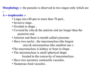
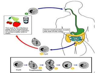
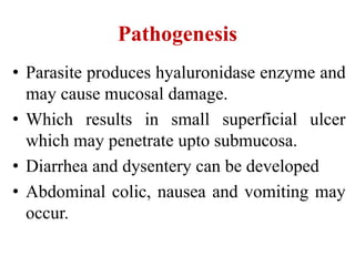
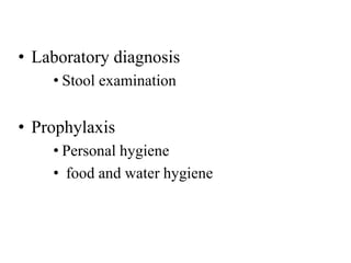
Balantidium coli
Balantidium coli
Largest protozoal parasite inhabiting large
intestine of man. Also found in pigs and
monkeys.
It is found world wide
Morphology :- the parasite is observed in two stages only which are
:-
A – trophozoite :-
• Large size (40 µm to more than 70 µm) .
• Invasive stage .
• Ovoidal in shape .
• Covered by cilia & the anterior end are longer than the
posterior end .
• Anterior end there is mouth called cytosome
• Have two nuclei , the macronucleus (the largest
one) & micronucleus (the smallest one ) .
•The macronucleus is kidney or bean in shape .
• The micronucleus is small spherical shape
located in the concavity of macronucleus .
• Have two secretory contractile vacuoles .
• Numerous food vacuoles .
B- Cyst :-
•Oval
•Thick outer wall
•Contain the macronucleus and contractile vacuoles are
visible in the cyst .
•The cilia are undetectable , only the roots may present .
Multiplication :- occur by two method
1- Asexual type by binary fission .
2- Sexual type by conjugation .
Life cycle :-
•No intermediate host is required .
•Mature cysts are passed with feces.
•Transmission :- by ingestion of contaminated food or
water (NOT in undercooked meat) with feces contain
the mature cyst .
•Excystation occurs in the small intestine, and the
trophozoites colonize in the large intestine.
•Trophozoites undergo encystation to produce infective
cysts.
Cysts are the infective stage of this parasite.
Pathogenesis
• Parasite produces hyaluronidase enzyme and
may cause mucosal damage.
• Which results in small superficial ulcer
which may penetrate upto submucosa.
• Diarrhea and dysentery can be developed
• Abdominal colic, nausea and vomiting may
occur.
• Laboratory diagnosis
• Stool examination
• Prophylaxis
• Personal hygiene
• food and water hygiene
8. balantidium coli

8. balantidium coli

  • 1.
  • 2.
    Balantidium coli Largest protozoalparasite inhabiting large intestine of man. Also found in pigs and monkeys. It is found world wide
  • 3.
    Morphology :- theparasite is observed in two stages only which are :- A – trophozoite :- • Large size (40 µm to more than 70 µm) . • Invasive stage . • Ovoidal in shape . • Covered by cilia & the anterior end are longer than the posterior end . • Anterior end there is mouth called cytosome • Have two nuclei , the macronucleus (the largest one) & micronucleus (the smallest one ) . •The macronucleus is kidney or bean in shape . • The micronucleus is small spherical shape located in the concavity of macronucleus . • Have two secretory contractile vacuoles . • Numerous food vacuoles .
  • 4.
    B- Cyst :- •Oval •Thickouter wall •Contain the macronucleus and contractile vacuoles are visible in the cyst . •The cilia are undetectable , only the roots may present .
  • 5.
    Multiplication :- occurby two method 1- Asexual type by binary fission . 2- Sexual type by conjugation . Life cycle :- •No intermediate host is required . •Mature cysts are passed with feces. •Transmission :- by ingestion of contaminated food or water (NOT in undercooked meat) with feces contain the mature cyst . •Excystation occurs in the small intestine, and the trophozoites colonize in the large intestine. •Trophozoites undergo encystation to produce infective cysts. Cysts are the infective stage of this parasite.
  • 7.
    Pathogenesis • Parasite produceshyaluronidase enzyme and may cause mucosal damage. • Which results in small superficial ulcer which may penetrate upto submucosa. • Diarrhea and dysentery can be developed • Abdominal colic, nausea and vomiting may occur.
  • 8.
    • Laboratory diagnosis •Stool examination • Prophylaxis • Personal hygiene • food and water hygiene