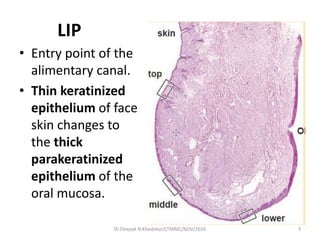
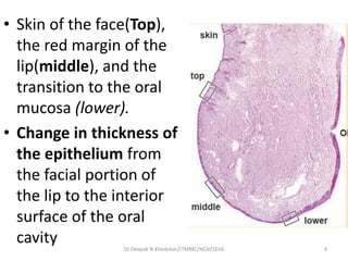
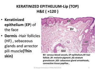
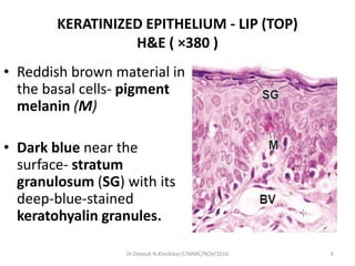
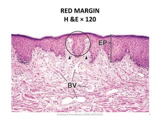
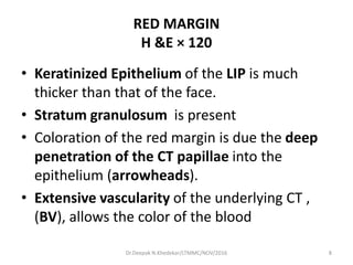
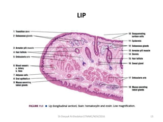
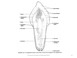
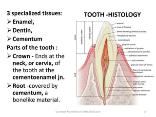
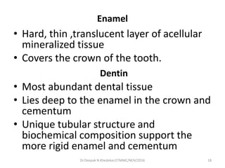
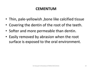
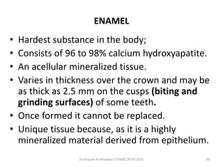
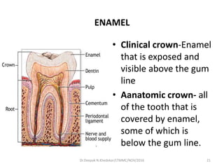
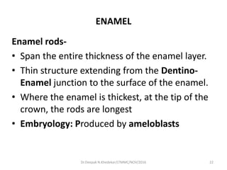
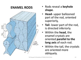
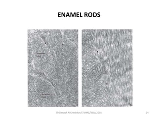
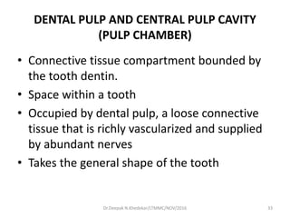
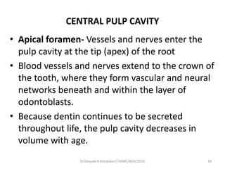
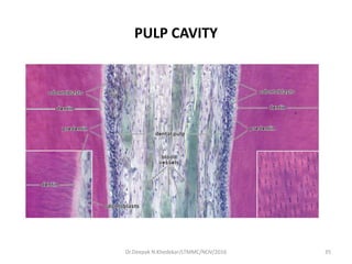
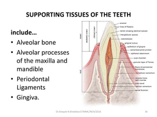
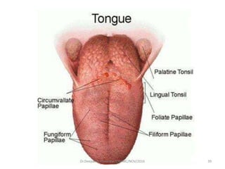
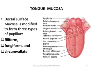
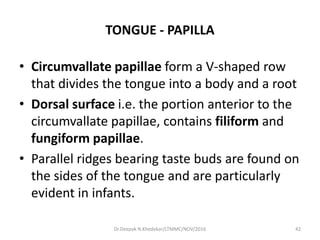
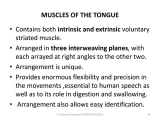
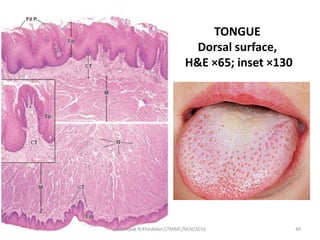
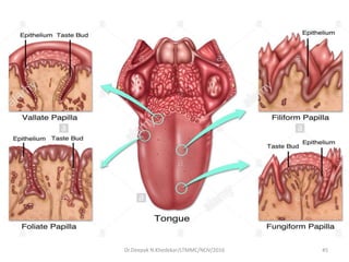
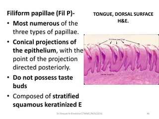
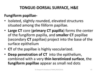
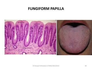
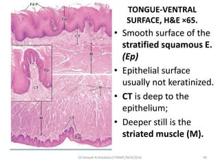
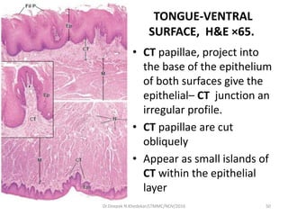
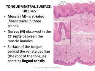
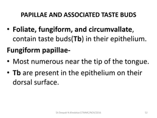
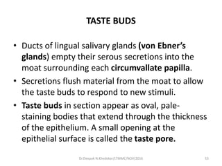
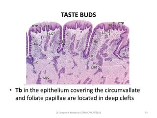
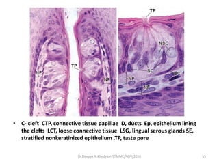
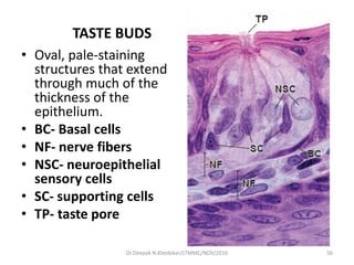
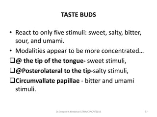
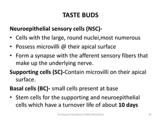
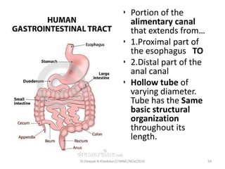
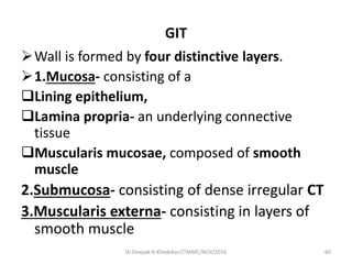
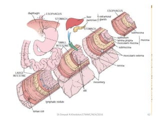
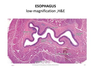
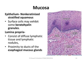
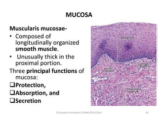
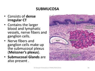
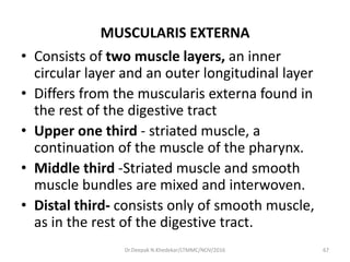
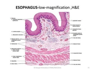
DIGESTIVE SYSTEM-1
DR.DEEPAK N.KHEDEKAR
DEPARTMENT OF ANATOMY
NOV 2016
1Dr.Deepak N.Khedekar/LTMMC/NOV/2016
LIP
2Dr.Deepak N.Khedekar/LTMMC/NOV/2016
LIP
• Entry point of the
alimentary canal.
• Thin keratinized
epithelium of face
skin changes to
the thick
parakeratinized
epithelium of the
oral mucosa.
3Dr.Deepak N.Khedekar/LTMMC/NOV/2016
• Skin of the face(Top),
the red margin of the
lip(middle), and the
transition to the oral
mucosa (lower).
• Change in thickness of
the epithelium from
the facial portion of
the lip to the interior
surface of the oral
cavity 4Dr.Deepak N.Khedekar/LTMMC/NOV/2016
KERATINIZED EPITHELIUM-Lip (TOP)
H&E ( ×120 )
• Keratinized
epithelium (EP) of
the face
• Dermis -Hair follicles
(HF) , sebaceous
glands and arrector
pili muscle(Thin
skin) BV- venous blood vessels, EP-epithelium,HF-hair
follicle ,M- melanin pigment ,SG-stratum
granulosum ,SGl- sebaceous gland arrowheads,
connective tissue papillae ,
5Dr.Deepak N.Khedekar/LTMMC/NOV/2016
KERATINIZED EPITHELIUM - LIP (TOP)
H&E ( ×380 )
• Reddish brown material in
the basal cells- pigment
melanin (M)
• Dark blue near the
surface- stratum
granulosum (SG) with its
deep-blue-stained
keratohyalin granules.
6Dr.Deepak N.Khedekar/LTMMC/NOV/2016
RED MARGIN
H &E × 120
7Dr.Deepak N.Khedekar/LTMMC/NOV/2016
RED MARGIN
H &E × 120
• Keratinized Epithelium of the LIP is much
thicker than that of the face.
• Stratum granulosum is present
• Coloration of the red margin is due the deep
penetration of the CT papillae into the
epithelium (arrowheads).
• Extensive vascularity of the underlying CT ,
(BV), allows the color of the blood
8Dr.Deepak N.Khedekar/LTMMC/NOV/2016
RED MARGIN
H &E × 380
• Sensitivity of the
red margin to
stimuli such as light
touch is due to the
presence of an
increased number
of sensory
receptors.
• Meissner’s
corpuscle, (MC)
seen in each of the
two deep papillae in
9Dr.Deepak N.Khedekar/LTMMC/NOV/2016
MUCOCUTANEOUS JUNCTION, H&E × 120
• Transition from the keratinized red margin to the
fairly thick stratified squamous parakeratinized
epithelium of the oral mucosa.
• Stratum granulosum suddenly ends.
10Dr.Deepak N.Khedekar/LTMMC/NOV/2016
MUCOCUTANEOUS JUNCTION, H&E × 380
• Beyond the site where
the stratum granulosum
cells disappear, nuclei are
seen in the superficial
cells up to the
surface(arrows).
• The epithelium is also
much thicker at this point
• and remains so
throughout the oral
cavity.
11Dr.Deepak N.Khedekar/LTMMC/NOV/2016
DEEPER STRUCTURES OF LIP
• Labial glands –tubuloacinar, mucus secreting
in deep CT
• Adipose cells
• Central core is formed by Skeletal muscle-
orbicularis oris
12Dr.Deepak N.Khedekar/LTMMC/NOV/2016
LIP
13Dr.Deepak N.Khedekar/LTMMC/NOV/2016
TOOTH
14Dr.Deepak N.Khedekar/LTMMC/NOV/2016
TOOTH
• Major component of the oral cavity
• Essential for the the digestive process.
• Embedded in and attached to the alveolar
processes of the maxilla and mandible.
(Gomphosis)
• Children have 10 deciduous (primary, milk)
teeth in each jaw, on each side:
• Adult has 32 Permanent (secondary) teeth
15Dr.Deepak N.Khedekar/LTMMC/NOV/2016
16Dr.Deepak N.Khedekar/LTMMC/NOV/2016
TOOTH -HISTOLOGY3 specialized tissues:
Enamel,
Dentin,
Cementum
Parts of the tooth :
Crown - Ends at the
neck, or cervix, of
the tooth at the
cementoenamel jn.
Root -covered by
cementum, a
bonelike material.
17Dr.Deepak N.Khedekar/LTMMC/NOV/2016
Enamel
• Hard, thin ,translucent layer of acellular
mineralized tissue
• Covers the crown of the tooth.
Dentin
• Most abundant dental tissue
• Lies deep to the enamel in the crown and
cementum
• Unique tubular structure and
biochemical composition support the
more rigid enamel and cementum
18Dr.Deepak N.Khedekar/LTMMC/NOV/2016
CEMENTUM
• Thin, pale-yellowish ,bone like calcified tissue
• Covering the dentin of the root of the teeth.
• Softer and more permeable than dentin.
• Easily removed by abrasion when the root
surface is exposed to the oral environment.
19Dr.Deepak N.Khedekar/LTMMC/NOV/2016
ENAMEL
• Hardest substance in the body;
• Consists of 96 to 98% calcium hydroxyapatite.
• An acellular mineralized tissue.
• Varies in thickness over the crown and may be
as thick as 2.5 mm on the cusps (biting and
grinding surfaces) of some teeth.
• Once formed it cannot be replaced.
• Unique tissue because, as it is a highly
mineralized material derived from epithelium.
20Dr.Deepak N.Khedekar/LTMMC/NOV/2016
ENAMEL
• Clinical crown-Enamel
that is exposed and
visible above the gum
line
• Aanatomic crown- all
of the tooth that is
covered by enamel,
some of which is
below the gum line.
21Dr.Deepak N.Khedekar/LTMMC/NOV/2016
ENAMEL
Enamel rods-
• Span the entire thickness of the enamel layer.
• Thin structure extending from the Dentino-
Enamel junction to the surface of the enamel.
• Where the enamel is thickest, at the tip of the
crown, the rods are longest
• Embryology: Produced by ameloblasts
22Dr.Deepak N.Khedekar/LTMMC/NOV/2016
ENAMEL RODS • Rods reveal a keyhole
shape.
• Head- upper ballooned
part of the rod, oriented
superiorly,
• Tail- lower part of the rod,
is directed inferiorly.
• Within the head, the
enamel crystals are
oriented parallel to the
long axis of each rod.
• Within the tail, the crystals
are oriented more
obliquely.
23Dr.Deepak N.Khedekar/LTMMC/NOV/2016
ENAMEL RODS
24Dr.Deepak N.Khedekar/LTMMC/NOV/2016
DENTIN
• Dentin is produced by neural crest–derived
odontoblasts of the adjacent mesenchyme.
• Calcified material that forms most of the tooth
substance.
• Lies deep to the enamel and cementum.
25Dr.Deepak N.Khedekar/LTMMC/NOV/2016
DENTIN
• Contains less hydroxyapatite than enamel,
about 70%, but more than is found in bone
and cementum.
• Like ameloblasts, odontoblasts are columnar
cells that contain a well-developed rER, a large
Golgi apparatus, and other organelles
associated with the synthesis and secretion of
large amounts of protein
Dr.Deepak N.Khedekar/LTMMC/NOV/2016 26
DENTINAL TUBULES
• Apical surface of the odontoblast is in contact
with the forming dentin
• Junctional complexes of the odontoblasts
separate the dentine from the pulp
• Odontoblast processes embedded in the dentin
in narrow channels called dentinal tubules .
• Tubules and processes continue to elongate as
the dentin continues to thicken by rhythmic
growth.
Dr.Deepak N.Khedekar/LTMMC/NOV/2016 27
DENTIN -GROWTH LINES
• Also known as Incremental lines of von
Ebner OR thicker lines of Owen
• Produces by Rhythmic growth of dentin
produces certain “growth lines” in the dentin
• Mark significant developmental times such as
birth (neonatal line)
• Study of growth lines has proved useful in
forensic medicine.
Dr.Deepak N.Khedekar/LTMMC/NOV/2016 28
CEMENTUM
• Avascular structure ,covers the root of the tooth.
• Thin layer of bonelike material
• Secreted by cementocytes , cells that closely
resemble osteocytes.
• Like bone, cementum is 65% mineral.
• Contain Lacunae and canaliculi consist of in the
the cementocytes and their processes,
29Dr.Deepak N.Khedekar/LTMMC/NOV/2016
CEMENTUM
• Consist of canaliculi which do not form an
interconnecting network.
• A layer of cementoblasts is seen on the outer
surface of the cementum, adjacent to the
periodontal ligament.
• Sharpey’s fibers -Collagen fibers that project
out of the matrix and embed in the bony matrix
of the socket wall form the bulk of the
periodontal ligament
30Dr.Deepak N.Khedekar/LTMMC/NOV/2016
CEMENTUM
• Elastic fibers are also a component of the
periodontal ligament allowing slight
movement of the tooth to occur naturally.
• Forms the basis of various orthodontic
procedures
• During corrective tooth movements, the
alveolar bone of the socket is resorbed and
resynthesized, but the cementum is not.
31Dr.Deepak N.Khedekar/LTMMC/NOV/2016
SHARPEYS FIBRES
32Dr.Deepak N.Khedekar/LTMMC/NOV/2016
DENTAL PULP AND CENTRAL PULP CAVITY
(PULP CHAMBER)
• Connective tissue compartment bounded by
the tooth dentin.
• Space within a tooth
• Occupied by dental pulp, a loose connective
tissue that is richly vascularized and supplied
by abundant nerves
• Takes the general shape of the tooth
33Dr.Deepak N.Khedekar/LTMMC/NOV/2016
CENTRAL PULP CAVITY
• Apical foramen- Vessels and nerves enter the
pulp cavity at the tip (apex) of the root
• Blood vessels and nerves extend to the crown of
the tooth, where they form vascular and neural
networks beneath and within the layer of
odontoblasts.
• Because dentin continues to be secreted
throughout life, the pulp cavity decreases in
volume with age.
34Dr.Deepak N.Khedekar/LTMMC/NOV/2016
PULP CAVITY
35Dr.Deepak N.Khedekar/LTMMC/NOV/2016
SUPPORTING TISSUES OF THE TEETH
include…
• Alveolar bone
• Alveolar processes
of the maxilla and
mandible
• Periodontal
Ligaments
• Gingiva.
36Dr.Deepak N.Khedekar/LTMMC/NOV/2016
PERIODONTAL LIGAMENT
• Fibrous connective tissue
• Joining the tooth to its surrounding bone.
• Provides for the following :
 Tooth attachment (fixation)
 Tooth support
 Bone remodeling (during movement of a
tooth)
37Dr.Deepak N.Khedekar/LTMMC/NOV/2016
DRIED SECTION OF TOOTH
Shows following features…
• Lines of schreger
• Lines of Retzius
• Interglobular spaces
• Granular layers of Tomes
Dr.Deepak N.Khedekar/LTMMC/NOV/2016 38
39Dr.Deepak N.Khedekar/LTMMC/NOV/2016
TONGUE
• Muscular organ projecting into the oral cavity.
• Covered with a mucous membrane that
• Consists of stratified squamous epithelium,
keratinized in parts
• Resting on a loose connective tissue.
• Parts : Root &Free part i.e body
• Surfaces: Dorsal & Ventral
40Dr.Deepak N.Khedekar/LTMMC/NOV/2016
TONGUE- MUCOSA
• Dorsal surface
Mucosa is modified
to form three types
of papillae:
filiform,
fungiform, and
circumvallate
Dr.Deepak N.Khedekar/LTMMC/NOV/2016 41
TONGUE - PAPILLA
• Circumvallate papillae form a V-shaped row
that divides the tongue into a body and a root
• Dorsal surface i.e. the portion anterior to the
circumvallate papillae, contains filiform and
fungiform papillae.
• Parallel ridges bearing taste buds are found on
the sides of the tongue and are particularly
evident in infants.
42Dr.Deepak N.Khedekar/LTMMC/NOV/2016
MUSCLES OF THE TONGUE
• Contains both intrinsic and extrinsic voluntary
striated muscle.
• Arranged in three interweaving planes, with
each arrayed at right angles to the other two.
• Arrangement is unique.
• Provides enormous flexibility and precision in
the movements ,essential to human speech as
well as to its role in digestion and swallowing.
• Arrangement also allows easy identification.
43Dr.Deepak N.Khedekar/LTMMC/NOV/2016
TONGUE
Dorsal surface,
H&E ×65; inset ×130
44Dr.Deepak N.Khedekar/LTMMC/NOV/2016
Dr.Deepak N.Khedekar/LTMMC/NOV/2016 45
TONGUE, DORSAL SURFACE
H&E.
Filiform papillae (Fil P)-
• Most numerous of the
three types of papillae.
• Conical projections of
the epithelium, with the
point of the projection
directed posteriorly.
• Do not possess taste
buds
• Composed of stratified
squamous keratinized E
46Dr.Deepak N.Khedekar/LTMMC/NOV/2016
TONGUE-DORSAL SURFACE, H&E
Fungiform papillae-
• Isolated, slightly rounded, elevated structures
situated among the filiform papillae.
• Large CT core (primary CT papilla) forms the center
of the fungiform papilla, and smaller CT papillae
(secondary CT papillae) project into the base of the
surface epithelium
• CT of the papillae is highly vascularized.
• Deep penetration of CT into the epithelium,
combined with a very thin keratinized surface, the
fungiform papillae appear as small red dots
47Dr.Deepak N.Khedekar/LTMMC/NOV/2016
FUNGIFORM PAPILLA
Dr.Deepak N.Khedekar/LTMMC/NOV/2016 48
TONGUE-VENTRAL
SURFACE, H&E ×65.
• Smooth surface of the
stratified squamous E.
(Ep)
• Epithelial surface
usually not keratinized.
• CT is deep to the
epithelium;
• Deeper still is the
striated muscle (M).
49Dr.Deepak N.Khedekar/LTMMC/NOV/2016
TONGUE-VENTRAL
SURFACE, H&E ×65.
• CT papillae, project into
the base of the epithelium
of both surfaces give the
epithelial– CT junction an
irregular profile.
• CT papillae are cut
obliquely
• Appear as small islands of
CT within the epithelial
layer
50Dr.Deepak N.Khedekar/LTMMC/NOV/2016
TONGUE-VENTRAL SURFACE,
H&E ×65
• Muscle (M)- is striated
,fibers travel in three
planes.
• Nerves (N) observed in the
CT septa between the
muscle bundles.
• Surface of the tongue
behind the vallate papillae
(the root of the tongue)
contains lingual tonsils
51Dr.Deepak N.Khedekar/LTMMC/NOV/2016
PAPILLAE AND ASSOCIATED TASTE BUDS
• Foliate, fungiform, and circumvallate,
contain taste buds(Tb) in their epithelium.
Fungiform papillae-
• Most numerous near the tip of the tongue.
• Tb are present in the epithelium on their
dorsal surface.
52Dr.Deepak N.Khedekar/LTMMC/NOV/2016
TASTE BUDS
• Ducts of lingual salivary glands (von Ebner’s
glands) empty their serous secretions into the
moat surrounding each circumvallate papilla.
• Secretions flush material from the moat to allow
the taste buds to respond to new stimuli.
• Taste buds in section appear as oval, pale-
staining bodies that extend through the thickness
of the epithelium. A small opening at the
epithelial surface is called the taste pore.
53Dr.Deepak N.Khedekar/LTMMC/NOV/2016
TASTE BUDS
• Tb in the epithelium covering the circumvallate
and foliate papillae are located in deep clefts
54Dr.Deepak N.Khedekar/LTMMC/NOV/2016
• C- cleft CTP, connective tissue papillae D, ducts Ep, epithelium lining
the clefts LCT, loose connective tissue LSG, lingual serous glands SE,
stratified nonkeratinized epithelium ,TP, taste pore
55Dr.Deepak N.Khedekar/LTMMC/NOV/2016
TASTE BUDS
• Oval, pale-staining
structures that extend
through much of the
thickness of the
epithelium.
• BC- Basal cells
• NF- nerve fibers
• NSC- neuroepithelial
sensory cells
• SC- supporting cells
• TP- taste pore
Dr.Deepak N.Khedekar/LTMMC/NOV/2016 56
TASTE BUDS
• React to only five stimuli: sweet, salty, bitter,
sour, and umami.
• Modalities appear to be more concentrated…
@ the tip of the tongue- sweet stimuli,
@Posterolateral to the tip-salty stimuli,
Circumvallate papillae - bitter and umami
stimuli.
57Dr.Deepak N.Khedekar/LTMMC/NOV/2016
TASTE BUDS
Neuroepithelial sensory cells (NSC)-
• Cells with the large, round nuclei,most numerous
• Possess microvilli @ their apical surface
• Form a synapse with the afferent sensory fibers that
make up the underlying nerve.
Supporting cells (SC)-Contain microvilli on their apical
surface.
Basal cells (BC)- small cells present at base
• Stem cells for the supporting and neuroepithelial
cells which have a turnover life of about 10 days
58Dr.Deepak N.Khedekar/LTMMC/NOV/2016
• Portion of the
alimentary canal
that extends from…
• 1.Proximal part of
the esophagus TO
• 2.Distal part of the
anal canal
• Hollow tube of
varying diameter.
Tube has the Same
basic structural
organization
throughout its
length.
Dr.Deepak N.Khedekar/LTMMC/NOV/2016 59
GIT
Wall is formed by four distinctive layers.
1.Mucosa- consisting of a
Lining epithelium,
Lamina propria- an underlying connective
tissue
Muscularis mucosae, composed of smooth
muscle
2.Submucosa- consisting of dense irregular CT
3.Muscularis externa- consisting in layers of
smooth muscle
60Dr.Deepak N.Khedekar/LTMMC/NOV/2016
BASIC LAYERS OF GIT
4.Serosa- a serous membrane consisting of a
simple squamous E., the mesothelium, and a
small amount of underlying connective tissue.
Adventitia consist of CT is found where the wall
of the tube is directly attached or fixed to
adjoining structures (i.e., body wall and
retroperitoneal organs).
61Dr.Deepak N.Khedekar/LTMMC/NOV/2016
62Dr.Deepak N.Khedekar/LTMMC/NOV/2016
ESOPHAGUS
low-magnification ,H&E
63Dr.Deepak N.Khedekar/LTMMC/NOV/2016
Mucosa
Epithelium- Nonkeratinized
stratified squamous
• Surface cells may exhibit
some keratohyalin
granules.
Lamina propria-
• Consist of diffuse lymphatic
tissue and lymphatic
nodules,
• Proximity to ducts of the
esophageal mucous glands
64Dr.Deepak N.Khedekar/LTMMC/NOV/2016
MUCOSA
Muscularis mucosae-
• Composed of
longitudinally organized
smooth muscle.
• Unusually thick in the
proximal portion.
Three principal functions of
mucosa:
Protection,
Absorption, and
Secretion
65Dr.Deepak N.Khedekar/LTMMC/NOV/2016
SUBMUCOSA
• Consists of dense
irregular CT
• Contains the larger
blood and lymphatic
vessels, nerve fibers and
ganglion cells.
• Nerve fibers and
ganglion cells make up
the submucosal plexus
(Meissner’s plexus).
• Submucosal Glands are
also present .
66Dr.Deepak N.Khedekar/LTMMC/NOV/2016
MUSCULARIS EXTERNA
• Consists of two muscle layers, an inner
circular layer and an outer longitudinal layer
• Differs from the muscularis externa found in
the rest of the digestive tract
• Upper one third - striated muscle, a
continuation of the muscle of the pharynx.
• Middle third -Striated muscle and smooth
muscle bundles are mixed and interwoven.
• Distal third- consists only of smooth muscle,
as in the rest of the digestive tract.
67Dr.Deepak N.Khedekar/LTMMC/NOV/2016
MUSCULARIS EXTERNA
• Nerve plx, the myenteric plx (Auerbach’s plx), is
present between the outer and inner muscle layers.
• Plx innervates the muscularis externa
Adventitia
• Esophagus is fixed to adjoining structures throughout.
• After entering the abdominal cavity, the short
remainder of the tube is covered by serosa, the
visceral peritoneum.
Mucosal and submucosal glands of the esophagus
secrete
mucus to lubricate and protect the luminal wall. 68Dr.Deepak N.Khedekar/LTMMC/NOV/2016
ESOPHAGUS
69Dr.Deepak N.Khedekar/LTMMC/NOV/2016
ESOPHAGEAL GLANDS PROPER
• Two types ,both secrete mucus,
Esophageal glands proper lie in the submucosa-
• Scattered along the length of the esophagus
• More concentrated in the upper half.
• Small, compound, tubuloalveolar glands
• Excretory duct is composed of stratified squamous
epithelium
• Mucus produces by it is slightly acidic and serves to
lubricate the luminal wall.
70Dr.Deepak N.Khedekar/LTMMC/NOV/2016
ESOPHAGEAL CARDIAC GLANDS
• Found in the lamina propria of the mucosa.
• Present in the terminal part of the esophagus, OR
in the beginning portion of the esophagus.
• Produce neutral mucus.
• Protect the esophagus from regurgitated gastric
contents. Under certain conditions, however,
they are not fully effective,and excessive reflux
results in pyrosis, a condition more commonly
known as heartburn. may progress to fully
developed (GERD).
71Dr.Deepak N.Khedekar/LTMMC/NOV/2016
MUSCLE OF THE ESOPHAGEAL WALL
• Innervated by both autonomic and somatic NS.
• Striated musculature in the upper is innervated
the vagus nerve, (from the nucleus ambiguus).
• Smooth muscle of the lower part is innervated by
visceral motor neurons of the vagus (from the
dorsal motor nucleus).
• Postsynaptic neurons are located in the wall of
the esophagus.
72Dr.Deepak N.Khedekar/LTMMC/NOV/2016
ESOPHAGUS-low-magnification ,H&E
73Dr.Deepak N.Khedekar/LTMMC/NOV/2016
THE END
Dr.Deepak N.Khedekar/LTMMC/NOV/2016 74

Histology - GIT 1

  • 1.
    DIGESTIVE SYSTEM-1 DR.DEEPAK N.KHEDEKAR DEPARTMENTOF ANATOMY NOV 2016 1Dr.Deepak N.Khedekar/LTMMC/NOV/2016
  • 2.
  • 3.
    LIP • Entry pointof the alimentary canal. • Thin keratinized epithelium of face skin changes to the thick parakeratinized epithelium of the oral mucosa. 3Dr.Deepak N.Khedekar/LTMMC/NOV/2016
  • 4.
    • Skin ofthe face(Top), the red margin of the lip(middle), and the transition to the oral mucosa (lower). • Change in thickness of the epithelium from the facial portion of the lip to the interior surface of the oral cavity 4Dr.Deepak N.Khedekar/LTMMC/NOV/2016
  • 5.
    KERATINIZED EPITHELIUM-Lip (TOP) H&E( ×120 ) • Keratinized epithelium (EP) of the face • Dermis -Hair follicles (HF) , sebaceous glands and arrector pili muscle(Thin skin) BV- venous blood vessels, EP-epithelium,HF-hair follicle ,M- melanin pigment ,SG-stratum granulosum ,SGl- sebaceous gland arrowheads, connective tissue papillae , 5Dr.Deepak N.Khedekar/LTMMC/NOV/2016
  • 6.
    KERATINIZED EPITHELIUM -LIP (TOP) H&E ( ×380 ) • Reddish brown material in the basal cells- pigment melanin (M) • Dark blue near the surface- stratum granulosum (SG) with its deep-blue-stained keratohyalin granules. 6Dr.Deepak N.Khedekar/LTMMC/NOV/2016
  • 7.
    RED MARGIN H &E× 120 7Dr.Deepak N.Khedekar/LTMMC/NOV/2016
  • 8.
    RED MARGIN H &E× 120 • Keratinized Epithelium of the LIP is much thicker than that of the face. • Stratum granulosum is present • Coloration of the red margin is due the deep penetration of the CT papillae into the epithelium (arrowheads). • Extensive vascularity of the underlying CT , (BV), allows the color of the blood 8Dr.Deepak N.Khedekar/LTMMC/NOV/2016
  • 9.
    RED MARGIN H &E× 380 • Sensitivity of the red margin to stimuli such as light touch is due to the presence of an increased number of sensory receptors. • Meissner’s corpuscle, (MC) seen in each of the two deep papillae in 9Dr.Deepak N.Khedekar/LTMMC/NOV/2016
  • 10.
    MUCOCUTANEOUS JUNCTION, H&E× 120 • Transition from the keratinized red margin to the fairly thick stratified squamous parakeratinized epithelium of the oral mucosa. • Stratum granulosum suddenly ends. 10Dr.Deepak N.Khedekar/LTMMC/NOV/2016
  • 11.
    MUCOCUTANEOUS JUNCTION, H&E× 380 • Beyond the site where the stratum granulosum cells disappear, nuclei are seen in the superficial cells up to the surface(arrows). • The epithelium is also much thicker at this point • and remains so throughout the oral cavity. 11Dr.Deepak N.Khedekar/LTMMC/NOV/2016
  • 12.
    DEEPER STRUCTURES OFLIP • Labial glands –tubuloacinar, mucus secreting in deep CT • Adipose cells • Central core is formed by Skeletal muscle- orbicularis oris 12Dr.Deepak N.Khedekar/LTMMC/NOV/2016
  • 13.
  • 14.
  • 15.
    TOOTH • Major componentof the oral cavity • Essential for the the digestive process. • Embedded in and attached to the alveolar processes of the maxilla and mandible. (Gomphosis) • Children have 10 deciduous (primary, milk) teeth in each jaw, on each side: • Adult has 32 Permanent (secondary) teeth 15Dr.Deepak N.Khedekar/LTMMC/NOV/2016
  • 16.
  • 17.
    TOOTH -HISTOLOGY3 specializedtissues: Enamel, Dentin, Cementum Parts of the tooth : Crown - Ends at the neck, or cervix, of the tooth at the cementoenamel jn. Root -covered by cementum, a bonelike material. 17Dr.Deepak N.Khedekar/LTMMC/NOV/2016
  • 18.
    Enamel • Hard, thin,translucent layer of acellular mineralized tissue • Covers the crown of the tooth. Dentin • Most abundant dental tissue • Lies deep to the enamel in the crown and cementum • Unique tubular structure and biochemical composition support the more rigid enamel and cementum 18Dr.Deepak N.Khedekar/LTMMC/NOV/2016
  • 19.
    CEMENTUM • Thin, pale-yellowish,bone like calcified tissue • Covering the dentin of the root of the teeth. • Softer and more permeable than dentin. • Easily removed by abrasion when the root surface is exposed to the oral environment. 19Dr.Deepak N.Khedekar/LTMMC/NOV/2016
  • 20.
    ENAMEL • Hardest substancein the body; • Consists of 96 to 98% calcium hydroxyapatite. • An acellular mineralized tissue. • Varies in thickness over the crown and may be as thick as 2.5 mm on the cusps (biting and grinding surfaces) of some teeth. • Once formed it cannot be replaced. • Unique tissue because, as it is a highly mineralized material derived from epithelium. 20Dr.Deepak N.Khedekar/LTMMC/NOV/2016
  • 21.
    ENAMEL • Clinical crown-Enamel thatis exposed and visible above the gum line • Aanatomic crown- all of the tooth that is covered by enamel, some of which is below the gum line. 21Dr.Deepak N.Khedekar/LTMMC/NOV/2016
  • 22.
    ENAMEL Enamel rods- • Spanthe entire thickness of the enamel layer. • Thin structure extending from the Dentino- Enamel junction to the surface of the enamel. • Where the enamel is thickest, at the tip of the crown, the rods are longest • Embryology: Produced by ameloblasts 22Dr.Deepak N.Khedekar/LTMMC/NOV/2016
  • 23.
    ENAMEL RODS •Rods reveal a keyhole shape. • Head- upper ballooned part of the rod, oriented superiorly, • Tail- lower part of the rod, is directed inferiorly. • Within the head, the enamel crystals are oriented parallel to the long axis of each rod. • Within the tail, the crystals are oriented more obliquely. 23Dr.Deepak N.Khedekar/LTMMC/NOV/2016
  • 24.
  • 25.
    DENTIN • Dentin isproduced by neural crest–derived odontoblasts of the adjacent mesenchyme. • Calcified material that forms most of the tooth substance. • Lies deep to the enamel and cementum. 25Dr.Deepak N.Khedekar/LTMMC/NOV/2016
  • 26.
    DENTIN • Contains lesshydroxyapatite than enamel, about 70%, but more than is found in bone and cementum. • Like ameloblasts, odontoblasts are columnar cells that contain a well-developed rER, a large Golgi apparatus, and other organelles associated with the synthesis and secretion of large amounts of protein Dr.Deepak N.Khedekar/LTMMC/NOV/2016 26
  • 27.
    DENTINAL TUBULES • Apicalsurface of the odontoblast is in contact with the forming dentin • Junctional complexes of the odontoblasts separate the dentine from the pulp • Odontoblast processes embedded in the dentin in narrow channels called dentinal tubules . • Tubules and processes continue to elongate as the dentin continues to thicken by rhythmic growth. Dr.Deepak N.Khedekar/LTMMC/NOV/2016 27
  • 28.
    DENTIN -GROWTH LINES •Also known as Incremental lines of von Ebner OR thicker lines of Owen • Produces by Rhythmic growth of dentin produces certain “growth lines” in the dentin • Mark significant developmental times such as birth (neonatal line) • Study of growth lines has proved useful in forensic medicine. Dr.Deepak N.Khedekar/LTMMC/NOV/2016 28
  • 29.
    CEMENTUM • Avascular structure,covers the root of the tooth. • Thin layer of bonelike material • Secreted by cementocytes , cells that closely resemble osteocytes. • Like bone, cementum is 65% mineral. • Contain Lacunae and canaliculi consist of in the the cementocytes and their processes, 29Dr.Deepak N.Khedekar/LTMMC/NOV/2016
  • 30.
    CEMENTUM • Consist ofcanaliculi which do not form an interconnecting network. • A layer of cementoblasts is seen on the outer surface of the cementum, adjacent to the periodontal ligament. • Sharpey’s fibers -Collagen fibers that project out of the matrix and embed in the bony matrix of the socket wall form the bulk of the periodontal ligament 30Dr.Deepak N.Khedekar/LTMMC/NOV/2016
  • 31.
    CEMENTUM • Elastic fibersare also a component of the periodontal ligament allowing slight movement of the tooth to occur naturally. • Forms the basis of various orthodontic procedures • During corrective tooth movements, the alveolar bone of the socket is resorbed and resynthesized, but the cementum is not. 31Dr.Deepak N.Khedekar/LTMMC/NOV/2016
  • 32.
  • 33.
    DENTAL PULP ANDCENTRAL PULP CAVITY (PULP CHAMBER) • Connective tissue compartment bounded by the tooth dentin. • Space within a tooth • Occupied by dental pulp, a loose connective tissue that is richly vascularized and supplied by abundant nerves • Takes the general shape of the tooth 33Dr.Deepak N.Khedekar/LTMMC/NOV/2016
  • 34.
    CENTRAL PULP CAVITY •Apical foramen- Vessels and nerves enter the pulp cavity at the tip (apex) of the root • Blood vessels and nerves extend to the crown of the tooth, where they form vascular and neural networks beneath and within the layer of odontoblasts. • Because dentin continues to be secreted throughout life, the pulp cavity decreases in volume with age. 34Dr.Deepak N.Khedekar/LTMMC/NOV/2016
  • 35.
  • 36.
    SUPPORTING TISSUES OFTHE TEETH include… • Alveolar bone • Alveolar processes of the maxilla and mandible • Periodontal Ligaments • Gingiva. 36Dr.Deepak N.Khedekar/LTMMC/NOV/2016
  • 37.
    PERIODONTAL LIGAMENT • Fibrousconnective tissue • Joining the tooth to its surrounding bone. • Provides for the following :  Tooth attachment (fixation)  Tooth support  Bone remodeling (during movement of a tooth) 37Dr.Deepak N.Khedekar/LTMMC/NOV/2016
  • 38.
    DRIED SECTION OFTOOTH Shows following features… • Lines of schreger • Lines of Retzius • Interglobular spaces • Granular layers of Tomes Dr.Deepak N.Khedekar/LTMMC/NOV/2016 38
  • 39.
  • 40.
    TONGUE • Muscular organprojecting into the oral cavity. • Covered with a mucous membrane that • Consists of stratified squamous epithelium, keratinized in parts • Resting on a loose connective tissue. • Parts : Root &Free part i.e body • Surfaces: Dorsal & Ventral 40Dr.Deepak N.Khedekar/LTMMC/NOV/2016
  • 41.
    TONGUE- MUCOSA • Dorsalsurface Mucosa is modified to form three types of papillae: filiform, fungiform, and circumvallate Dr.Deepak N.Khedekar/LTMMC/NOV/2016 41
  • 42.
    TONGUE - PAPILLA •Circumvallate papillae form a V-shaped row that divides the tongue into a body and a root • Dorsal surface i.e. the portion anterior to the circumvallate papillae, contains filiform and fungiform papillae. • Parallel ridges bearing taste buds are found on the sides of the tongue and are particularly evident in infants. 42Dr.Deepak N.Khedekar/LTMMC/NOV/2016
  • 43.
    MUSCLES OF THETONGUE • Contains both intrinsic and extrinsic voluntary striated muscle. • Arranged in three interweaving planes, with each arrayed at right angles to the other two. • Arrangement is unique. • Provides enormous flexibility and precision in the movements ,essential to human speech as well as to its role in digestion and swallowing. • Arrangement also allows easy identification. 43Dr.Deepak N.Khedekar/LTMMC/NOV/2016
  • 44.
    TONGUE Dorsal surface, H&E ×65;inset ×130 44Dr.Deepak N.Khedekar/LTMMC/NOV/2016
  • 45.
  • 46.
    TONGUE, DORSAL SURFACE H&E. Filiformpapillae (Fil P)- • Most numerous of the three types of papillae. • Conical projections of the epithelium, with the point of the projection directed posteriorly. • Do not possess taste buds • Composed of stratified squamous keratinized E 46Dr.Deepak N.Khedekar/LTMMC/NOV/2016
  • 47.
    TONGUE-DORSAL SURFACE, H&E Fungiformpapillae- • Isolated, slightly rounded, elevated structures situated among the filiform papillae. • Large CT core (primary CT papilla) forms the center of the fungiform papilla, and smaller CT papillae (secondary CT papillae) project into the base of the surface epithelium • CT of the papillae is highly vascularized. • Deep penetration of CT into the epithelium, combined with a very thin keratinized surface, the fungiform papillae appear as small red dots 47Dr.Deepak N.Khedekar/LTMMC/NOV/2016
  • 48.
  • 49.
    TONGUE-VENTRAL SURFACE, H&E ×65. •Smooth surface of the stratified squamous E. (Ep) • Epithelial surface usually not keratinized. • CT is deep to the epithelium; • Deeper still is the striated muscle (M). 49Dr.Deepak N.Khedekar/LTMMC/NOV/2016
  • 50.
    TONGUE-VENTRAL SURFACE, H&E ×65. •CT papillae, project into the base of the epithelium of both surfaces give the epithelial– CT junction an irregular profile. • CT papillae are cut obliquely • Appear as small islands of CT within the epithelial layer 50Dr.Deepak N.Khedekar/LTMMC/NOV/2016
  • 51.
    TONGUE-VENTRAL SURFACE, H&E ×65 •Muscle (M)- is striated ,fibers travel in three planes. • Nerves (N) observed in the CT septa between the muscle bundles. • Surface of the tongue behind the vallate papillae (the root of the tongue) contains lingual tonsils 51Dr.Deepak N.Khedekar/LTMMC/NOV/2016
  • 52.
    PAPILLAE AND ASSOCIATEDTASTE BUDS • Foliate, fungiform, and circumvallate, contain taste buds(Tb) in their epithelium. Fungiform papillae- • Most numerous near the tip of the tongue. • Tb are present in the epithelium on their dorsal surface. 52Dr.Deepak N.Khedekar/LTMMC/NOV/2016
  • 53.
    TASTE BUDS • Ductsof lingual salivary glands (von Ebner’s glands) empty their serous secretions into the moat surrounding each circumvallate papilla. • Secretions flush material from the moat to allow the taste buds to respond to new stimuli. • Taste buds in section appear as oval, pale- staining bodies that extend through the thickness of the epithelium. A small opening at the epithelial surface is called the taste pore. 53Dr.Deepak N.Khedekar/LTMMC/NOV/2016
  • 54.
    TASTE BUDS • Tbin the epithelium covering the circumvallate and foliate papillae are located in deep clefts 54Dr.Deepak N.Khedekar/LTMMC/NOV/2016
  • 55.
    • C- cleftCTP, connective tissue papillae D, ducts Ep, epithelium lining the clefts LCT, loose connective tissue LSG, lingual serous glands SE, stratified nonkeratinized epithelium ,TP, taste pore 55Dr.Deepak N.Khedekar/LTMMC/NOV/2016
  • 56.
    TASTE BUDS • Oval,pale-staining structures that extend through much of the thickness of the epithelium. • BC- Basal cells • NF- nerve fibers • NSC- neuroepithelial sensory cells • SC- supporting cells • TP- taste pore Dr.Deepak N.Khedekar/LTMMC/NOV/2016 56
  • 57.
    TASTE BUDS • Reactto only five stimuli: sweet, salty, bitter, sour, and umami. • Modalities appear to be more concentrated… @ the tip of the tongue- sweet stimuli, @Posterolateral to the tip-salty stimuli, Circumvallate papillae - bitter and umami stimuli. 57Dr.Deepak N.Khedekar/LTMMC/NOV/2016
  • 58.
    TASTE BUDS Neuroepithelial sensorycells (NSC)- • Cells with the large, round nuclei,most numerous • Possess microvilli @ their apical surface • Form a synapse with the afferent sensory fibers that make up the underlying nerve. Supporting cells (SC)-Contain microvilli on their apical surface. Basal cells (BC)- small cells present at base • Stem cells for the supporting and neuroepithelial cells which have a turnover life of about 10 days 58Dr.Deepak N.Khedekar/LTMMC/NOV/2016
  • 59.
    • Portion ofthe alimentary canal that extends from… • 1.Proximal part of the esophagus TO • 2.Distal part of the anal canal • Hollow tube of varying diameter. Tube has the Same basic structural organization throughout its length. Dr.Deepak N.Khedekar/LTMMC/NOV/2016 59
  • 60.
    GIT Wall is formedby four distinctive layers. 1.Mucosa- consisting of a Lining epithelium, Lamina propria- an underlying connective tissue Muscularis mucosae, composed of smooth muscle 2.Submucosa- consisting of dense irregular CT 3.Muscularis externa- consisting in layers of smooth muscle 60Dr.Deepak N.Khedekar/LTMMC/NOV/2016
  • 61.
    BASIC LAYERS OFGIT 4.Serosa- a serous membrane consisting of a simple squamous E., the mesothelium, and a small amount of underlying connective tissue. Adventitia consist of CT is found where the wall of the tube is directly attached or fixed to adjoining structures (i.e., body wall and retroperitoneal organs). 61Dr.Deepak N.Khedekar/LTMMC/NOV/2016
  • 62.
  • 63.
  • 64.
    Mucosa Epithelium- Nonkeratinized stratified squamous •Surface cells may exhibit some keratohyalin granules. Lamina propria- • Consist of diffuse lymphatic tissue and lymphatic nodules, • Proximity to ducts of the esophageal mucous glands 64Dr.Deepak N.Khedekar/LTMMC/NOV/2016
  • 65.
    MUCOSA Muscularis mucosae- • Composedof longitudinally organized smooth muscle. • Unusually thick in the proximal portion. Three principal functions of mucosa: Protection, Absorption, and Secretion 65Dr.Deepak N.Khedekar/LTMMC/NOV/2016
  • 66.
    SUBMUCOSA • Consists ofdense irregular CT • Contains the larger blood and lymphatic vessels, nerve fibers and ganglion cells. • Nerve fibers and ganglion cells make up the submucosal plexus (Meissner’s plexus). • Submucosal Glands are also present . 66Dr.Deepak N.Khedekar/LTMMC/NOV/2016
  • 67.
    MUSCULARIS EXTERNA • Consistsof two muscle layers, an inner circular layer and an outer longitudinal layer • Differs from the muscularis externa found in the rest of the digestive tract • Upper one third - striated muscle, a continuation of the muscle of the pharynx. • Middle third -Striated muscle and smooth muscle bundles are mixed and interwoven. • Distal third- consists only of smooth muscle, as in the rest of the digestive tract. 67Dr.Deepak N.Khedekar/LTMMC/NOV/2016
  • 68.
    MUSCULARIS EXTERNA • Nerveplx, the myenteric plx (Auerbach’s plx), is present between the outer and inner muscle layers. • Plx innervates the muscularis externa Adventitia • Esophagus is fixed to adjoining structures throughout. • After entering the abdominal cavity, the short remainder of the tube is covered by serosa, the visceral peritoneum. Mucosal and submucosal glands of the esophagus secrete mucus to lubricate and protect the luminal wall. 68Dr.Deepak N.Khedekar/LTMMC/NOV/2016
  • 69.
  • 70.
    ESOPHAGEAL GLANDS PROPER •Two types ,both secrete mucus, Esophageal glands proper lie in the submucosa- • Scattered along the length of the esophagus • More concentrated in the upper half. • Small, compound, tubuloalveolar glands • Excretory duct is composed of stratified squamous epithelium • Mucus produces by it is slightly acidic and serves to lubricate the luminal wall. 70Dr.Deepak N.Khedekar/LTMMC/NOV/2016
  • 71.
    ESOPHAGEAL CARDIAC GLANDS •Found in the lamina propria of the mucosa. • Present in the terminal part of the esophagus, OR in the beginning portion of the esophagus. • Produce neutral mucus. • Protect the esophagus from regurgitated gastric contents. Under certain conditions, however, they are not fully effective,and excessive reflux results in pyrosis, a condition more commonly known as heartburn. may progress to fully developed (GERD). 71Dr.Deepak N.Khedekar/LTMMC/NOV/2016
  • 72.
    MUSCLE OF THEESOPHAGEAL WALL • Innervated by both autonomic and somatic NS. • Striated musculature in the upper is innervated the vagus nerve, (from the nucleus ambiguus). • Smooth muscle of the lower part is innervated by visceral motor neurons of the vagus (from the dorsal motor nucleus). • Postsynaptic neurons are located in the wall of the esophagus. 72Dr.Deepak N.Khedekar/LTMMC/NOV/2016
  • 73.
  • 74.