Call Girls in Dwarka Mor Delhi Contact Us 9654467111
Alveolar bone.pptx
1. ALVEOLAR BONE
PRESENTED TO;
DR. DEEPAK GROVER (PROFESSOR)
DR. DEEPAK BALA (READER)
DR TANVI OHRI (READER)
DR. SHIKHA,DR. JASPREET
DR. SARVANI, DR VIKRAM DR.SONAM
PRESENTED BY
MALTI RANI
PERIODONTOLOGY AND IMPLANTOLOGY
GUIDED BY
DR. DEEPAK GOVER
1
2. CONTENTS
INTRODUCTION
DEFINITION
DEVELOPMENT AND GROWTH
COMPOSITION AND CLASSIFICATION
PARTS OF ALVEOLAR PROCESS
CELLS AND INTERCELLULAR MATRIX
REMODELLING & COUPLING
FUNCTIONS
BLOOD SUPPLY
NERVEVSUPPLY
LYMPHATIC DRAINAGE
AGE CHANGES
CLINICAL CONSIDERATIONS
CONCLUSIONS
REFERENCES
2
3. INTRODUCTION
Bone is a dynamic specialized, highly vascular rigid mineralized connective
tissue, used to designate both an organ and tissue.
3
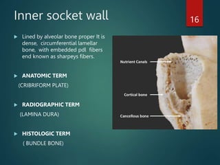
The alveolar bone is composed of
the ridges of the jaw that support
the teeth.
The roots of the teeth are contained
in deep depressions, the alveolar
sockets in the bone.
4. DEFINITON
The alveolar process is defined as the parts of the maxilla and the
mandible that form and support the sockets of the teeth. The alveolar
process develops in conjunction with the development and eruption of
the teeth.
LINDHE
The alveolar process is the portion of the maxilla and mandible that
forms and supports the tooth sockets (alveoli). It forms when the tooth
erupts to provide the osseous attachment to the forming periodontal
ligament; it disappears gradually after the tooth is lost.
CARRANZA
4
5. Development & GROWTH OF BONE
At the late bell stage, bony septa and bony bridge start to form, and
separate the individual tooth germs from another from dental follicle ,
keeping individual tooth germs in clearly outlined bony compartment.
(BERKOVITZ)
Initially this bone forms a thin egg shell to support, termed as the tooth
crypt around each tooth germ.
5
tooth crypt
6. The alveolar bone develops around each tooth follicle during odontogenesis.
When a deciduous tooth is shed, its alveolar bone is resorbed.
The succadeneous permanent tooth moves into place and develops its own
alveolar bone from its own dental follicle.
As the tooth root forms and the surrounding tissues develop and mature, alveolar
bone merges with the separately developing basal bone, and the two become one
continuous structure.
alveolar bone origin and growth occur by intramembranous ossification
6
9. INFANTS ALVEOLAR PROCESS -THE GUM PADS
The alveolar ridge from birth until the eruption of first primary teeth is
referred to as 'gum pads’.
Gum pads are horse shoe shaped; pink, firm structures seen along maxilla
and mandible.
Gum pads. The gum pad develops into two parts the lingual and labio
buccal portions which are separated by a dental groove.
9
14. PARTS OF ALVEOLAR PROCESS
1. An external cortical plate.
2. The inner socket wall
3. Cancellous trabeculae.
4.The interdental septum
5. Alveolar Crest
14
15. OUTER CORTICAL PLATE
Circumferential lamella –forms the outer perimeter
2) Concentric lamellae -(make up bulk of compact bone and forms the basic
metabolic unit of bone, the osteon)
3) Interstitial lamella inter-spread between adjacent concentric lamellae and
fill the spaces between them, actually fragments of pre-existing concentric
lamellae and can be of many shapes)
15
16. Inner socket wall
Lined by alveolar bone proper It is
dense, circumferential lamellar
bone, with embedded pdl fibers
end known as sharpeys fibers.
ANATOMIC TERM
(CRIBRIFORM PLATE)
RADIOGRAPHIC TERM
(LAMINA DURA)
HISTOLOGIC TERM
( BUNDLE BONE)
16
17. CANCELLOUS BONE 17
ANATOMIC TERM
(SPONGY BONE)
RADIOGRAPHIC TERM
(TRABECULAR BONE)
HISTOLOGIC TERM
(cancellous bone)
Trabeculae lined in path of
comperssive and tensile forces to
provide maximum resistance to
occlusal forces with minimum bone
substance (Glickman et.al 1970)
Increase in thickness and number with
increase forces .
18. Presence of trabeculae enclosing
irregular marrow spaces lined
with a layer of thin, flattened
endosteal cells.
Variation in trabeculae pattern
depending upon occlusal forces
and genetically .
Matrix consists of regularly
arranged lamellae separated by
incremental and resorption lines
18
19. Interdental septum
Consists of cancellous bone bordered by alveolar bone
proper of approximating teeth and facial and lingual
cortical plates
Narrow sopta- only cribriform plate in anterior teeth.
19
20. Mesiodistal angulation of IDS is parallel to line drawn
between CEJ of approximating teeth (Ritchey et al,
1953)
Shape and size of IDS depends on
1) Size and convexity of crowns of approximating teeth
2) Postion of teeth
3) Degree of eruption
20
21. INTERRADICULAR SEPTA
INTERRADICULAR SEPTA The bone between the roots of multirooted teeth.
Both of them contain perforating canals of Zukerkandl & Hirschfeld [nutrient
canals).
BASAL BONE It is the osseous tissue of the mandible and the maxilla except
the alveolar process.
Anatomically, there is no distinct boundary that exists between the body of
the maxilla / mandible and their alveolar process.
21
22. Nutrient canal
Nutrient canals are Radio-Lucent lines of fairly uniform width with hyperostotic
borders
Running vertically from the inferior dental canal to the apex of tooth or into the
interdental Medulla of incisors. Space Visible in 5% patients, esp. in blacks, males,
older persons, and individuals with high blood pressure or advanced periodontal,
disease.
22
Nutrient
canal
Nutrient
canals
23. Alveolar crest
Formed when the inner and outer cortical plates meet.
The margin is thin& knife edged in vestibular surfaces of anterior and
rounded/beaded in posterior teeth.
Most prominent border of interdental septum
23
24. Bundle bone
Bundle bone is a histologic term for the portion of the alveolar
bone proper bone that surrounds teeth and into which terminal
ends of principle fibers of periodontal ligaments are embedded.
24
25. CELLS AND INTERCELLULAR MATRIX
MAINLY DIVIDE IN TWO TYPE
25
Bone
formative
Bone
destructive
osteoprogenitors
Pre-osteoblasts
osteoblasts
osteocytes
bone lining cells
Osteoclast
26. Osteoprogenitor cells, which originate from MSCs and differentiate into
osteoblasts.
Osteoprogenitor cells are located on the endosteal and periosteal surface
the bone and inner surface of the Haversian canals
Some osteoprogenitor cells transform into osteoblasts after being divided
by mitosis.
Osteoprogenitor cells are activated during the bone remodeling process o
regeneration of injury.
26
27. Osteoblast
The bone matrix that is laid down by osteoblast in form of osteoid.
differentiated from pluripotent follicle cells
Numbers decrease with age
Uninuclear cells
ACTIVE cuboidal, INACTIVE flattened
Secrets collagen and non collagenous protein
When entrapped in matrix known as osteocytes.
Have alkaline phosphatase in matrix
27
29. Osteocytes
Origin : Entrapped osteoblast in calcified matrix
The osteocytes extend processes into canaliculi that radiate from the
lacunae.
The canaliculi form an anastomosing system through the intercellular
matrix of the bone, which brings oxygen and nutrients to the
osteocytes through the blood and removes metabolic waste products.
Osteocytic osteolysis: osteocytes capable of resorption.
Three functional state of osteocytes quiescent, formative and
resorptive In osteocytic osteolysis
29
30. Osteoclast
Origin from haemopoietic cells
Many monocyte fuses to form giant multinucleated osteoclastic cell
Mobile and capable of migration
Cells are with folded membranes,
ruffled border (active form) enzymes like carbonic anhydrase tartrate
resistant acid phosphatase proton pump ATPase .
Lies in Howship’s lacunae.
Clear zone site of adhesion of osteoclast to bone
30
32. Bone lining cells
When bone is in resting stage osteoblast cells becomes flattened
Function maintain homeostasis and endure bone vitality
32
33. Periosteum and endosteum
Periosteum contains osteogenic cells that regulate the
outer shape of bone and work in coordination with
inner cortical 'endosteum' (tissue lining the internal
bone cavities) to regulate cortical thickness and size.
Periosteum consists of an outer fibrous layer and an
inner cellular layer (cambium).
33
34. Bone matrix
The organic matrix consists mainly of collagen type I (90%), with
small amounts of noncollagenous proteins such as osteocalcin,
osteonectin, bone morphogenetic protein, phosphoproteins, and
proteoglycans.
Osteopontin and bone sialoprotein are cell-adhesion
proteins that appear to be important for the adhesion of both
osteoclasts and osteoblasts.
In addition, paracrine factors, including
cytokines, chemokines, and growth factors, have been implicated in
the local control of mesenchymal condensations that occur at the onset of
organogenesis.
34
35. Collagen protein
Comprises the major(80-90%) organic component in mineralised
bone tissue -Type I collagen greater than 95% is the principal
collagen in the mineralised bone and together with type V (greater
than 5%) collagen
The type III collagen is present as mixed fibres with type I collagen
with sharpeys fibres that insert from the PDL into the lamellar bone
lining the alveolus to provide a stable connection with the tooth .
(Huang YH;1991)
35
36. Non collagen protiens
Numerous non collagen proteins such as
osteocalcin
Osteonectin,
osteopontin,
matrix glycoprotein
sialoproteins,
proteoglycans
2 substrate adhesion molecules, fibronectin and tenascin,
have been identified on the periosteal and endosteal surfaces
of alveolar bone.
36
37. Osteocalcin( Gla protiens)
37
• Also known as bone gamma
carboxyglutamic acid containing
protein Which are vitamin k dependent
• The presence of Gla residues in
osteocalcin allows it to bind to calcium
ions, contributing to proper bone
mineralization and strength.
• Gla-containing proteins like osteocalcin
are involved in the regulation of bone
turnover and the maintenance of bone
health.
• More than15%of the non collagenous
protein.
• Extracellular matrix proteins,
• Modulate apatite crystal growth.
• Osteocalcin regulates mineral
maturation.
• Expressed by fully differenciated cells
and produced by osteoblast .
38. Osteonectin
Osteonectin (ON), also called SPARC or BM40, is a non-
collagenous calcium binding protein of bone matrix
expressed in mineralized tissues including endothelial
cells and fibroblasts.
SPARC has been characterized as a counter adhesive
protein that modulates interactions of cells with the
extracellular matrix. (Murphy-Ullrich JE;1995)
Recent studies have also shown SPARC has a role in
signalling functions
38
secreted protein, acidic and rich in
cysteine)SPARC
39. Osteopontin
BSP-1
Glycosylated phosphoprotiens
Significant amount at mineralising front.
Role in cell adhesion
also known as sialoprotien
These proteins are thought to contribute to the regulation of
mineralization and to tissue cohesion at sites of increased biomechanical
strain.
39
40. Matrix glycoproteins and proteoglycans
Ground substance that fills the spaces between fibers
and cells.
(mainly hyaluronic acid and chondroitin sulfate) and
glycoproteins (mainly fibronectin)
40
41. INORGANIC COMPONENT 41
HYDROXYAPETITE
It is also known as tricalcium phosphate and calcium hydroxyapatite. The uni
cell ( the smallest unit containing some form of symmetry) consists of two
triangular prismatic subcells.
FORM (NEEDLE LIKE).
SIZE 11nm to 177nm
Distributed both within the spaces between and on the surface of the
collagen fibrils
OTHER IONS :
MAGNESIUM ,SODIUM (CATIONS ).
CARBONATES,CITRATES,FLUORIDE(ANIONS).
42. BONE REMODELLING( turnover)
DEFINITION "Bone remodeling refers to the coordinated process of resorption
and formation that occurs in bone, leading to changes in its shape, structure,
and mass. It is a continuous, lifelong process that maintains bone homeostasis
and adapts bone to mechanical and functional demands.“
CARRANZA
REMODELING: Remodeling is the major pathway of bone changes In
shape,
resistance to forces, repair of wounds, and
calcium and phosphate homeostasis in the body.
Regulation of bone remodelling is a complex process involving hormones and
local factors acting in a autocrine and paracrine manner on the generation and
activity of differentiated bone cells - Sodek et al
Bone-99% of body calcium ions Major source of calcium release when blood
Monitored by parathyroid gland
42
43. 43
Bone is constantly undergoing remodeling involving the resorption of bone on a
particular surface followed by a phase of bone formation.
In normal adults , there is a balance between the amount of bone formed by
osteoblasts.(Frost;1964)
44. Bone coupling
COUPLING' refers to interdependency of osteoclasts and
osteoblasts in remodelling.
44
Parfitt (1982) stated that the termination of bone resoption & initiation of
bone formation occurs through a coupling mechanism
45. Incremental lines of bone
1- Resting lines :-are smooth straight lines consist of intercellular
substance with less collagen and relatively more ground substance, and
because of this they stained dark blue with H+E stain, they represent the
resting period of the osteoblast during bone formation.
2- Reversal lines :-The reversal lines are scalloped, not smooth, They
represent the out line of Howship's lacunae with their convexity toward the
old bone. Following bone resorption, new osteoblast differentiation and
deposited bone tissue leaving these lines separating between old and new
bone.
45
46. Factors affecting bone remodelling
Increase Bone resorption
Parathyroid hormone
Glucocorticoids
Thyroid Hormone
Vitamin D metabolites in high dose
Decrease Bone Resorption
Calcitonin
Gonadal steroids
Increase Bone Formation
Growth hormone
Vitamin D metabolites
Gonadal steroids
Decrease Bone Formation
Glucocorticoids
46
47. The primary factors that stimulate bone resorption through
osteoclasts include PTH, vit D3, IL-1, IL-6, TNF α where as calcitonin,
TGF β, estrogen and IF inhibit osteoclastic bone resorption.
PTH, PTHrP, vit D3, TGF α and pro inflammatory like IL-1, TNF α all
promote differentiation of osteoclasts.the pro inflammatory
cytokines can act through the OPG/OPGL/RANK regulatory pathway
which may be a key target of factors that effect osteoclast
generation and activity IL-6 is produced by osteoblastic cells in
response to PTH and vit D3 and is a prominent cytokine produced
by osteoclast
The prostaglandins exert a local effect on osteoclast and their
precursors often mediating the effect of growth factors and
cytokines such as EGF and TGF β.
Estrogen is believed to suppress the production of bone resorbing
cytokines ,including IL-1 and IL-6, while TGF β, and IF- inhibit
proliferation and differentiation of committed precursor into mature
osteoclast activity, but its effect is transient
47
48. 48
HAUSMANN CONCEPTS OF BONE LOSS: by two ways
Stimulates gingival cells, causing them to release mediators, which
in turn Induce bone progenitor cells to differentiate into osteoclasts
2.Direct action of plaque on bone through non cellular mechanisms
Cause gingival cells to release agents that can act as cofactors in
bone resorption
Cause gingival cells to release agents the destroy bone by direct
chemical action without osteoclasts
1.Direct action of plaque on bone progenitor cells osteoclasts
49. FUNCTIONS
The primary function of alveolar bone is to hold the teeth firmly
in position and to transfer the occlusal forces to the basal bone.
It is a dynamic tissue and adapts to withstand the occlusal
forces put on the teeth.
It provides vascular supply to PDL and cementum.
It houses and protects the permanent teeth while supporting the
deciduous teeth.
49
50. BLOOD SUPPLY OF ALVEOLAR BONE
The inferior alveolar artery is the main blood supply to the mandible
bone.
The perfusion territory of the inferior alveolar artery includes the
lower jaw gingivae and teeth. The branches of the inferior alveolar
artery perfuse regions, including the chin, neck, and lower lip.
50
53. AGE CHANGES
Similar to those occurring in remainder of skeletal system
Decreased vascularity
Reduction in metabolic rate and healing capacity(implants. extraction
sockets, bone grafts)
Bone resorption may be increased or decreased
More irregular periodontal surface
Thinning of cortical plates
Rarefication of bone
Reduction in no of trabeculae
Lacunar resorption more prominent
Susceptibility to fracture
Thickening of collagen fibers
53
54. BONE DESTRUCTION CAUSED BY PERIDONTAL DISEASE
Fenestration and dehiscence
Horizontal bone loss
Vertical bone loss
Osseous craters
Buttressing bone/Bulbous bone contours
Reversed architecture
Ledges
Furcation involvement
54
Clinical considerations
56. FENESTRATION AND DEHISCENCE 56
Occurrence (20% ;facial >lingual ; anterior
>posterior).
FENESTRATION -Isolated areas in which the root is
denuded of bone and the root surface is covered
only by periosteum and overlying gingiva are
known as fenestrations
Marginal bone is intact
DEHISCENCE
Isolated areas in which the denuded areas extend
through the marginal bone are known
FENESTRATION
DEHISCENCE
57. HORIZONTAL BONE LOSS
Most common pattern of bone loss
Bone is reduced in height
Bone margins remain approximately perpendicular to the tooth
surface
Interdental septa and facial and lingual plates are affected, but not
to an equal degree around the same teeth
57
58. VERTICAL OR ANGULAR BONE LOSS
Occur in a oblique direction
Base of defect is located apical to the surrounding
bone
In most situations, angular defects are accompanied by
infrabony pockets
58
59. GOLDMAN AND COHEN CLASSIFICATION ON THE BASIS OF
NUMBER OF WALLS PRESENT- 59
• Three walled defect or
intrabony defect
• Two walled defect
• One walled or hemiseptal
defect
• Combined osseous
defect- number of walls in
the apical portion is often
greater than in its occlusal
portion
60. OSSEOUS CRATERS
Concavities in the crest of the interdental bone are confined within facial
and lingual walls
Posterior> anterior
Following reasons have been suggested for the high frequency of
interdental craters
Interdental areas are more to accumulation of plaque and are more
difficult to clean
The normal flat or even concave faciolingual shape of the interdental
septum in lower molars may favour crater formation
Vascular pathway from the gingiva to the centre of the crest may provide
a pathway for inflammation
Diagnosis is done by TRANSGINGIVAL PROBING
60
61. Buttressing bone
Buttressing bone formation has been described as the
development of thickened or exostosis of buccal alveolar bone in
response to heavy occlusal forces.
Bone formation sometimes occurs in an attempt to buttress bony
trabeculae weakened by resorption.
When it occurs within the jaw, it is termed central buttressing bone
formation.
When it occurs on the external surface, it is referred to as peripheral
61
63. FURCATION INVOVEMENT
The term furcation involvement refers
to the invasion of the bifurcation and
trifurcation of multirooted teeth by
periodontal disease
ETILOGY
Plaque
Trauma from occlusion
Presence of enamel projections
Presence of accessory pulpal canals
63
64. REVERSED ARCHITECTURE
Produced by loss of
interdental bone, including
the facial and lingual plates,
without concomitant loss of
radicular bone
Common in maxilla
64
65. REFERENCES
Carranza’s clinical periodontology Volume I 10TH edition
Orban’s oral histology and embryology 13th edition
TenCate AR. Oral histology, development, structure and function 5TH edition
Oral anatomy and histology and embryology by B.K.B. Berkovitz, G.R. Holland,
B.J. Maxmohan 4th edition
Carl.E .Misch, Hamzah A .Abbas, contemporary implant dentistry ,3RD edition,
Mosby ElsevierFOUNDATION OF PERIODONTICS FOR THE DENTAL HYGIENIST;
JILL S ,NIELD –GEHRIG,DONALD E. WILLMAN .
TEXTBOOK OF ORAL AND MAXILLOFACIAL ANATOMY HISTOLOGY AND
EMBROLOGY ,D.F. WILSON
JANE LINDHE ; 4TH EDITION
65
Molecular sieve or filter: The negatively charged glycosaminoglycan (GAG) chains of proteoglycans create a molecular sieve effect, selectively controlling the diffusion of molecules within the ECM. This helps regulate the transport of nutrients, growth factors, and signaling molecules to cells.
Hyaluronic acid is a polymer of disaccharides, which are composed of D-glucuronic acid and N-acetyl-D-glucosamine, linked via alternating β-(1→4) and β-(1→3) glycosidic bonds.
Fibronectin (FBN) is an extracellular matrix (ECM) component that, through binding integrin receptors of the cell surface, acts as a key player of the communication between the intra and the extracellular environment, thus controlling cell behavior
Activation: The remodeling process begins with the activation of specialized cells called osteoclasts. Activation can be triggered by various factors, including hormonal signals, mechanical loading, and inflammatory cytokines. Osteoclasts are responsible for bone resorption.
Resorption: Activated osteoclasts attach to the bone surface and secrete enzymes and acids that break down the mineralized matrix, releasing calcium and other minerals. This process is known as resorption. Osteoclasts create small cavities called resorption lacunae or Howship's lacunae in the bone.
Reversal: After resorption, the reversal phase follows, during which the activity of osteoclasts decreases, and the bone surface is prepared for the next phase.
Formation: Osteoblasts, which are bone-building cells, are recruited to the resorbed surface. They deposit new bone matrix composed of collagen fibers and other proteins. This process is known as formation.
Mineralization: The deposited bone matrix undergoes mineralization as calcium and other minerals are incorporated into it, making the newly formed bone hard and mineralized.
Quiescence: Some osteoblasts become embedded within the mineralized matrix and differentiate into osteocytes, which are mature bone cells. Osteocytes reside in small spaces called lacunae within the bone and remain in a relatively quiescent state but can still play important roles in bone maintenance and mechanosensing.
interactions between osteoclasts, osteoblasts, and osteocytes in bone remodeling. Osteoblasts and osteocytes express RANKL and osteoprotegerin (OPG). RANKL binding to RANK receptor on the osteoclasts leads to differentiation and activation of osteoclasts. OPG acts as a decoy receptor for RANKL and thus inhibits osteoclast differentiation. Osteocytes inhibit bone formation and promote bone resorption via Sclerostin (SOST). Osteoclasts regulate migration and activity of osteoblasts through factors secreted from osteoclasts themselves and bone resorption cavities