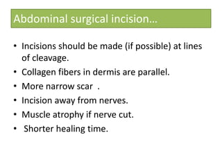
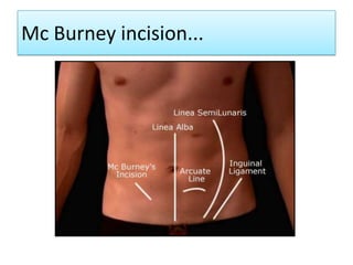
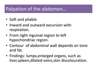
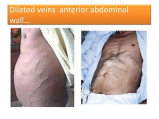
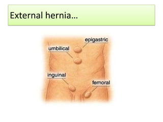
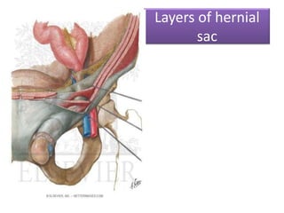
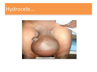
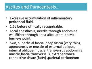
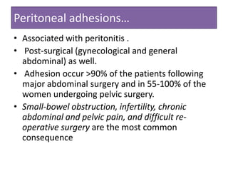
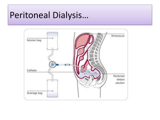
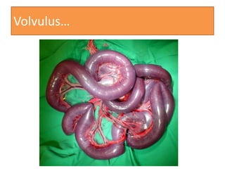
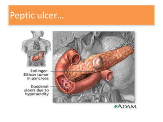
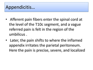
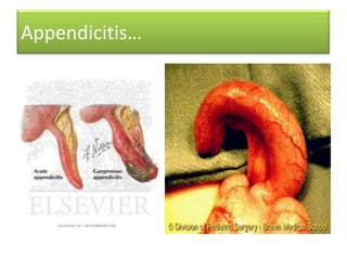
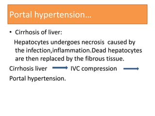
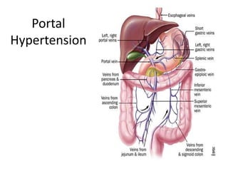
Abdomen
Clinical Anatomy
Dr.Deepak N.Khedekar;M.B.B.S,M.D.
Asst. professor

Dept of Anatomy
LTMMC,SION,MUMBAI.
Feb 2012
Clinical anatomy…
•
•
•
•
•
•
•

Congenital
Truama
Infection
Inflammation
Tumour
Procedure: 1.Therapeutic
2.Diagnostic
Abdominal wall…
Abdominal surgical incision…
• Incisions should be made (if possible) at lines
of cleavage.
• Collagen fibers in dermis are parallel.
• More narrow scar .
• Incision away from nerves.
• Muscle atrophy if nerve cut.
• Shorter healing time.
Mc Burney incision...
Palpation of the abdomen…
• Soft and pliable
• Inward and outward excursion with
respiration.
• From right inguinal region to left
hypochondriac region.
• Contour of abdominal wall depends on tone
and fat.
• Findings: lumps,enlarged organs, such as
liver,spleen,dilated veins,skin discolouration.
Dilated veins anterior abdominal
wall…
Palpation of the superficial inguinal
ring…
• Looking for indirect hernia .
• Male: Palpate spermatic cord in upper part of
the scrotum.
• Firm cordlike structure (posterior part) is vas
deference.
• Patient coughs to increase intra-abdominal
pressure.
• Female: smaller and difficult to palpate.
• Transmits round ligament
Umbilicus…
• Meeting point of CVS,GUS,GIS. during
development. Hotbed of embryology
• Skin around umbilicus supplied by T10
spinal segment.
• Surgical incision should never be crossed
umbilicus.
• Congenital defects include
Omphalocele,hernia,fistula
Omphalocele…
Hernia…
I

• Classified as …External
•
…Internal
• Inguinal hernia can be classified as ….Direct
….Indirect
Indirect further classified as …. Complete
….incomplete.
External hernia…
Layers of hernial
sac
Hydrocele…
Hydrocele…
• Congenital-Persistent processus vaginalis
(congenital)
• Fluid-filled sac tunica vaginalis surrounds testicle.
• 1 in 10 male infants at birth.
• Typically not painful or harmful
• Other causes- infection,truama,tumour.
• Differential diagnosis is hematocele,pyocele.
Varicocele…
Varicocele…
• Pampiniform plexus (testicles) becomes
dilated ,tortuous,thickened,lengthening.
• On inspection -bag filled with worms
• Common in adolescents.
• Mostly left side due to connection with
renal vein vs. right side, which joins with IVC.
• Could be caused by primary kidney disease.
• Pt Presents with infertility (plexus not cooling)
Testicular cancer …
• Spreads upward via lumbar lymph to L1 .
• Later spreads locally to superficial inguinal
lymph
Torsion of the Testicle…
• Rotation around spermatic cord.
• Often associated with excessively large tunica
vaginalis.
• Active young men and children.
• severe pain, could lead to testicular necrosis.
Torsion of testis…
Abdominal quadrants and regions…
Peritonitis…
• Inflamed parietal peritoneum is extremely
sensitive to stretching .
• Pressure applied to abdominal wall with one
finger.
• Rebound tenderness
Ascites…
Ascites and Paracentesis…
• Excessive accumulation of inflammatory
peritoneal fluid.
• 1.5L before clinically recognizable.
• Local anesthesia, needle through abdominal
wallEither through linea alba.lateral to Mc
burneys point.
• Skin, superficial fascia, deep fascia (very thin),
aponeurosis or muscle of external oblique,
internal oblique muscle, transversus abdominis
muscle,fascia transversalis, extraperitoneal
connective tissue (fatty) ,parietal peritoneum
Peritoneal adhesions…
• Associated with peritonitis .
• Post-surgical (gynecological and general
abdominal) as well.
• Adhesion occur >90% of the patients following
major abdominal surgery and in 55-100% of the
women undergoing pelvic surgery.
• Small-bowel obstruction, infertility, chronic
abdominal and pelvic pain, and difficult reoperative surgery are the most common
consequence
Peritoneal Dialysis…
Volvulus…
Stomach…
• Gastric ulcer commonly seen on lesser
curvature.
• Gastric carcinoma commonly occurs at pyloric
antrum,greater curvature
• Zollinger ellison syndrome.
• Vagotomy
• Gastrin-bearing somach mucosa(Antral
gastrecomy)
Ulcer, Gasrtectomy…
Peptic ulcer…
Duodenal (Peptic) ulcers…
• An ulcer of the posterior wall of the first part
of the duodenum may penetrate the wall and
erode the relatively large gastroduodenal
artery causing severe haemorrhage.
• As the stomach empties its contents into the
duodenum, the acid chyme is squirted against
the anterolateral wall of the first part of
the duodenum.
Peptic ulcer…
Appendicitis…
• Afferent pain fibers enter the spinal cord at
the level of the T10c segment, and a vague
referred pain is felt in the region of the
umbilicus .
• Later, the pain shifts to where the inflamed
appendix irritates the parietal peritoneum.
Here the pain is precise, severe, and localized
Appendicitis…
Portal hypertension…
• Cirrhosis of liver:
Hepatocytes undergoes necrosis caused by
the infection,inflammation.Dead hepatocytes
are then replaced by the fibrous tissue.
Cirrhosis liver
IVC compression
Portal hypertension.
Portal
Hypertension
Colitis…
• Pain, tenderness in the abdomen, fever,
swelling of the colon tissue, bleeding,
erythema (redness) of the surface of the
colon, rectal bleeding, and ulcerations of the
colon .
• Associated with Crohn’s disease(Ulcerative
colitis)
Colostomy…
• Cancer of the large bowel.
• Minimally invasive laparoscopic surgery.
• Lymph vessels and nodes removed
•
Ileostomy…
• Small intestine brought out to skin; collected
in pouch.
• Usually done in groin on right side.
• Ulcerative colitis; Hirschprung's disease.
Colonoscopy…
• Colorectal cancer is a leading cause of death in
the Western world b.
• Bowels washed, patient sedated, tube
inserted.
•
Rupture of spleen…
• Associated with truama, mononucleosis.
• Requires immediate medical and surgical
attention.
• Severe blood loss (shocK)
• Left side, T9-T11.
• Removed rather than repaired
• Sx-Total splenectomy
Pancreatic cancer .
• Near Head of bile duct; obstructive jaundice
(yellow skin tint)
• Whipple procedure (pancreatoduodenectom
• Stents could do well.
Impaction of gallstones.
• Usually asymptomatic .
• Sometimes very painful.
• Eating fatty foods contracts gallbladder
against stones.
• May result in infected bile into the pancreatic
duct leading to pancreatitis.
• Sx.-cholecystectomy.
Cirrhosis of liver …
• Scarring of liver tissue as hepatocytes die off.
• Associated with chronic alcoholism and
hepatitis.
• Portal hypertension develops.
Portal hypertension…
• Anastomotic backup of surrounding veins(Gastroesophogeal, Anorectal, Paraumbilical,
Retroperitoneal)
• leads ascities,caput medusae,spider naevi
• Rx-Can insert a shunt from the portal vein
back to IVC.
• Blood flow through sinusoids impeded
Renal and Ureteric calculi (stones)…
•
•
•
•
•
•

Consolidation of dissolved minerals in urine.
Episodic flank pain and hematuria.
Pain -loin to groin radiating pain
Most pass with timed.
Surgical navigation removal.
Non Sx- Extracorporeal Shock
Wave Lithotripsy.(ESWL)
Abdominal aortic aneurysm…
• Localized enlargement of the aorta as a result
of congenital or acquired weakness.
• can be detected left of midline.
• Common site is just above the bifurcation
of the aorta to common iliac arteries.
• Unrecognized rupture of an aneurysm has a
90% mortality rate.
Abdomen, clinical anatomy

Abdomen, clinical anatomy

  • 1.
    Abdomen Clinical Anatomy Dr.Deepak N.Khedekar;M.B.B.S,M.D. Asst.professor Dept of Anatomy LTMMC,SION,MUMBAI. Feb 2012
  • 3.
  • 4.
  • 5.
    Abdominal surgical incision… •Incisions should be made (if possible) at lines of cleavage. • Collagen fibers in dermis are parallel. • More narrow scar . • Incision away from nerves. • Muscle atrophy if nerve cut. • Shorter healing time.
  • 6.
  • 7.
    Palpation of theabdomen… • Soft and pliable • Inward and outward excursion with respiration. • From right inguinal region to left hypochondriac region. • Contour of abdominal wall depends on tone and fat. • Findings: lumps,enlarged organs, such as liver,spleen,dilated veins,skin discolouration.
  • 8.
    Dilated veins anteriorabdominal wall…
  • 9.
    Palpation of thesuperficial inguinal ring… • Looking for indirect hernia . • Male: Palpate spermatic cord in upper part of the scrotum. • Firm cordlike structure (posterior part) is vas deference. • Patient coughs to increase intra-abdominal pressure. • Female: smaller and difficult to palpate. • Transmits round ligament
  • 10.
    Umbilicus… • Meeting pointof CVS,GUS,GIS. during development. Hotbed of embryology • Skin around umbilicus supplied by T10 spinal segment. • Surgical incision should never be crossed umbilicus. • Congenital defects include Omphalocele,hernia,fistula
  • 11.
  • 12.
    Hernia… I • Classified as…External • …Internal • Inguinal hernia can be classified as ….Direct ….Indirect Indirect further classified as …. Complete ….incomplete.
  • 13.
  • 14.
  • 15.
  • 16.
    Hydrocele… • Congenital-Persistent processusvaginalis (congenital) • Fluid-filled sac tunica vaginalis surrounds testicle. • 1 in 10 male infants at birth. • Typically not painful or harmful • Other causes- infection,truama,tumour. • Differential diagnosis is hematocele,pyocele.
  • 17.
  • 18.
    Varicocele… • Pampiniform plexus(testicles) becomes dilated ,tortuous,thickened,lengthening. • On inspection -bag filled with worms • Common in adolescents. • Mostly left side due to connection with renal vein vs. right side, which joins with IVC. • Could be caused by primary kidney disease. • Pt Presents with infertility (plexus not cooling)
  • 19.
    Testicular cancer … •Spreads upward via lumbar lymph to L1 . • Later spreads locally to superficial inguinal lymph
  • 20.
    Torsion of theTesticle… • Rotation around spermatic cord. • Often associated with excessively large tunica vaginalis. • Active young men and children. • severe pain, could lead to testicular necrosis.
  • 21.
  • 22.
  • 23.
    Peritonitis… • Inflamed parietalperitoneum is extremely sensitive to stretching . • Pressure applied to abdominal wall with one finger. • Rebound tenderness
  • 24.
  • 25.
    Ascites and Paracentesis… •Excessive accumulation of inflammatory peritoneal fluid. • 1.5L before clinically recognizable. • Local anesthesia, needle through abdominal wallEither through linea alba.lateral to Mc burneys point. • Skin, superficial fascia, deep fascia (very thin), aponeurosis or muscle of external oblique, internal oblique muscle, transversus abdominis muscle,fascia transversalis, extraperitoneal connective tissue (fatty) ,parietal peritoneum
  • 26.
    Peritoneal adhesions… • Associatedwith peritonitis . • Post-surgical (gynecological and general abdominal) as well. • Adhesion occur >90% of the patients following major abdominal surgery and in 55-100% of the women undergoing pelvic surgery. • Small-bowel obstruction, infertility, chronic abdominal and pelvic pain, and difficult reoperative surgery are the most common consequence
  • 27.
  • 28.
  • 29.
    Stomach… • Gastric ulcercommonly seen on lesser curvature. • Gastric carcinoma commonly occurs at pyloric antrum,greater curvature • Zollinger ellison syndrome. • Vagotomy • Gastrin-bearing somach mucosa(Antral gastrecomy)
  • 30.
  • 31.
  • 32.
    Duodenal (Peptic) ulcers… •An ulcer of the posterior wall of the first part of the duodenum may penetrate the wall and erode the relatively large gastroduodenal artery causing severe haemorrhage. • As the stomach empties its contents into the duodenum, the acid chyme is squirted against the anterolateral wall of the first part of the duodenum.
  • 33.
  • 34.
    Appendicitis… • Afferent painfibers enter the spinal cord at the level of the T10c segment, and a vague referred pain is felt in the region of the umbilicus . • Later, the pain shifts to where the inflamed appendix irritates the parietal peritoneum. Here the pain is precise, severe, and localized
  • 35.
  • 36.
    Portal hypertension… • Cirrhosisof liver: Hepatocytes undergoes necrosis caused by the infection,inflammation.Dead hepatocytes are then replaced by the fibrous tissue. Cirrhosis liver IVC compression Portal hypertension.
  • 37.
  • 38.
    Colitis… • Pain, tendernessin the abdomen, fever, swelling of the colon tissue, bleeding, erythema (redness) of the surface of the colon, rectal bleeding, and ulcerations of the colon . • Associated with Crohn’s disease(Ulcerative colitis)
  • 39.
    Colostomy… • Cancer ofthe large bowel. • Minimally invasive laparoscopic surgery. • Lymph vessels and nodes removed •
  • 40.
    Ileostomy… • Small intestinebrought out to skin; collected in pouch. • Usually done in groin on right side. • Ulcerative colitis; Hirschprung's disease.
  • 41.
    Colonoscopy… • Colorectal canceris a leading cause of death in the Western world b. • Bowels washed, patient sedated, tube inserted. •
  • 42.
    Rupture of spleen… •Associated with truama, mononucleosis. • Requires immediate medical and surgical attention. • Severe blood loss (shocK) • Left side, T9-T11. • Removed rather than repaired • Sx-Total splenectomy
  • 43.
    Pancreatic cancer . •Near Head of bile duct; obstructive jaundice (yellow skin tint) • Whipple procedure (pancreatoduodenectom • Stents could do well.
  • 44.
    Impaction of gallstones. •Usually asymptomatic . • Sometimes very painful. • Eating fatty foods contracts gallbladder against stones. • May result in infected bile into the pancreatic duct leading to pancreatitis. • Sx.-cholecystectomy.
  • 45.
    Cirrhosis of liver… • Scarring of liver tissue as hepatocytes die off. • Associated with chronic alcoholism and hepatitis. • Portal hypertension develops.
  • 46.
    Portal hypertension… • Anastomoticbackup of surrounding veins(Gastroesophogeal, Anorectal, Paraumbilical, Retroperitoneal) • leads ascities,caput medusae,spider naevi • Rx-Can insert a shunt from the portal vein back to IVC. • Blood flow through sinusoids impeded
  • 47.
    Renal and Uretericcalculi (stones)… • • • • • • Consolidation of dissolved minerals in urine. Episodic flank pain and hematuria. Pain -loin to groin radiating pain Most pass with timed. Surgical navigation removal. Non Sx- Extracorporeal Shock Wave Lithotripsy.(ESWL)
  • 48.
    Abdominal aortic aneurysm… •Localized enlargement of the aorta as a result of congenital or acquired weakness. • can be detected left of midline. • Common site is just above the bifurcation of the aorta to common iliac arteries. • Unrecognized rupture of an aneurysm has a 90% mortality rate.