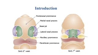
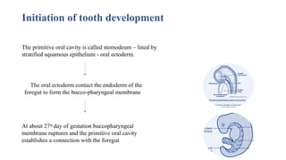
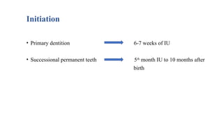
Initiation of toothdevelopment
The primitive oral cavity is called stomodeum – lined by
stratified squamous epithelium - oral ectoderm.
The oral ectoderm contact the endoderm of the
foregut to form the bucco-pharyngeal membrane.
At about 27th day of gestation buccopharyngeal
membrane ruptures and the primitive oral cavity
establishes a connection with the foregut
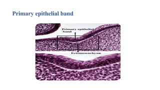
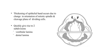
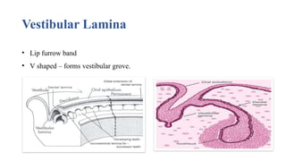
• Thickening ofepithelial band occurs due to
change in orientation of mitotic spindle &
cleavage plane of dividing cells.
• Quickly give rise to 2
subdivisions
ˉ vestibular lamina
ˉ dental lamina
8.
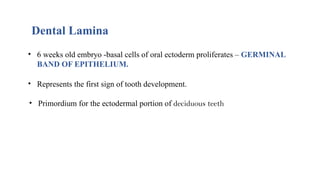
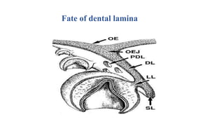
Dental Lamina
• 6weeks old embryo -basal cells of oral ectoderm proliferates – GERMINAL
BAND OF EPITHELIUM.
• Represents the first sign of tooth development.
• Primordium for the ectodermal portion of deciduous teeth
9.
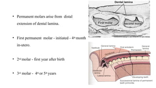
• Permanent molarsarise from distal
extension of dental lamina.
• First permanent molar - initiated - 4th month
in-utero.
• 2nd molar - first year after birth
• 3rd molar - 4th or 5th years
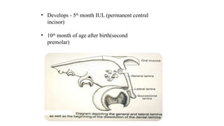
10.
• Develops -5th
month IUL (permanent central
incisor)
• 10th
month of age after birth(second
premolar)
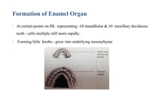
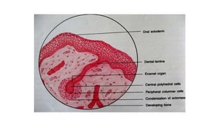
Formation of EnamelOrgan
At certain points on DL representing -10 mandibular & 10 maxillary deciduous
teeth - cells multiply still more rapidly.
Forming little knobs - grow into underlying mesenchyme
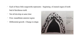
17.
Each ofthese little outgrowths represents – beginning of enamel organ of tooth
bud of deciduous teeth
Not all develop at same time
First- mandibular anterior region
Differential growth - Change in shape
18.
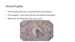
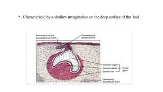
Dental Papilla
• Onthe inside of the cap - ectomesenchymal cells increases.
• Tissue appears - more dense than the surrounding mesenchyme
• Represents the beginning of the dental papilla
19.
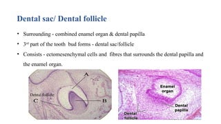
Dental sac/ Dentalfollicle
• Surrounding - combined enamel organ & dental papilla
• 3rd
part of the tooth bud forms - dental sac/follicle
• Consists - ectomesenchymal cells and fibres that surrounds the dental papilla and
the enamel organ.
Dental follicle
20.
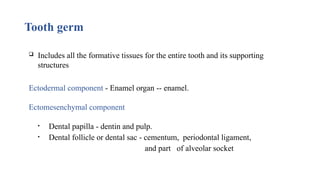
Tooth germ
Includesall the formative tissues for the entire tooth and its supporting
structures
Ectodermal component - Enamel organ -- enamel.
Ectomesenchymal component
• Dental papilla - dentin and pulp.
• Dental follicle or dental sac - cementum, periodontal ligament,
and part of alveolar socket
21.
• Development oftooth – interaction of cells.
• Upto 12 days- first arch epithelium - retains ability.
• When combined with neural crest cells in other regions.
• But transferred to neural crest cells as revealed in various recombinant
experiments.
22.
• Like anyother organ development –numerous & complex gene expressions
– occurs to control – development process- through molecular signals.
• Experimental studies to understand the genetic control and molecular
signalling have been done on mice as it is amenable for genetic
manipulations to produce “ knock-out mice” or “null mice”
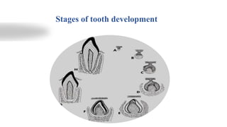
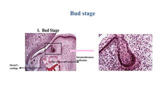
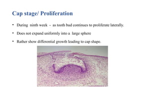
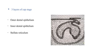
Cap stage/ Proliferation
•During ninth week - as tooth bud continues to proliferate laterally.
• Does not expand uniformly into a large sphere
• Rather show differential growth leading to cap shape.
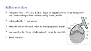
Stellate reticulum
Polygonalcells – b/w OEE & IEE - begin to separate due to water being drawn
into the enamel organ from the surrounding dental papilla.
polygonal cells star shaped
Maintain contact with each other by their cytoplasmic process
star shaped cells – forms cellular network- hence the name SR.
Shock absorber
31.
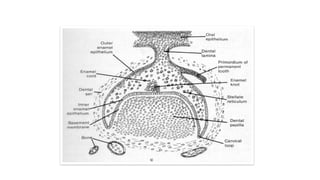
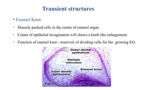
Transient structures
• EnamelKnot:
• Densely packed cells in the center of enamel organ.
• Center of epithelial invagination will shows a knob like enlargement.
• Function of enamel knot - reservoir of dividing cells for the growing EO.
32.
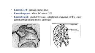
• Enamel cord: Vertical enamel knot
• Enamel septum : when EC meets OEE
• Enamel navel : small depression - attachment of enamel cord to outer
dental epithelium (resembles umbilicus)
33.
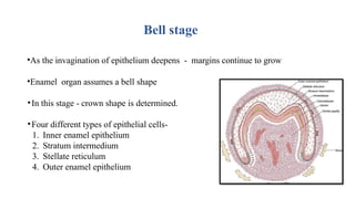
Bell stage
•As theinvagination of epithelium deepens - margins continue to grow
•Enamel organ assumes a bell shape
•In this stage - crown shape is determined.
•Four different types of epithelial cells-
1. Inner enamel epithelium
2. Stratum intermedium
3. Stellate reticulum
4. Outer enamel epithelium
34.
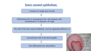
Consists of asingle layer of cells
Differentiate prior to amelogenesis into tall columnar cells-
Ameloblasts(4-5 in diameter; 40 high)
The cells of the inner enamel epithelium exert an organizing influence on
mesenchymal cells in the dental papilla
later differentiate into odontoblasts.
Inner enamel epithelium
35.
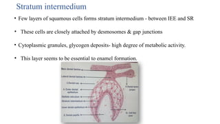
Stratum intermedium
• Fewlayers of squamous cells forms stratum intermedium - between IEE and SR
• These cells are closely attached by desmosomes & gap junctions
• Cytoplasmic granules, glycogen deposits- high degree of metabolic activity.
• This layer seems to be essential to enamel formation.
36.
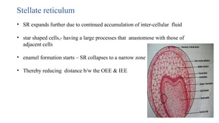
Stellate reticulum
• SRexpands further due to continued accumulation of inter-cellular fluid
• star shaped cells,- having a large processes that anastomose with those of
adjacent cells
• enamel formation starts – SR collapses to a narrow zone
• Thereby reducing distance b/w the OEE & IEE
37.
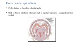
Outer enamel epithelium
•Cells - flatten to form low cuboidal cells
• OEE is thrown into folds which are rich in capillary network – source of nutrition
for EO
38.
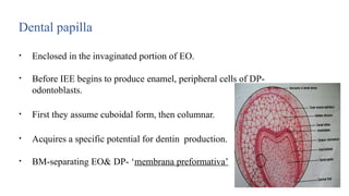
Dental papilla
• Enclosedin the invaginated portion of EO.
• Before IEE begins to produce enamel, peripheral cells of DP-
odontoblasts.
• First they assume cuboidal form, then columnar.
• Acquires a specific potential for dentin production.
• BM-separating EO& DP- ‘membrana preformativa’
39.
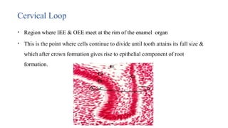
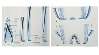
Cervical Loop
• Regionwhere IEE & OEE meet at the rim of the enamel organ
• This is the point where cells continue to divide until tooth attains its full size &
which after crown formation gives rise to epithelial component of root
formation.
40.
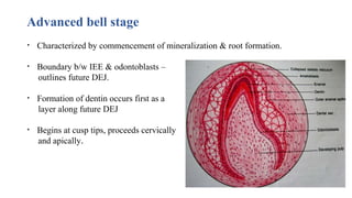
Advanced bell stage
•Characterized by commencement of mineralization & root formation.
• Boundary b/w IEE & odontoblasts –
outlines future DEJ.
• Formation of dentin occurs first as a
layer along future DEJ
• Begins at cusp tips, proceeds cervically
and apically.
41.
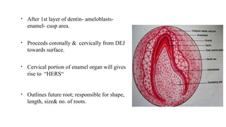
• After 1stlayer of dentin- ameloblasts-
enamel- cusp area.
• Proceeds coronally & cervically from DEJ
towards surface.
• Cervical portion of enamel organ will gives
rise to “HERS“
• Outlines future root; responsible for shape,
length, size& no. of roots.
42.
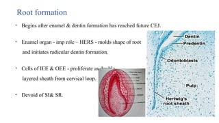
Root formation
• Beginsafter enamel & dentin formation has reached future CEJ.
• Enamel organ - imp role – HERS - molds shape of root
and initiates radicular dentin formation.
• Cells of IEE & OEE - proliferate as double
layered sheath from cervical loop.
• Devoid of SI& SR.
44.
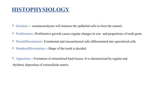
HISTOPHYSIOLOGY
Initiation :-ectomesenchyme will instructs the epithelial cells to form the enamel.
Proliferation:- Proliferative growth causes regular changes in size and proportions of tooth germ.
Histodifferentiation:- Ectodermal and mesenchymal cells differentiated into specialized cells
Morphodifferentiation :- Shape of the tooth is decided
Apposition :- Formation of mineralized hard tissues. It is characterized by regular and
rhythmic deposition of extracellular matrix.
45.
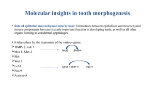
Molecular insights intooth morphogenesis
• Role of epithelial mesenchymail interactions: Interactions between epithelium and mesenchymal
tissues components have particularly important function in developing teeth, as well as all other
organs forming as ectodermal appendages.
• It takes place by the expression of the various genes.
BMP- 2, 4 & 7
Msx 1, Msx 2
Shh
Wnt 7
Lef-1
Pax-9
Activin-A
Msx1 BMP-4
Fgf-8 + BMP-4 Pax-9
46.
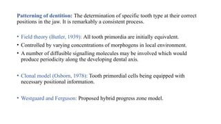
Patterning of dentition:The determination of specific tooth type at their correct
positions in the jaw. It is remarkably a consistent process.
• Field theory (Butler, 1939): All tooth primordia are initially equivalent.
• Controlled by varying concentrations of morphogens in local environment.
• A number of diffusible signalling molecules may be involved which would
produce periodicity along the developing dental axis.
• Clonal model (Osborn, 1978): Tooth primordial cells being equipped with
necessary positional information.
• Westgaard and Ferguson: Proposed hybrid progress zone model.
47.
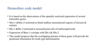
Homeobox code model
•It is based on the observations of the spatially restricted expression of several
homeobox genes.
• Msx-1 &Msx-2 restricted to distal midline mesenchymal regions of incisors &
canines.
• Dlx-1 &Dlx-2 restricted to mesenchymal cells of multicuspid teeth.
• Expression of Barx-1 overlaps with Dlx-1& Dlx-2.
• This model proposes that the overlapping domain of these genes will provide the
positional information for tooth type determination.
48.
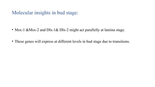
Molecular insights inbud stage:
• Msx-1 &Msx-2 and Dlx-1& Dlx-2 might act parallelly at lamina stage.
• These genes will express at different levels in bud stage due to transitions.
49.
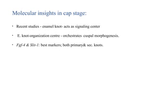
Molecular insights incap stage:
• Recent studies - enamel knot- acts as signaling center
• E. knot-organization centre - orchestrates cuspal morphogenesis.
• Fgf-4 & Slit-1: best markers; both primary& sec. knots.
50.
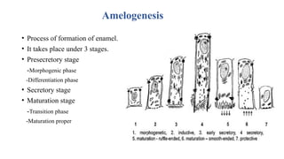
Amelogenesis
• Process offormation of enamel.
• It takes place under 3 stages.
• Presecretory stage
-Morphogenic phase
-Differentiation phase
• Secretory stage
• Maturation stage
-Transition phase
-Maturation proper
51.
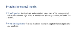
Proteins in enamelmatrix:
Amelogenins: Predominant and comprises about 80% of the young enamel
matrix and contains high levels of amino acids proline, glutamine, histidine and
leucine.
Non-amelogenins: Tuftelin, sheathlin, enamelin, sulphated enamel proteins
and amelotin.
52.
Dentinogenesis
• The processof formation of dentin.
• Stages of dentin development:
-Differentiation of odontoblasts
-Deposition of organic matrix
-Mineralization
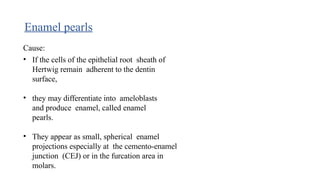
Enamel pearls
Cause:
• Ifthe cells of the epithelial root sheath of
Hertwig remain adherent to the dentin
surface,
• they may differentiate into ameloblasts
and produce enamel, called enamel
pearls.
• They appear as small, spherical enamel
projections especially at the cemento-enamel
junction (CEJ) or in the furcation area in
molars.
55.
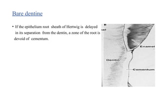
Bare dentine
• Ifthe epithelium root sheath of Hertwig is delayed
in its separation from the dentin, a zone of the root is
devoid of cementum.
56.
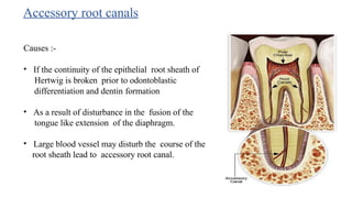
Accessory root canals
Causes:-
• If the continuity of the epithelial root sheath of
Hertwig is broken prior to odontoblastic
differentiation and dentin formation
• As a result of disturbance in the fusion of the
tongue like extension of the diaphragm.
• Large blood vessel may disturb the course of the
root sheath lead to accessory root canal.
57.
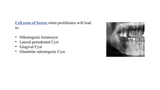
Cell rests ofSerres when proliferates will lead
to:
• Odontogenic keratocyst
• Lateral periodontal Cyst
• Gingival Cyst
• Glandular odontogenic Cyst
References
Orban OralHistology And Embryology 12th Edition
Tencate’s Oral Histology Antonio Nanci 8th Edition
Oral Development And Histology James Avery Third Edition.
Inderbeer Singh: Human Embryology
#24 Epitheium of dental lamina is sep frm the underlying ectomesenchyme by a basement mrm, round or ovoid swllingd
#33 Shape of the crown determined by the pressure by stellate reticulum, the folding of enamel organ to cause diuff crown sgapes due to mitosis nd diff
#47 3 diff conclusions. 1)no specific gene for each tooth type 2)code is both +ve and _ve so the absence of gene is a s imp 3)code is overlapping nd can thus provide positional inf
#52 Odontoblast differentiation is characterized by the recurrent expression of Msx genes& other molecues&the expression of tgf-beta 1 nd 2, fgf 3 and other growth factors.morphologic changes: cell inc in size, vt nuclei in proximal, abundant golgi apparatus vt. Elongation and polarization of the cell is accompanied by the intracellular skeletal proteins such as the actin, vimentin,most of the cell processes directed towards the IEE, as differentiation proceed,s the no.of processes reduces and one large process will be seen. Cell to cell junctions b/w the odontoblasts and subodontoblasts will be seen more in numerous
Deposition- DPP, DSP
Mineralization- Odontoblast produces matrix that becomes mineralized. It controls the release nd transfer of calcium ions