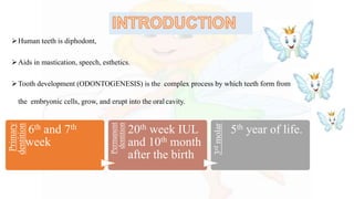
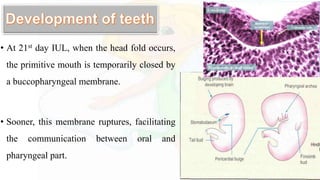
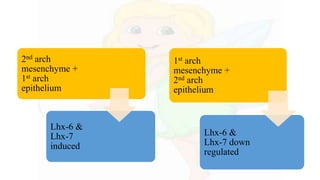
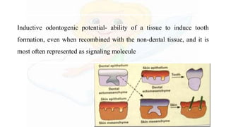
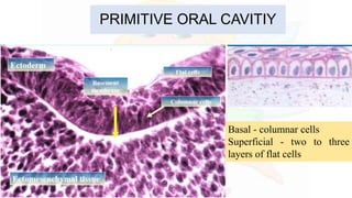
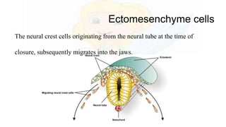
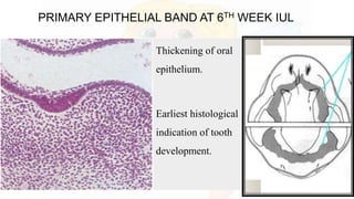
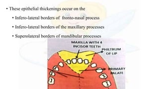
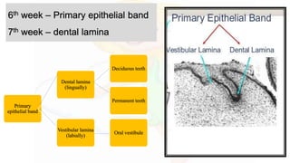
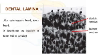
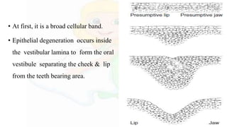
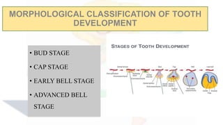
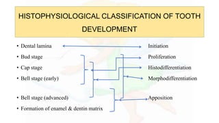
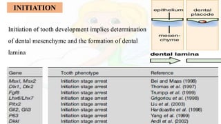
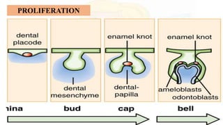
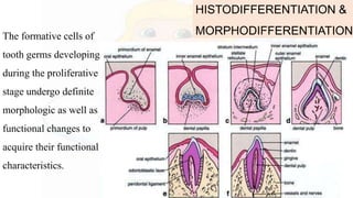
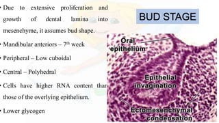
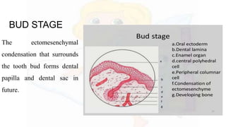
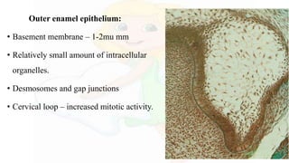
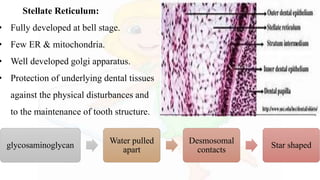
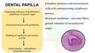
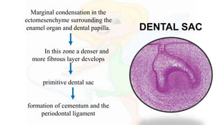
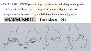
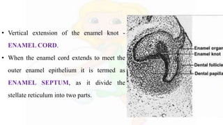
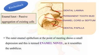
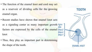
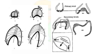
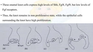
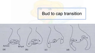
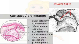
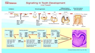
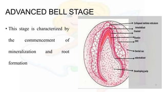
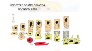
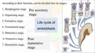
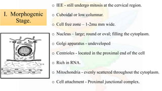
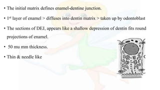
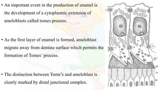
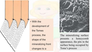
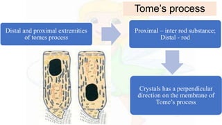
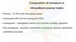
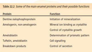
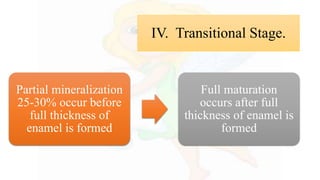
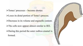
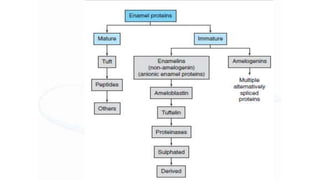
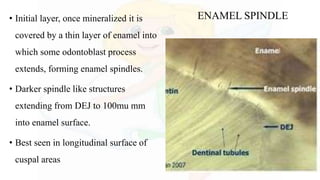
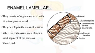
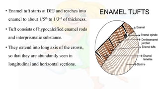
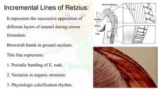
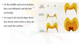
Coronal and radicular pulp
Apical foramen
Accessory canal
Functions of dental pulp
Components of dental pulp
Functions of pulpal extracellular matrix
Organization of cells in the pulp
The principle cells of the pulp
The pathways of collagen synthesis
Matrix and ground substances
Vasculature and lymphatic supply
Innervation of Dentin- pulp complex
Disorders of the dental pulp
Advances in pulp vitality testing
I prepared this presentation during the first year of my MDS. This will give you a basic idea and necessary information about the pulp of the teeth and its histology. Hope you guys find it useful.
BE UPDATE TO IT,, AS IT IS 3 years back from 2017
Kindly mail me if you feel, needy of this presentation
you can find my mail id @ slide share,,, if not mail me @
sukesh3567@gmail.com.
Good luck
A Brief Description about the development of teeth. Understanding the process of tooth development is of particular importance for the dentist; as developmental disturbances may occur at any stage of this process resulting in different types of tooth anomalies.
Tooth development can be classified either based on morphology or histology
Morphological stages:
Bud stage
Cap stage
Bell stage:
* Early
* Advanced
Physiological stages:
Initiation
Proliferation
Histodifferentiation
Morphodifferentiation
Apposition
Model Attribute Check Company Auto PropertyCeline George
In Odoo, the multi-company feature allows you to manage multiple companies within a single Odoo database instance. Each company can have its own configurations while still sharing common resources such as products, customers, and suppliers.
Unit 8 - Information and Communication Technology (Paper I).pdfThiyagu K
This slides describes the basic concepts of ICT, basics of Email, Emerging Technology and Digital Initiatives in Education. This presentations aligns with the UGC Paper I syllabus.
Welcome to TechSoup New Member Orientation and Q&A (May 2024).pdfTechSoup
In this webinar you will learn how your organization can access TechSoup's wide variety of product discount and donation programs. From hardware to software, we'll give you a tour of the tools available to help your nonprofit with productivity, collaboration, financial management, donor tracking, security, and more.
2024.06.01 Introducing a competency framework for languag learning materials ...Sandy Millin
http://sandymillin.wordpress.com/iateflwebinar2024
Published classroom materials form the basis of syllabuses, drive teacher professional development, and have a potentially huge influence on learners, teachers and education systems. All teachers also create their own materials, whether a few sentences on a blackboard, a highly-structured fully-realised online course, or anything in between. Despite this, the knowledge and skills needed to create effective language learning materials are rarely part of teacher training, and are mostly learnt by trial and error.
Knowledge and skills frameworks, generally called competency frameworks, for ELT teachers, trainers and managers have existed for a few years now. However, until I created one for my MA dissertation, there wasn’t one drawing together what we need to know and do to be able to effectively produce language learning materials.
This webinar will introduce you to my framework, highlighting the key competencies I identified from my research. It will also show how anybody involved in language teaching (any language, not just English!), teacher training, managing schools or developing language learning materials can benefit from using the framework.
Biological screening of herbal drugs: Introduction and Need for
Phyto-Pharmacological Screening, New Strategies for evaluating
Natural Products, In vitro evaluation techniques for Antioxidants, Antimicrobial and Anticancer drugs. In vivo evaluation techniques
for Anti-inflammatory, Antiulcer, Anticancer, Wound healing, Antidiabetic, Hepatoprotective, Cardio protective, Diuretics and
Antifertility, Toxicity studies as per OECD guidelines
Synthetic Fiber Construction in lab .pptxPavel ( NSTU)
Synthetic fiber production is a fascinating and complex field that blends chemistry, engineering, and environmental science. By understanding these aspects, students can gain a comprehensive view of synthetic fiber production, its impact on society and the environment, and the potential for future innovations. Synthetic fibers play a crucial role in modern society, impacting various aspects of daily life, industry, and the environment. ynthetic fibers are integral to modern life, offering a range of benefits from cost-effectiveness and versatility to innovative applications and performance characteristics. While they pose environmental challenges, ongoing research and development aim to create more sustainable and eco-friendly alternatives. Understanding the importance of synthetic fibers helps in appreciating their role in the economy, industry, and daily life, while also emphasizing the need for sustainable practices and innovation.
Read| The latest issue of The Challenger is here! We are thrilled to announce that our school paper has qualified for the NATIONAL SCHOOLS PRESS CONFERENCE (NSPC) 2024. Thank you for your unwavering support and trust. Dive into the stories that made us stand out!
Honest Reviews of Tim Han LMA Course Program.pptxtimhan337
Personal development courses are widely available today, with each one promising life-changing outcomes. Tim Han’s Life Mastery Achievers (LMA) Course has drawn a lot of interest. In addition to offering my frank assessment of Success Insider’s LMA Course, this piece examines the course’s effects via a variety of Tim Han LMA course reviews and Success Insider comments.
June 3, 2024 Anti-Semitism Letter Sent to MIT President Kornbluth and MIT Cor...Levi Shapiro
Letter from the Congress of the United States regarding Anti-Semitism sent June 3rd to MIT President Sally Kornbluth, MIT Corp Chair, Mark Gorenberg
Dear Dr. Kornbluth and Mr. Gorenberg,
The US House of Representatives is deeply concerned by ongoing and pervasive acts of antisemitic
harassment and intimidation at the Massachusetts Institute of Technology (MIT). Failing to act decisively to ensure a safe learning environment for all students would be a grave dereliction of your responsibilities as President of MIT and Chair of the MIT Corporation.
This Congress will not stand idly by and allow an environment hostile to Jewish students to persist. The House believes that your institution is in violation of Title VI of the Civil Rights Act, and the inability or
unwillingness to rectify this violation through action requires accountability.
Postsecondary education is a unique opportunity for students to learn and have their ideas and beliefs challenged. However, universities receiving hundreds of millions of federal funds annually have denied
students that opportunity and have been hijacked to become venues for the promotion of terrorism, antisemitic harassment and intimidation, unlawful encampments, and in some cases, assaults and riots.
The House of Representatives will not countenance the use of federal funds to indoctrinate students into hateful, antisemitic, anti-American supporters of terrorism. Investigations into campus antisemitism by the Committee on Education and the Workforce and the Committee on Ways and Means have been expanded into a Congress-wide probe across all relevant jurisdictions to address this national crisis. The undersigned Committees will conduct oversight into the use of federal funds at MIT and its learning environment under authorities granted to each Committee.
• The Committee on Education and the Workforce has been investigating your institution since December 7, 2023. The Committee has broad jurisdiction over postsecondary education, including its compliance with Title VI of the Civil Rights Act, campus safety concerns over disruptions to the learning environment, and the awarding of federal student aid under the Higher Education Act.
• The Committee on Oversight and Accountability is investigating the sources of funding and other support flowing to groups espousing pro-Hamas propaganda and engaged in antisemitic harassment and intimidation of students. The Committee on Oversight and Accountability is the principal oversight committee of the US House of Representatives and has broad authority to investigate “any matter” at “any time” under House Rule X.
• The Committee on Ways and Means has been investigating several universities since November 15, 2023, when the Committee held a hearing entitled From Ivory Towers to Dark Corners: Investigating the Nexus Between Antisemitism, Tax-Exempt Universities, and Terror Financing. The Committee followed the hearing with letters to those institutions on January 10, 202
How to Make a Field invisible in Odoo 17Celine George
It is possible to hide or invisible some fields in odoo. Commonly using “invisible” attribute in the field definition to invisible the fields. This slide will show how to make a field invisible in odoo 17.
2. • INTRODUCTION
• DEVELOPMENT OF TEETH
• CLASSIFICAION OF TOOTH DEVELOPMENT
• MORPHOLOICAL STAGES
• BUD STAGE
• CAP STAGE
• BELL STAGE
• AMELOGENESIS
• REFERENCES
3. Human teeth is diphodont,
Aids in mastication, speech, esthetics.
Tooth development (ODONTOGENESIS) is the complex process by which teeth form from
the embryonic cells, grow, and erupt into the oral cavity.
Primary
dentition
6th and 7th
week
Permanent
dentition
20th week IUL
and 10th month
after the birth
3rdmolar
5th year of life.
4. • At 21st day IUL, when the head fold occurs,
the primitive mouth is temporarily closed by
a buccopharyngeal membrane.
• Sooner, this membrane ruptures, facilitating
the communication between oral and
pharyngeal part.
5. ORAL / ABORAL AXIS
• Controlled by the signaling molecule FGF-8, which is expressed in the
oral epithelium from embryonic day 8.
• This induces the expression of genes necessary for tooth development
such as homeobox genes Lhx-6, Lhx-8 in the oral mesenchyme.
6. TOOTH ORIGIN
• The tooth originates
from ectoderm of 1st
pharyngeal arch and
ectomesenchyme of the
neural crest.
7. EPITHELIAL MESENCHYMAL INTERACTION
• It is a series of programmed, sequential and
reciprocal (complex and multiphasic)
communications between the epithelium and
mesenchyme (two heterotypic cell populations)
resulting in the differentiation of one or both the
cell populations involved.
8. MECHANISM OF EPITHELIAL-MESENCHYMAL INTERACTION
Cell
signal to
each
other
through
signaling
molecule
s
Transmit
information
to adjacent
cells
Activation of
transcription
factors
Enter
nucleus
Regulate
s gene
expressio
n
New
proteins
are
produce
d
Change
in target
cell
10. Inductive odontogenic potential- ability of a tissue to induce tooth
formation, even when recombined with the non-dental tissue, and it is
most often represented as signaling molecule
12. Ectomesenchyme cells
The neural crest cells originating from the neural tube at the time of
closure, subsequently migrates into the jaws.
13. PRIMARY EPITHELIAL BAND AT 6TH WEEK IUL
Thickening of oral
epithelium.
Earliest histological
indication of tooth
development.
14. • These epithelial thickenings occur on the
• Infero-lateral borders of fronto-nasal process
• Infero-lateral borders of the maxillary processes
• Superolateral borders of mandibular processes
17. TOOTH GERM POSITIONING
• FGF-8 has been proposed to act antagonistically with BMP-4 to specify the sites of
tooth initiation.
- regulated by PITx-2
• Wnt – maintains boundaries between tooth forming areas and non-tooth forming
areas
Lef-1 > expressed in dental epithelium thickenings.
>also the ectopic expression of Lef-1 in the oral epithelium also results
in ectopic tooth formation.
18. - 5 months IUL – 10 months after birth
- 4th month, 1st year & 4th year
DENTAL LAMINA
19. EPITHELIAL CELL REST
OF SERRE
• Dental lamina functions – 5 years
• Remnants of dental lamina persists as
epithelial islands within gingiva called
epithelial cell rest of serre.
20. VESTIBULAR LAMINA
LIP FURROW BAND/ VESTIBULAR
RIDGE/ LABIO-GINGIVAL LAMINA.
The vestibular lamina is formed as ectodermal
proliferation in the underlying
ectomesenchyme.
21. • At first, it is a broad cellular band.
• Epithelial degeneration occurs inside
the vestibular lamina to form the oral
vestibule separating the cheek & lip
from the teeth bearing area.
22. • BUD STAGE
• CAP STAGE
• EARLY BELL STAGE
• ADVANCED BELL
STAGE
23. HISTOPHYSIOLOGICAL CLASSIFICATION OF TOOTH
DEVELOPMENT
• Dental lamina Initiation
• Bud stage Proliferation
• Cap stage Histodifferentiation
• Bell stage (early) Morphodifferentiation
• Bell stage (advanced) Apposition
• Formation of enamel & dentin matrix
24. INITIATION
Initiation of tooth development implies determination
of dental mesenchyme and the formation of dental
lamina
27. BUD STAGE
• Due to extensive proliferation and
growth of dental lamina into
mesenchyme, it assumes bud shape.
• Mandibular anteriors – 7th week
• Peripheral – Low cuboidal
• Central – Polyhedral
• Cells have higher RNA content than
those of the overlying epithelium.
• Lower glycogen
29. • Four major signaling pathways and their inhibitors control tooth
formation:
• BMP
• FGF
• SHH
• WNT
• Overexpression of BMP or functional inactivation of FGF or SHH –
arrest at bud stage.
30. Lef-1 > first expressed in dental epithelium thickenings.
> during bud formation it shifts its location to express in
condensing mesenchyme.
>in Lef-1 knock-out mice, all the dental development is
arrested at the bud stage.
32. CAP STAGE
• As the tooth bud continues to proliferate, the rate of mitosis is
increased in peripheral areas.
• A shallow deep invagination on the deeper surface of the bud
occurs, resulting in formation of cap.
• Outer enamel epithelium
• Inner enamel epithelium
• Stellate reticulum
33. Outer enamel epithelium:
• Basement membrane – 1-2mu mm
• Relatively small amount of intracellular
organelles.
• Desmosomes and gap junctions
• Cervical loop – increased mitotic activity.
35. DENTAL PAPILLA
Organizing influence of proliferative
epithelium of enamel organ
Ectomesenchymal cells proliferate
Budding of capillaries and mitotic
activity
dental papilla – odontoblasts,
primordial of pulp
Fibroblast (primary) and mesenchymal
cells with communicating cytoplasmic
process.
Basement membrane – reticulate fibres,
ground substance of mesenchymal
origin.
36. Marginal condensation in the
ectomesenchyme surrounding the
enamel organ and dental papilla.
In this zone a denser and
more fibrous layer develops
primitive dental sac
formation of cementum and the
periodontal ligament
DENTAL SAC
37. • The ENAMEL KNOT projects in part towards the underlying dental papilla, so
that the centre of the epithelial invagination shows a slightly knob like
enlargement that is bordered by the labial and lingual enamel grooves.
ENAMEL KNOT Hans Ahrens, 1913
38. • Vertical extension of the enamel knot -
ENAMEL CORD.
• When the enamel cord extends to meet the
outer enamel epithelium it is termed as
ENAMEL SEPTUM, as it divide the
stellate reticulum into two parts.
39. • The outer enamel epithelium at the point of meeting shows a small
depression and this is termed ENAMEL NOVEL, as it resembles
the umbilicus.
Enamel knot - Passive
aggregation of existing cells
40. • The function of the enamel knot and cord may act
as a reservoir of dividing cells for the growing
enamel organ.
• Recent studies have shown that enamel knot acts
as a signaling center as many important growth
factors are expressed by the cells of the enamel
knot
• Thus, they play an important part in determining
the shape of the tooth.
41.
42. • These enamel knot cells express high levels of Shh, Fgf4, Fgf9, but low levels of
Fgf receptors.
• Thus, the knot remains in non proliferative state, while the epithelial cells
surrounding the knot have high proliferation.
45. • As the undersurface of cap deepens, the enamel
organ assumes a bell shape.
• IEE interacts with the adjacent mesenchymal cells
and determines the shape of the crown.
• During the late bell stage the morphology of the
crown is determined.
EARLY BELL STAGE
46. • It was thought that the shape of crown is due to pressure exerted by the
growing dental papilla cells on the IEE. Though, this pressure however
was shown to be opposed equally by the pressure exerted by the fluid
present in stellate reticulum.
• At present, the concept of folding of enamel organ to cause different
crown shapes is shown to be due to intrinsic growth caused by
differential rates of mitotic division within the IEE.
47. • The IEE cells which lie in the future cusp tip or incisor region stop dividing
earlier and begin to differentiate first.
• Pressure exerted by the continuous cell division on these cells from other enamel organ
cause these cells to be pushed out into the enamel organ in the form of a cusp tip.
• The cells in another future cusp area begin to differentiate & by a similar way results in a
cusp tip form. The area between two cusp tips (i.e) the cuspal slope extends and
therefore the cusp tips to the depth of the sulcus.
Inner enamel epithelium
48. - layers of cells between IEE and SR.
- high mitotic activity (cytoplasmic
organelles, acid muco, glycogen
deposits).
- Important for tooth formation esp
enamel since its not seen in root
formation.
Stratum intermedium
49. • SI function: not understood, but its believed to plat a role in enamel
production either itself though control of fluid diffusion into and out
of ameloblasts or by actual contribution of necessary formative
elements or enzymes
50. • DENTAL LAMINA;
The dental lamina joining the tooth germ to oral
epithelial fragments, eventually separating the
developing tooth from oral epithelium.
It extends lingually and termed successional
dental lamina as it gives rise to enamel organ of
permanent successors of deciduous teeth.
The enamel organ of deciduous teeth in bell
stage show successional lamina and their permanent
successor teeth in bud stage.
51. CERVICAL LOOP
• OEE & IEE are continuous; the region
where the IEE & OEE meet is known as
zone of reflexion or cervical loop.
• This point is where the IEE continue to
divide until the tooth crown attains its full
size and which, after crown formation, gives
rise to epithelial component of root
formation.
52.
53. ADVANCED BELL STAGE
• This stage is characterized by
the commencement of
mineralization and root
formation
55. Life cycle of
ameloblasts
According to their function, can be divided into six stages:
1. Morphogenic stage
2. Organizing stage.
3. Formative stage.
4. Transitional stage
5. Maturative stage.
6. Protective stage.
7. Desmolytic stage.
Pre secretory
stage
Post
maturative
stage
56. o IEE - still undergo mitosis at the cervical region.
o Cuboidal or low columnar.
o Cell free zone – 1-2mu mm wide.
o Nucleus – large; round or oval; filling the cytoplasm.
o Golgi apparatus - undeveloped
o Centrioles - located in the proximal end of the cell
o Rich in RNA.
o Mitochondria - evenly scattered throughout the cytoplasm.
o Cell attachment - Proximal junctional complex.
I. Morphogenic
Stage.
57. • This stage has 2 principal features:
• Resorption of basal lamina
• Differentiation to ameloblast
II. Differentiation
“organization” Stage.
58. • Pre-ameloblast – produces some enamel
proteins, even before the basal lamina is lost
- expresses dentin sialoprotein,
an odontoblast product transiently.
- phagocytosed by developing
odontoblasts.
- suggesting that they
participate in epithelial mesenchymal
interaction.
II. Differentiation
“organization” Stage.
60. Elongated up to 40 microns.
Nucleus – round; shifts to proximal end
Golgi complex - increases in volume; migrates
distally;
Mitochondria – clusters in infra-nuclear
cytoplasm
RER – significantly increased
Second junctional complex develops at distal
extremity of the cell.
Ameloblasts
62. • With the formation of first layer of dentin the nutrition to ameloblasts from
the dental papillary cells is blocked.
• To compensate, the stellate reticulum collapses and the invagination of
OEE by blood vessels lying outside.
63. • Golgi complex – extensive & well developed;
- forms cylindrical tubules around
the RER; occupies major supra nuclear
compartment.
• mitochondria clustered in the proximal region.
• RER increased in number.
III. Secretory “Formative” Stage.
65. • The initial matrix defines enamel-dentine junction.
• 1st layer of enamel > diffuses into dentin matrix > taken up by odontoblast
• The sections of DEJ, appears like a shallow depression of dentin fits round
projections of enamel.
• 50 mu mm thickness.
• Thin & needle like
66. • An important event in the production of enamel is
the development of a cytoplasmic extension of
ameloblasts called tomes process.
• As the first layer of enamel is formed, ameloblast
migrate away from dentine surface which permits the
formation of Tomes’ process.
• The distinction between Tome’s and ameloblast is
clearly marked by distal junctional complex.
67. The mineralizing surface
presents a honeycomb
appearance, the pits in the
surface being occupied by
Tome’s process
• With the
development of
the Tomes
process, the
shape of the
mineralizing front
changes to a
68. Tome’s process
Distal and proximal extremities
of tomes process
Proximal – inter rod substance;
Distal - rod
Crystals has a perpendicular
direction on the membrane of
Tome’s process
69. Composition of immature or
mineralized enamel matrix
• Proteins – 25-30% early developing enamel.
• Amelogenin (90%) & non-amelogenin (10%)
• Amelogenins – hydrophobic, protein rich in proline, histidine, glutamine.
• Non-amelogenin – enamelin, ameloblastin, enamelysin, amelotin, odontoblast
ameloblast associated.
70.
71. IV. Transitional Stage.
Partial mineralization
25-30% occur before
full thickness of
enamel is formed
Full maturation
occurs after full
thickness of enamel is
formed
72. Tomes’ processes – become shorter.
Loses its distal portion of Tome’s process.
Decrease in its volume and organelle content.
The cells now appear almost similar to IEE.
During this period the outer rodless enamel is
formed.
73. • The number of ameloblasts reduced.
• 50% ameloblast – apoptosis
• RER, golgi complex reduced - autophagocytosis
74. Ameloblasts are
slightly reduced in length
closely attached to enamel matrix.
25% of the cells die during this phase.
cytoplasmic vacuoles containing material
resembling enamel matrix.
These structures indicate an absorptive function of
these cells.
V. Maturative Stage.
75. Ameloblasts develop cycles of modulation of alternating ruffled
and smooth bordered end which is expressed like a wave.
Associated with
introduction of inorganic
materials.
Distal tight proximal
junctions.
Associated with
removal of protein and
water.
Distal leaky proimal
junction.
V. Maturative Stage.
76. Maturation seems to begin at the dentinal end of the rod
• Each rod mature from the depth to the surface
• The sequence of maturing rod is from cusps or incisal edge
toward the cervical line
• Ameloblasts undergo a transition that not only reduces their secretion of enamel
proteins, but also initiates the secretion of kallikrein 4 (KLK4), a serine protease
that degrades the organic matrix, facilitating its removal from the extracellular
compartment
77.
78. • Ameloblast ceased to be arranged in a well-defined
layer.
• No longer distinguished from the cells of stratum
intermedium and OEE.
• These cells are called as Reduced Enamel
Epithelium.
VI. Protective Stage.
79. • If the connective tissue comes in contact with
the enamel, enamel may be either resorbed or
covered by a layer of cementum.
• REE – secrets some material – establish the
dentino gingival junction.
80. VII. Desmolytic Stage.• The REE proliferates and seems to induce
atrophy of the connective tissue, separating it
from the oral epithelium.
• The fusion of two epithelium occurs as
pathway for tooth eruption
• Premature degeneration of the reduced
enamel epithelium may prevent the eruption
of a tooth.
81. • Initial layer, once mineralized it is
covered by a thin layer of enamel into
which some odontoblast process
extends, forming enamel spindles.
• Darker spindle like structures
extending from DEJ to 100mu mm
into enamel surface.
• Best seen in longitudinal surface of
cuspal areas
ENAMEL SPINDLE
82. ENAMEL LAMELLAE
• They consist of organic material with
little inorganic mineral.
• They develop in the areas of tension.
• When the rod crosses such planes, a
short segment of rod remains
uncalcified.
83. ENAMEL TUFTS• Enamel tuft starts at DEJ and reaches into
enamel to about 1/5th to 1/3rd of thickness.
• Tuft consists of hypocalcified enamel rods
and interprismatic substance.
• They extend into long axis of the crown,
so that they are abundantly seen in
longitudinal and horizontal sections.
84. Incremental Lines of Retzius:
It represents the successive apposition of
different layers of enamel during crown
formation.
Brownish bands in ground sections.
This line represents:
1. Periodic bending of E. rods.
2. Variation in organic structure.
3. Physiologic calcification rhythm.
85. • At the middle and cervical parts,
they run obliquely and deviate
occlusally;
• At cusp tis & incisal edges these
line forms semi-circle as they do
not reach the surface
86. Perikymata
• They are transverse, wave like
grooves, continuous around tooth.
• Usually lies parallel to each other
and to CEJ.
88. REFERENCES
• Tencates Oral histology, 3rd edition
• Orbans Oral histology and Embryology, 13th edition
• Essentials of Oral histology & Embryology, James K Avery, 3rd edition
• Oral anatomy, histology and embryology, B.K.B. Berkovitz,
G.R.Holland, B.J.Moxham, 4th edition
Editor's Notes
Entire primary dentition is initiated between 6th and 7th week of IUL.
Successional permanent teeth initiated between 20th week in utero and 10th month after birth.
Permanent molars initiated in 5th year of life
formed from the prochordal plate.
Occurs on the oral surface of the first branchial arch. At 27th day of gestation
The oral-aboral axis of developing jaw makes sure that teeth will develop only within oral cavity.
The development of teeth is characterized by extensive interaction between the epithelial and mesenchymal cells.
Epithelial mesenchymal interaction is an hallmark of odontogenesis.
In odontogenesis, this interaction can be observed in initiation of tooth bud as well as for histodifferentiation of IEE cells and dental papilla cells.
Many other organs like hair, lungs, salivary glands develops by such interactions
Mechanism of epithelial mesenchyme interaction.
The cell signal to each other that is from epithelium to mesenchyme or from mesenchyme to epithelium, through several signaling molecules and transmits information to adjacent cells leading to the activation of transcription factors. This activated transcription factors enters the nucleus and regulated the gene expression resulting in the formation of new proteins that alters the morphology or functions of the target cell.
Experiments demonstrates that the expression of LIM homeobox gene resides within the epithelium of 1st branchial arch.
However, after 12th day of development, the first arch epithelium loses its odontogenic potential, which then is assumed by the ectomesenchyme, so that the ectomesenchyme can elicit tooth formation from a variety of epithelia.
Studies reveal that when dental epithelium interact with skin mesenchyme it results in formation of skin
When skin epithelium is interacted with dental mesenchyme, it resulted in formation of tooth.
After the rupture of buccopharyngeal membrane, primitive oral cavity is lined by stratified squamous epithelium, composed of basal columnar and superficial two to three layers of flat cells, separated from connective tissue through a basement membrane.
The ectomesenchymal cells responsible for tooth development originates from the neural tube at the time of closure migrating into jaws and are responsible for the formation of hard and soft tissues of tooth.
Two or three weeks later, at about 37th day of gestation, certain areas of basal cells of oral ectoderm proliferate in the underlying connective tissue under the influence of ectomesenchyme and thickens, with resultant formation of horse-shoe shaped primary epithelial band.
Frontonasal process, providing four separate sites of origin of tooth tissue for maxillary anteriors at about 6 weeks.
So the anterior maxillary teeth are to be derived from the frontonasal process, while, maxillary posterior teeth are derived from maxillary process.
In contrast, mandibular dental arch develops from only the teo primary odontogenic sites in lower jaw.
At about 7 weeks, the free margin of primary epithelial band is divided into dental and vestibular lamina. Dental lamina forms first and soon after, vestibular lamina forms.
Due to increased mitotic activity in certain points of primary epithelial band, the ectoderm invaginates into the underlying mesenchyme.
This dental lamina is also known as tooth band or odontogenic band. It determines the location of tooth bud to develop.
Expressed in the oral portion of mesenchyme in the first branchial arch. It determines the positions where the tooth germs will form.
Pituitary homeobox
Wingless homologue in vertebrates WNT
Lymphoid enhancer factor
The expression of Lef is important in tooth germ positioning.
All the deciduous teeth arises from dental lamina, and later the permanent successors arise from successional lamina, initiated between 5 months iul to 10 months after birth.
Accessional lamina is the distal extension of dental lamina into maxillary tuberosity and mandibular ramus. The permanent molars develops from this accessional lamina during 4th month, 1st year & 4th year after birth corresponds to 1st, 2nd & 3rd permanent molars.
Fate of dental lamina – after 5 years; remains as cell rest of serres.
After 5 years of its activity, the epithelial remnants will degenerate and undergoes apoptosis.
The remnants if proliferates leads to pathologic cyst and tumor like Odontogenic keratocyst, lateral periodontal cyst, gingival cyst, glandular odontogenic cyst, ameloblastoma, calcifying epithelial odontogenic tumor
It is the buccal process of primary epithelial band.
Subsequently, the epithelial cells degenerates and hollows to form a sulcus in both upper and lower jaw, called oral vestibule.
The oral vestibule is a free space lined by a mucosa comprising a stratified squamous epithelium, that separates lips and cheek from alveolar process.
After the formation of dental lamina, tooth development is classified according to the shape of enamel organ.
Except for initiation, other stages overlaps and are continuous, with various morphologic stages of odontogenesis.
Msh like genes in vertebrates
Distaless homeobox gene
Enhanced proliferative activity proceeds at the point of initiation and results successively in bud, cap and bell stages.
Histodifferentiation is a process in which a cell changes its particular character to a more specialized type. In odontogenesis, it includes the differentiation of mesenchymal cells into dentin secreting odontoblast and of epithelial cells into enamel secreting ameloblast.
The morphologic pattern, i.e., basic form and relative size of the tooth, is established by morphodifferentiation, by differential growth.
The advanced bell stage marks active histodifferentiation and also an important stage of morphodifferentiation, outlining the dentinoenamel junction.
Apposition refers to formation & deposition of dental hard tissues.
Tooth buds are located close to the oral epithelium ; separated from the underlying mesenchyme by a basement membrane and surrounded by a mesenchymal condensation.
Mandibular anterior tooth buds appear about 7th week and are the first tooth bud to form.
By 8th week, all the tooth buds are present at varying stages.
As the tooth bud continues to proliferate, the cellular density in the ectomesenchyme increases, that forms future dental papilla and sac. The enamel organ consists of peripherally located columnar cells and central polyhedral cells.
Wingless homologue in vertebrates;
Arrest of tooth development may lead to anodontia.
Specially, when the Wnt signaling is stimulated in the oral epithelium, it results in production of dozens of extra teeth.
Ectodin and ectodysplasin are two other signaling molecules. The loss of these molecules inhibits BMP signaling and leads to the formation of supernumerary teeth.
Lymphoid enhancer factor
runx- runt related transcription factor.
Msh like genes in vertebrates
10-11 WEEK
Differential growth results in increase in the size of the enamel organ & change in its shape to exhibit a cap shape.
The OEE cells lines the outer concave surface of the cap and the cells are low cuboidal.
Golgi apparatus, endoplasmic reticulum, mitochondria.
The junction of IEE and OEE forms the cervical loop.
The polygonal cells located in the centre of the enamel organ.
It synthesis and secrete glycosaminoglycans into the extracellular compartment between the epithelial cells. Glycosaminoglycans are hydrophilic and exerts high osmotic pressure.
As a result water being drawn out into the enamel organ from the surrounding papilla, the central cells are forced apart.
Since these cells retain their connection with adjacent cells through desmosomal contacts they become star shaped. Thus, they are termed stellate reticulum.
Dental papilla:
Under the influence of proliferating epithelium of enamel organ, the ectomesenchymal cells undergoes mitotic activity and active budding of capillaries and condenses to form dental papilla which is later differentiates to pulp and also primordia of pulp.
Peripheral cells adjacent to the IEE enlarges and later differentiate into odontoblast.
Dental sac: Marginal condensation of ectomesenchymal cells, surrounding the enamel organ and dental papilla is called dental sac.
In late bud stage, the cells in the center of the enamel organ becomes undivided and, are densely packed and form the enamel knot.
He states that, the enamel knot is a main factor, responsible for determining the tooth crown shape in multicuspid tooth.
A vertical extension of cells arises from the enamel knot and meets the outer enamel epithelium. This vertical extension before joining the OEE is called enamel cord. After the enamel cord meets the OEE, it divides the stellate reticulum into 2 parts. Hence, it is termed as enamel septum.
Concluded that……..due to……
These are temporary (transitory) structures that disappears before enamel formation begins.
The enamel cord disappears in the late bell stage is associate with apoptosis.
In anterior teeth only one enamel knot is formed.
In addition to primary enamel knot, secondary enamel knots appear in molar bud. The number of secondary enamel knots are associated with the number of cusps to be formed.
Defects in enamel knot formation have been shown to be directly responsible for defects in molar cusp morphology.
Shh knockout – formation of numerous cusps; fusion of molars.
Fgf knockout – missing of some cusps; smaller than normal tooth.
Loss of ectodysplasin leads to small enamel knot and resulting molar teeth with flattened cusp.
Apart from this, the enamel knots are said to be responsible for bud to cap transition. The central stops dividing and thus only the peripheral cells proliferate into the mesenchyme resulting in the formation of cap stage.
Convex- cuboidal. Concave- columnar. OEE IEE separated by basement memb. Hemidesmosomes. Double attachment of DL- funnel shaped depression called as enamel niche.
Stellate- central polygonal cells. Expand by taking in water from the DP by glycosaminoglycans. Processes extending to adjacent cells forming a network.
Dp- gives rise to dentin and the primordial pulp. Grows concomitantly along with the enamel organ. Actively budding of capillaries and mitotic figures. Peripheral cells enlarge and form odontoblasts.
Nerve and vascular supply.
By 12-14 week further morpho differentiation and histodifferentiation lead to development of bell stage.
Crown shape is determined in this stage.
The cessation of mitotic division within the cells of IEE determines the shape of the crown.
In bell stage 4 types of epithelial cells can be observed. OEE, stellate reticulum, stratum intermedium, IEE.
Dental sac- first circular capsule- development of tooth, forms periodontal fibres.
This fragments of dental lamina may be activated to form supernumerary teeth
As the invagination deepens, and margins continue to grow, assume a bells shape.
CUSP tips: pressure exerted by continuous cell div on other areas of enamel organ- cells are pushed together. Cusp slopes: gradual proliferation till the sulcus.
This picture shows signaling networks that regulate tooth morphogenesis.
The signals marked with upper case letters in boxes are expressed in epithelium and the below one are expressed in mesenchyme.
In epithelium the signals are expressed mostly in signaling centres. (dental placode, enamel knots)
In experiments, knock out of ---- signals lead to arrest of tooth development in bud stage; knock out of ----- lead to arrest of tooth development in cap stage.
Histophysiologically, this stage is called apposition, commences at 18th week of development.
During this stage boundary between the enamel epithelium and odontoblast outlines the future dentino-enamel junction.
2 major cell types are involved in dental hard tissue formation; mesenchyme derived odontoblast and epithelium derived ameloblast.
Today I will explain differentiation of IEE to ameloblasts and the formation of enamel.
Pre secretory stage includes morphogenic and organizing stage
Before the differentiation of IEE to ameloblast.
Early bell stage.
The cell end facing stratum intermedium is termed as proximal end and the end towards the dental papilla is distal end.
Basement membrane; separating it from cell free zone of dental papilla.
Previously the pre- ameloblast were considered as non secreting cells. But now it has been clearly demonstrated that… …separating pre-ameloblast and pre-odontoblast is lost.
At the same time clear cell-free zone between the dental papilla and IEE disappears, probably because of the elongation of epithelial cells toward dental papilla.
Thus, the epithelial cells comes in close contact with the connective tissue cells of dental papilla, which differentiate into odontoblast.
Once, the first layer of dentine matrix is laid down, the pre-ameloblasts releases enzymes and degrades the basement membrane; and then reabsorbs the degradation products by endocytosis. With the loss of basement membrane, the pre-ameloblast and odontoblast cells are in intimate contact with each other, this allows inductive signals to occur between them.
Late bell stage; during histo-differentiation phase the cells are known as pre-ameloblast.
After differentiation the cells cant undergo mitosis. The differentiation of IEE cells begins at the cusp tips of molars and in incisal edges of anterior teeth. The process then sweeps down the slopes of the teeth.
N – towards stratum intermedium
GC – migrates distally from its proximal portion to occupy a major portion of supranuclear cytoplasm.
ER significantly increased in number and other cytoplasmic organells (centrioles) move towards proximal end of the cell. Arranged parallel to the long axis of the cell.
When this reverse polarity happens, the embryonic base becomes functional apex and the ambryonic apex becomes functional base.
This stage starts after the first layer of dentin is laid down.
SR- collapses once enamel formation starts reducing the distance between the nutrient capillaries in the OEE and the centrally located ameloblasts.
Cell attachment.. Proximal and distal junctional complexes.
Golgi apparatus; developed and condensed occupying a major part of the central core.
The synthesis of enamel protein occurs in ribosomes and are translocated to RER and are passed to Golgi complex to be condensed and packed into membrane bound secretory granules. These granules migrate to distal end of cells to secrete their content against newly formed mantle dentine.
The DEJ appears as scalloped line, with the convexities facing dentin.
This, initial layer of enamel is aprismatic due to absence of tome’s process.
Cone-shaped tome’s process.
Tomes’ process contains primarily secretory granules & small vesicles
cell body contains abundant synthetic organelles.
If the ameloblast are pulled away from the cell, the mineralizing surface
This is responsible for prismatic structure of enamel.
The composition of rods and interrods are similar; differs only by means of the orientation of rods.
The rods are formed from 1 ameloblast whereas the 4 ameloblasts contributes to interrod
Sem of surface of developing enamel.
Proteins account for.
The enamel proteins are unique proteins differing from any other protein in the body.
Although the precise functions of the enamel proteins
have yet to be determined, mutations in their genes are associated with
disturbances in enamel structure.
The period in which ameloblasts change from secretory to maturation form is transitional phase.
When the entire thickness of enamel has been formed and structured, it then acquires a significant amount of additional mineral coincident with the bulk removal of enamel proteins and water to yield a unique layer consisting of more than 95% mineral.
Because the rods form in relation to the distl portion of Tome’s, that no longer exists the finel few increments of enamel do not contain rods.
The enamel layer, thus, composed of, rod/ interod enamel sandwiched between the rodless(aprismatic) initial and final layer.
Enamel maturation (full mineralization) occurs after most of the thickness of the enamel matrix has been formed in the occlusal or incisal area.
While at the cervical parts of the crown, enamel matrix formation is still progressing at this time.
Two events takes during this stage;
The ruffle ended ameloblast are associated with….
Whereas the smooth ended ameloblast are ……
Following the process of maturation, the majority of the amelogenin proteins are degraded and removed so that, in mature enamel, the remaining 1% by weight of protein is comprised mainly of non-amelogenin proteins.
cells of the SI, SR and OEE reorganize so that, individual cell layers is, difficult to distinguish, forming a stratified called REE.
This epithelium is no longer involved in the secretion and maturation of enamel, continue to cover and protects the enamel.
REE protects the mature enamel by separating it from the connective tissue until the tooth erupts.
After complete formation and mineralization of enamel - the ameloblasts secrete a material between distal ends of cells and the enamel surface.
identical to basal lamina (Hemidesmosomes).
It providing a firm attachment for ameloblasts and enamel surface which establish the dentinogingival junction.
At the time of eruption the REE releases desmolytic enzymes that are able to destroy connective tissue by desmolysis.
Before enamel formation occurs, some newly forming odontoblastic process extends between the adjoining ameloblasts and gets entrapped between them resulting in enamel spindles.
Best seen in transmitted light.
Direction of spindle is right angle to dentine.
They resemble a tuft of grass when viewed in ground section.
They do not arise from single small area.
The striae of retzius are incremental growth lines or bands seen in tooth enamel
The incremental lines represents as a series of transverse depressions on enamel surface called perikymata.
There are about 30 perikymata each mm in the region of CEJ.