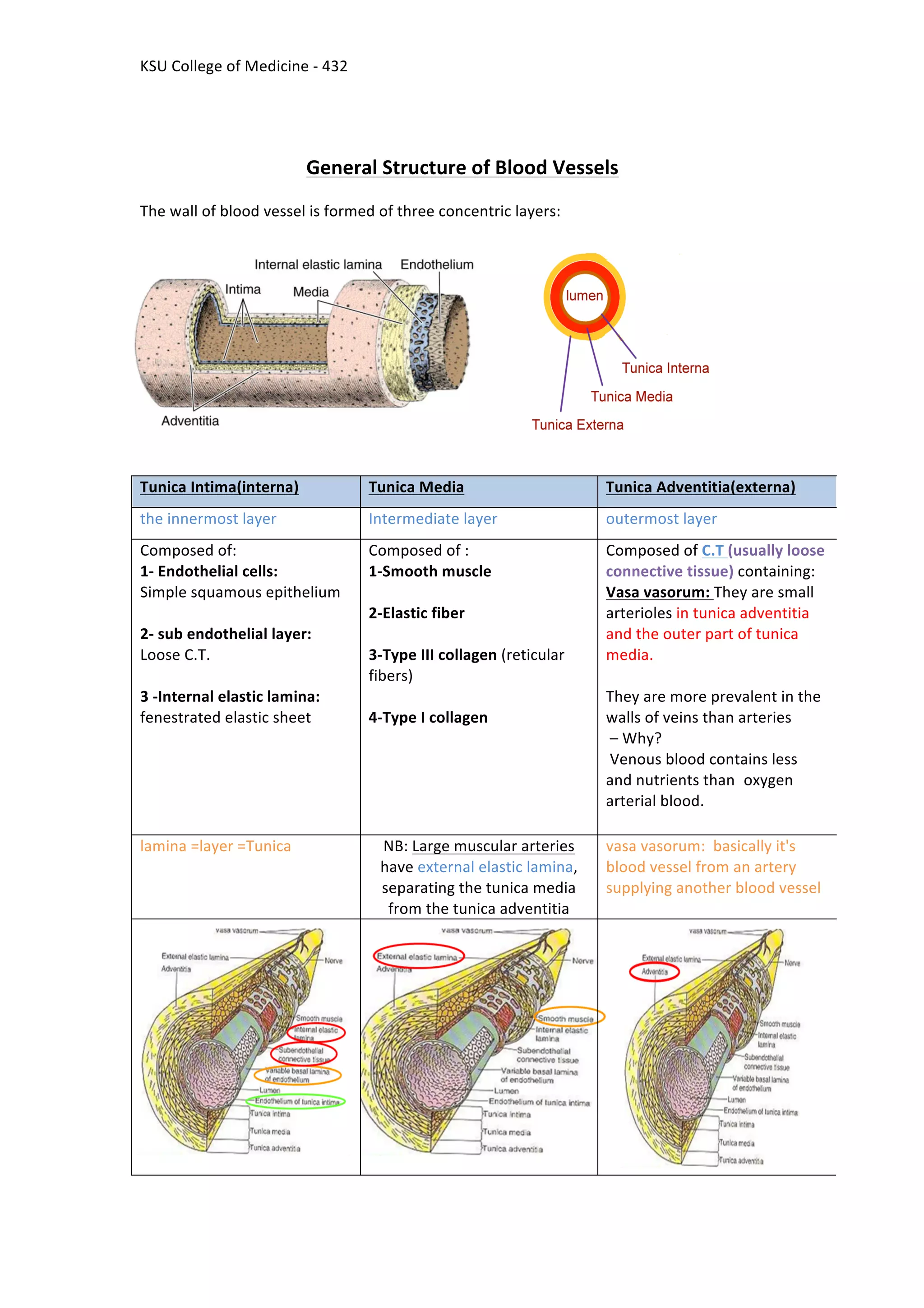
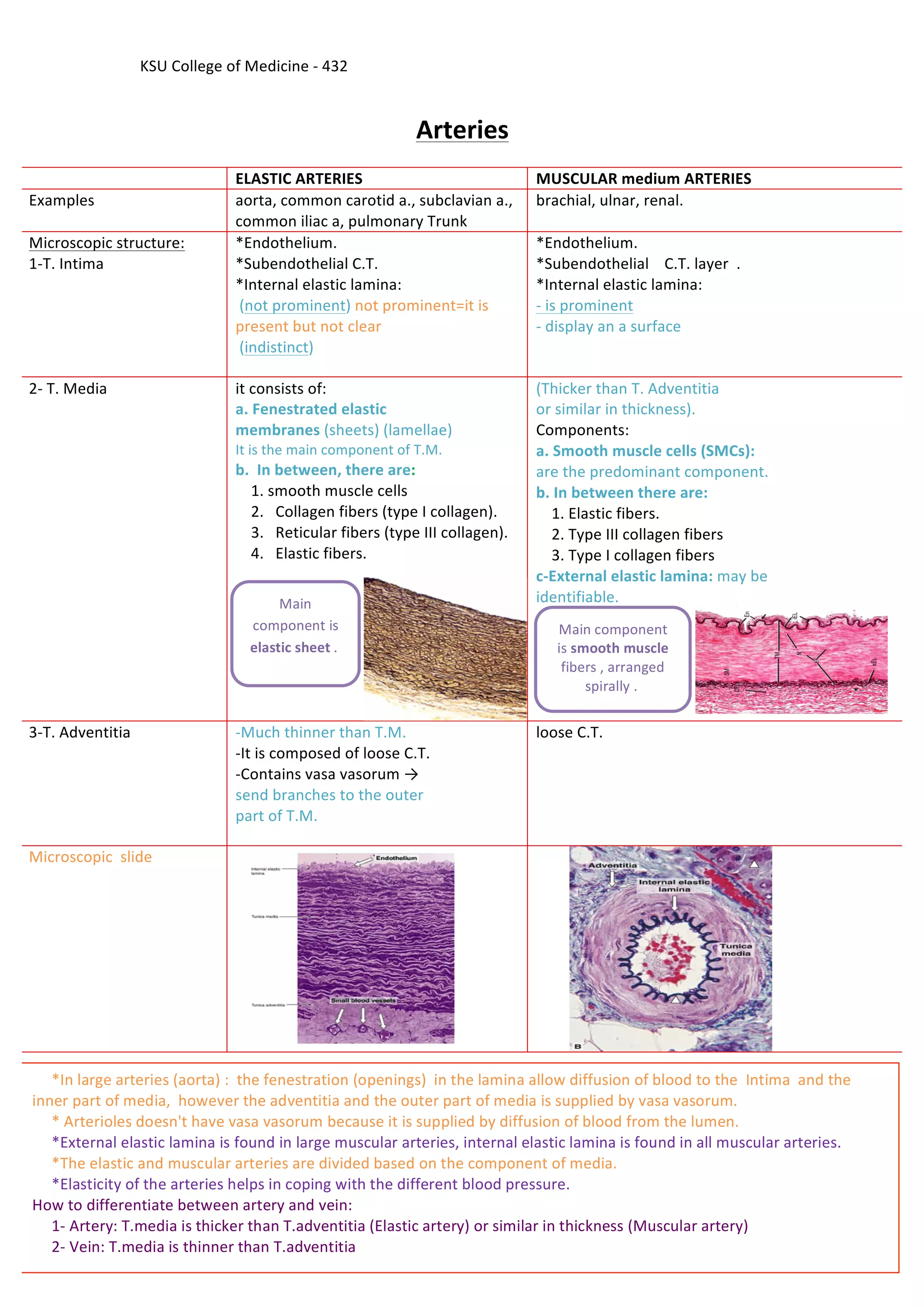
KSU	
  College	
  of	
  Medicine	
  -­‐	
  432
Cardiovascular Block
Histology Team
Lecture (2)
Histology of Blood Vessels
By: Reem	
  Al	
  Otaibi	
  
Lama	
  Al	
  Otaibi	
  
Rana	
  Al	
  Ohaly	
  
Mohammed	
  Adel	
  
By	
  the	
  end	
  of	
  this	
  lecture,	
  the	
  student	
  should	
  be	
  able	
  to	
  identify	
  and	
  describe	
  
the	
  microscopic	
  structure	
  of	
  the	
  wall	
  of	
  the	
  blood	
  vessels	
  including:	


  a.	
  Elastic	
  arteries.


  b.	
  Muscular	
  (medium-­‐sized)	
  arteries.


  c.	
  Medium-­‐sized	
  veins.


  d.	
  Blood	
  capillaries.
	
  
KSU	
  College	
  of	
  Medicine	
  -­‐	
  432
Blood	
  Vessels	
  
Viens	
  
-­‐Venules	


-­‐Small	
  viens	


-­‐Medium-­‐sized	
  veins	


-­‐Large	
  veins	
  
Blood	
  Capiliaries	
  Artries	
  
-­‐ElasMc	
  artry	


-­‐	
  Muscular	
  (distribuMng)
(medium-­‐sized)	
  artry	


-­‐Arterioles	
  
Black	
  &blue:	
  slides	
  
Orange:	
  explanation	
  
Purple	
  :	
  additional	
  informations	
  
Red:	
  important	
  
KSU	
  College	
  of	
  Medicine	
  -­‐	
  432
General	
  Structure	
  of	
  Blood	
  Vessels
The	
  wall	
  of	
  blood	
  vessel	
  is	
  formed	
  of	
  three	
  concentric	
  layers:
(externa)Tunica	
  AdventitiaTunica	
  Media(interna)Tunica	
  Intima
outermost	
  layerIntermediate	
  layerthe	
  innermost	
  layer
(usually	
  loose	
  	
  C.T	
  Composed	
  of
connective	
  tissue)	
  containing:
They	
  are	
  small	
  Vasa	
  vasorum:	
  
arterioles	
  in	
  tunica	
  adventitia	
  
and	
  the	
  outer	
  part	
  of	
  tunica	
  
media.
They	
  are	
  more	
  prevalent	
  in	
  the	
  
walls	
  of	
  veins	
  than	
  arteries
	
  –	
  Why?
	
  Venous	
  blood	
  contains	
  less	
  
oxygenand	
  nutrients	
  than	
  
arterial	
  blood.
Composed	
  of	
  :
1-­‐Smooth	
  muscle
2-­‐Elastic	
  fiber
3-­‐Type	
  III	
  collagen	
  (reticular	
  
fibers)
4-­‐Type	
  I	
  collagen
Composed	
  of:
1-­‐	
  Endothelial	
  cells:
Simple	
  squamous	
  epithelium
2-­‐	
  sub	
  endothelial	
  layer:
Loose	
  C.T.
3	
  -­‐Internal	
  elastic	
  lamina:
fenestrated	
  elastic	
  sheet
vasa	
  vasorum:	
  	
  basically	
  it's	
  
blood	
  vessel	
  from	
  an	
  artery	
  
supplying	
  another	
  blood	
  vessel	


  Large	
  muscular	
  arteriesNB:	
  
have	
  external	
  elastic	
  lamina,	
  
separating	
  the	
  tunica	
  media	
  
from	
  the	
  tunica	
  adventitia
lamina	
  =layer	
  =Tunica
KSU	
  College	
  of	
  Medicine	
  -­‐	
  432
Arteries
MUSCULAR	
  medium	
  ARTERIESELASTIC	
  ARTERIES
brachial,	
  ulnar,	
  renal.aorta,	
  common	
  carotid	
  a.,	
  subclavian	
  a.,	
  
common	
  iliac	
  a,	
  pulmonary	
  Trunk
Examples
*Endothelium.
*Subendothelial	


  C.T.	
  layer	
  	
  .
*Internal	
  elastic	
  lamina:
prominents	
  i	
  -­‐
-­‐	
  display	
  an	
  a	
  surface
*Endothelium.
*Subendothelial	
  C.T.	
  
*Internal	
  elastic	
  lamina:
not	
  prominent=it	
  is	
  	
  )not	
  prominent(	
  
present	
  but	
  not	
  clear	


)indistinct(	
  
Microscopic	
  structure:
1-­‐T.	
  Intima
(Thicker	
  than	
  T.	
  Adventitia
or	
  similar	
  in	
  thickness).
Components:
a.	
  Smooth	
  muscle	
  cells	
  (SMCs):
are	
  the	
  predominant	
  component.
b.	
  In	
  between	
  there	
  are:


  1.	
  Elastic	
  fibers.


  2.	
  Type	
  III	
  collagen	
  fibers


  3.	
  Type	
  I	
  collagen	
  fibers
c-­‐External	
  elastic	
  lamina:	
  may	
  be	
  
identifiable.
it	
  consists	
  of:
a.	
  Fenestrated	
  elastic
membranes	
  (sheets)	
  (lamellae)
It	
  is	
  the	
  main	
  component	
  of	
  T.M.
b.	
  	
  In	
  between,	
  there	
  are:


  1.	
  smooth	
  muscle	
  cells


  2.	
  	
  	
  Collagen	
  fibers	
  (type	
  I	
  collagen).


  3.	
  	
  	
  Reticular	
  fibers	
  (type	
  III	
  collagen).


  4.	
  	
  	
  Elastic	
  fibers.	


2-­‐	
  T.	
  Media
loose	
  C.T.-­‐Much	
  thinner	
  than	
  T.M.
-­‐It	
  is	
  composed	
  of	
  loose	
  C.T.
-­‐Contains	
  vasa	
  vasorum	
  →	
  
send	
  branches	
  to	
  the	
  outer	
  
part	
  of	
  T.M.
3-­‐T.	
  Adventitia
Microscopic	
  	
  slide


  *In	
  large	
  arteries	
  (aorta)	
  :	
  	
  the	
  fenestration	
  (openings)	
  	
  in	
  the	
  lamina	
  allow	
  diffusion	
  of	
  blood	
  to	
  the	
  	
  Intima	
  	
  and	
  the	
  
inner	
  part	
  of	
  media,	
  	
  however	
  the	
  adventitia	
  and	
  the	
  outer	
  part	
  of	
  media	
  is	
  supplied	
  by	
  vasa	
  vasorum.	


  *	
  Arterioles	
  doesn't	
  have	
  vasa	
  vasorum	
  because	
  it	
  is	
  supplied	
  by	
  diffusion	
  of	
  blood	
  from	
  the	
  lumen.	


  *External	
  elastic	
  lamina	
  is	
  found	
  in	
  large	
  muscular	
  arteries,	
  internal	
  elastic	
  lamina	
  is	
  found	
  in	
  all	
  muscular	
  arteries.	


  *The	
  elastic	
  and	
  muscular	
  arteries	
  are	
  divided	
  based	
  on	
  the	
  component	
  of	
  media.	


  *Elasticity	
  of	
  the	
  arteries	
  helps	
  in	
  coping	
  with	
  the	
  different	
  blood	
  pressure.	
  
How	
  to	
  differentiate	
  between	
  artery	
  and	
  vein:	


  1-­‐	
  Artery:	
  T.media	
  is	
  thicker	
  than	
  T.adventitia	
  (Elastic	
  artery)	
  or	
  similar	
  in	
  thickness	
  (Muscular	
  artery)	


  2-­‐	
  Vein:	
  T.media	
  is	
  thinner	
  than	
  T.adventitia	


Main	
  component	
  
is	
  smooth	
  muscle	
  
fibers	
  ,	
  arranged	
  
spirally	
  .	
  	
  
Main	
  
component	
  is	
  
elastic	
  sheet	
  .	
  	
  
KSU	
  College	
  of	
  Medicine	
  -­‐	
  432
	
  SIZED	
  VEIN-­‐EDIUMM
-­‐Thickness	
  of	
  the	
  wall:	
  thinner	
  than	
  the	
  accompanying	
  artery	
  
*usually	
  forms	
  valves	
  :T.	
  Intima	


  *no	
  internal	
  elastic	
  lamina	
  
T.	
  Media	
  
Thinner	
  than	
  T.	
  Adventitia	


  Consists	
  of:	


  *Fewer	
  SMCs	


  *Types	
  I	
  &	
  III	
  Collagen	
  fibers	
  
T.	
  Adventitia	
  
thicker	
  than	
  T.	
  Media	


VALVES	
  OF	
  VEINS	
  
-­‐Valve	
  of	
  a	
  vein	
  is	
  composed	
  of	
  2	
  leaflets	
  
-­‐Each	
  leaflet	
  has	
  a	
  thin	
  fold	
  of	
  the	
  T.	
  Intima.
-­‐Components:


  *Endothelium	


  *core	
  of	
  C.T.
	
  
*	
  	
  veins	
  are	
  supplied	
  by	
  vasa	
  vasorum	
  .	
  
*varicose	
  veins	
  happens	
  because	
  the	
  veins	
  doesn't	
  contain	
  a	
  lot	
  of	
  elastic	
  fibers	
  So	
  it	
  will	
  result	
  in	
  dilatation	
  of	
  the	
  
veins.	
  	
  
*veins	
  contain	
  valves	
  because	
  the	
  blood	
  pass	
  against	
  the	
  gravity.	
  	
  
*vein	
  lumen	
  is	
  large	
  and	
  the	
  wall	
  is	
  thin.	


KSU	
  College	
  of	
  Medicine	
  -­‐	
  432
BLOOD	
  CAPILLARIES	
  
TYPES	
  
CONTINUS	
  
FENESTRATED	
  
WITH	
  DIAPHRAM	
  
WITHOUT	
  
DIAPHRAM	
  
SINUSODIAL	
  
YSIZED	
  ARTER-­‐MEDIUM
AND	
  VEIN
BLOOD	
  CAPILLARIES
	
  
-­‐	
  Diameter:	
  usually	
  8-­‐10	
  µm.	
  
	
  the	
  capillaries	
  wall	
  is	
  thin	
  to	
  allow	
  gas	
  and	
  nutrient	
  exchange	
  	
  
Microscopic	
  structure-­‐


  1-­‐	
  Single	
  layer	
  of	
  squamous	
  endothelial	
  cells


  2-­‐Basal	
  lamina:	
  surrounds	
  the	
  external	
  surface	
  
	
  of	
  the	
  endothelial	
  cells.


  3-­‐Pericytes(peri=	
  around):	


  *Have	
  processes.


  *Share	
  the	
  basal	
  lamina	
  of	
  the	
  endothelial	
  cells.	
  
the	
  Pericytes	
  are	
  	
  not	
  part	
  of	
  the	
  capillary	
  wall	
  	
  
*Function	
  of	
  Pericytes	
  :	
  
1-­‐	
  act	
  as	
  massager	
  to	
  the	
  blood	
  capillary	
  	
  to	
  facilitate	
  the	
  diffusion	
  of	
  materials	


2-­‐	
  	
  converting	
  	
  to	
  smooth	
  muscle	
  	
  in	
  case	
  of	
  injury	
  to	
  nearby	
  arteriole	
  or	
  venules	


KSU	
  College	
  of	
  Medicine	
  -­‐	
  432
Fenestrated	
  Blood	
  Capillaries	
  
SinusoidalFenestrated	
  without	
  
diaphragm
Fenestrated	
  with	
  diaphragmContinuousTypes
*Their	
  
endothelia
	
  cells	
  have
fenestrae	
  without	
  
diaphragms.
*They	
  possess	
  
discontinuous	
  
endothelial	
  
cells.
*They	
  possess	
  
discontinuous	
  
basal	
  lamina.
*Macrophages	
  
may	
  be	
  located	
  
in	
  or	
  along	
  the	
  
outside	
  of	
  the	
  
endothelial	
  
wall.
*The	
  walls	
  of	
  their	
  
endothelial	
  cells	
  	
  
have	
  pores	
  
(fenestrae).
*These	
  	
  
pores	
  are	
  
	
  NOT
covered	
  
	
  by	
  diaphragm.
*The	
  walls	
  of	
  their	
  
endothelial	
  cells	
  	
  
have	
  pores	
  
(fenestrae).	
  
*These	
  pores	
  are	
  
covered	
  by	
  
diaphragm.


*No	
  pores	
  
*No	
  
fenestrae	
  
in	
  their	
  
walls.
Microscopic
	
  structure
Slide
Red	
  bone	
  marrow,	
  
liver,	
  spleen	
  and	
  certain	
  
endocrine	
  glands.
In	
  renal	
  glomerulus.In	
  intestine,	
  
pancreas
	
  and	
  endocrine	
  	
  glands
In	
  
muscles,
	
  Nervous	
  T.,	
  C.T	
  
Distribution
Diameter:	
  irregular	
  
(30-­‐40	
  µm)
It	
  is	
  in	
  muscles	
  and	
  
bain,	
  because	
  it's	
  
continuous	
  to	
  prevent	
  
toxic	
  materials	
  from	
  
going	
  inside	
  the	
  brain.
Extra	
  notes
Please send us any questions or mistakes on : 432histologyteam@gmail.com

(2) histology of blood vessels

  • 1.
    KSU  College  of  Medicine  -­‐  432 Cardiovascular Block Histology Team Lecture (2) Histology of Blood Vessels By: Reem  Al  Otaibi   Lama  Al  Otaibi   Rana  Al  Ohaly   Mohammed  Adel   By  the  end  of  this  lecture,  the  student  should  be  able  to  identify  and  describe   the  microscopic  structure  of  the  wall  of  the  blood  vessels  including:                        a.  Elastic  arteries.                b.  Muscular  (medium-­‐sized)  arteries.                c.  Medium-­‐sized  veins.                d.  Blood  capillaries.  
  • 2.
    KSU  College  of  Medicine  -­‐  432 Blood  Vessels   Viens   -­‐Venules     -­‐Small  viens     -­‐Medium-­‐sized  veins     -­‐Large  veins   Blood  Capiliaries  Artries   -­‐ElasMc  artry     -­‐  Muscular  (distribuMng) (medium-­‐sized)  artry     -­‐Arterioles   Black  &blue:  slides   Orange:  explanation   Purple  :  additional  informations   Red:  important  
  • 3.
    KSU  College  of  Medicine  -­‐  432 General  Structure  of  Blood  Vessels The  wall  of  blood  vessel  is  formed  of  three  concentric  layers: (externa)Tunica  AdventitiaTunica  Media(interna)Tunica  Intima outermost  layerIntermediate  layerthe  innermost  layer (usually  loose    C.T  Composed  of connective  tissue)  containing: They  are  small  Vasa  vasorum:   arterioles  in  tunica  adventitia   and  the  outer  part  of  tunica   media. They  are  more  prevalent  in  the   walls  of  veins  than  arteries  –  Why?  Venous  blood  contains  less   oxygenand  nutrients  than   arterial  blood. Composed  of  : 1-­‐Smooth  muscle 2-­‐Elastic  fiber 3-­‐Type  III  collagen  (reticular   fibers) 4-­‐Type  I  collagen Composed  of: 1-­‐  Endothelial  cells: Simple  squamous  epithelium 2-­‐  sub  endothelial  layer: Loose  C.T. 3  -­‐Internal  elastic  lamina: fenestrated  elastic  sheet vasa  vasorum:    basically  it's   blood  vessel  from  an  artery   supplying  another  blood  vessel        Large  muscular  arteriesNB:   have  external  elastic  lamina,   separating  the  tunica  media   from  the  tunica  adventitia lamina  =layer  =Tunica
  • 4.
    KSU  College  of  Medicine  -­‐  432 Arteries MUSCULAR  medium  ARTERIESELASTIC  ARTERIES brachial,  ulnar,  renal.aorta,  common  carotid  a.,  subclavian  a.,   common  iliac  a,  pulmonary  Trunk Examples *Endothelium. *Subendothelial        C.T.  layer    . *Internal  elastic  lamina: prominents  i  -­‐ -­‐  display  an  a  surface *Endothelium. *Subendothelial  C.T.   *Internal  elastic  lamina: not  prominent=it  is    )not  prominent(   present  but  not  clear       )indistinct(   Microscopic  structure: 1-­‐T.  Intima (Thicker  than  T.  Adventitia or  similar  in  thickness). Components: a.  Smooth  muscle  cells  (SMCs): are  the  predominant  component. b.  In  between  there  are:        1.  Elastic  fibers.        2.  Type  III  collagen  fibers        3.  Type  I  collagen  fibers c-­‐External  elastic  lamina:  may  be   identifiable. it  consists  of: a.  Fenestrated  elastic membranes  (sheets)  (lamellae) It  is  the  main  component  of  T.M. b.    In  between,  there  are:        1.  smooth  muscle  cells        2.      Collagen  fibers  (type  I  collagen).        3.      Reticular  fibers  (type  III  collagen).        4.      Elastic  fibers.         2-­‐  T.  Media loose  C.T.-­‐Much  thinner  than  T.M. -­‐It  is  composed  of  loose  C.T. -­‐Contains  vasa  vasorum  →   send  branches  to  the  outer   part  of  T.M. 3-­‐T.  Adventitia Microscopic    slide          *In  large  arteries  (aorta)  :    the  fenestration  (openings)    in  the  lamina  allow  diffusion  of  blood  to  the    Intima    and  the   inner  part  of  media,    however  the  adventitia  and  the  outer  part  of  media  is  supplied  by  vasa  vasorum.            *  Arterioles  doesn't  have  vasa  vasorum  because  it  is  supplied  by  diffusion  of  blood  from  the  lumen.            *External  elastic  lamina  is  found  in  large  muscular  arteries,  internal  elastic  lamina  is  found  in  all  muscular  arteries.              *The  elastic  and  muscular  arteries  are  divided  based  on  the  component  of  media.              *Elasticity  of  the  arteries  helps  in  coping  with  the  different  blood  pressure.   How  to  differentiate  between  artery  and  vein:              1-­‐  Artery:  T.media  is  thicker  than  T.adventitia  (Elastic  artery)  or  similar  in  thickness  (Muscular  artery)              2-­‐  Vein:  T.media  is  thinner  than  T.adventitia     Main  component   is  smooth  muscle   fibers  ,  arranged   spirally  .     Main   component  is   elastic  sheet  .    
  • 5.
    KSU  College  of  Medicine  -­‐  432  SIZED  VEIN-­‐EDIUMM -­‐Thickness  of  the  wall:  thinner  than  the  accompanying  artery   *usually  forms  valves  :T.  Intima                                                                                                                            *no  internal  elastic  lamina   T.  Media   Thinner  than  T.  Adventitia            Consists  of:                                *Fewer  SMCs                              *Types  I  &  III  Collagen  fibers   T.  Adventitia   thicker  than  T.  Media     VALVES  OF  VEINS   -­‐Valve  of  a  vein  is  composed  of  2  leaflets   -­‐Each  leaflet  has  a  thin  fold  of  the  T.  Intima. -­‐Components:        *Endothelium            *core  of  C.T.   *    veins  are  supplied  by  vasa  vasorum  .   *varicose  veins  happens  because  the  veins  doesn't  contain  a  lot  of  elastic  fibers  So  it  will  result  in  dilatation  of  the   veins.     *veins  contain  valves  because  the  blood  pass  against  the  gravity.     *vein  lumen  is  large  and  the  wall  is  thin.      
  • 6.
    KSU  College  of  Medicine  -­‐  432 BLOOD  CAPILLARIES   TYPES   CONTINUS   FENESTRATED   WITH  DIAPHRAM   WITHOUT   DIAPHRAM   SINUSODIAL   YSIZED  ARTER-­‐MEDIUM AND  VEIN BLOOD  CAPILLARIES   -­‐  Diameter:  usually  8-­‐10  µm.    the  capillaries  wall  is  thin  to  allow  gas  and  nutrient  exchange     Microscopic  structure-­‐          1-­‐  Single  layer  of  squamous  endothelial  cells          2-­‐Basal  lamina:  surrounds  the  external  surface    of  the  endothelial  cells.        3-­‐Pericytes(peri=  around):                                  *Have  processes.                                *Share  the  basal  lamina  of  the  endothelial  cells.   the  Pericytes  are    not  part  of  the  capillary  wall     *Function  of  Pericytes  :   1-­‐  act  as  massager  to  the  blood  capillary    to  facilitate  the  diffusion  of  materials       2-­‐    converting    to  smooth  muscle    in  case  of  injury  to  nearby  arteriole  or  venules    
  • 7.
    KSU  College  of  Medicine  -­‐  432 Fenestrated  Blood  Capillaries   SinusoidalFenestrated  without   diaphragm Fenestrated  with  diaphragmContinuousTypes *Their   endothelia  cells  have fenestrae  without   diaphragms. *They  possess   discontinuous   endothelial   cells. *They  possess   discontinuous   basal  lamina. *Macrophages   may  be  located   in  or  along  the   outside  of  the   endothelial   wall. *The  walls  of  their   endothelial  cells     have  pores   (fenestrae). *These     pores  are    NOT covered    by  diaphragm. *The  walls  of  their   endothelial  cells     have  pores   (fenestrae).   *These  pores  are   covered  by   diaphragm.             *No  pores   *No   fenestrae   in  their   walls. Microscopic  structure Slide Red  bone  marrow,   liver,  spleen  and  certain   endocrine  glands. In  renal  glomerulus.In  intestine,   pancreas  and  endocrine    glands In   muscles,  Nervous  T.,  C.T   Distribution Diameter:  irregular   (30-­‐40  µm) It  is  in  muscles  and   bain,  because  it's   continuous  to  prevent   toxic  materials  from   going  inside  the  brain. Extra  notes Please send us any questions or mistakes on : 432histologyteam@gmail.com