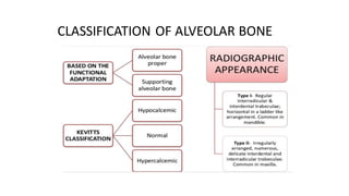
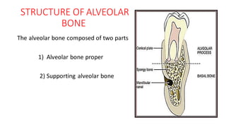
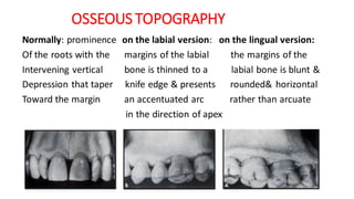
SlideShare a Scribd company logo
ALVEOLAR BONE
Dr. Sangeetha
GUIDED BY
Dr NEETA BHAVSAR
HEAD OF DEPARTMENT
DEPARTMENTOF PERIODONTIA
CONTENTS
• INTRODUCTION
• DEVELOPMENT OF ALVEOLAR BONE
• FUNCTIONS
• COMPOSITION OF BONE
• CLASSIFICATION OF BONE
• GROSS MORPHOLOGY
• HISTOLOGY
• BONE FORMATION
• BONE RESORPTION
• BONE REMODELLING
• BONE LOSS IN VARIOUS CONDITIONS
• CONCLUSION
• REFERENCES
INTRODUCTION
DEFINITION - Alveolar bone is the portion of maxilla and mandible that forms
and support the tooth socket.
• Together with root cementum and periodontal ligament the alveolar bone
constitutes the attachment apparatus of the tooth
• Since alveolar processes develop and undergo remodeling with tooth
formation and eruption, they are tooth dependent bony structures.
• Therefore the size , shape , location, function of teeth determine their
morphology
• Alveolar bone also called the FUNCTIONAL BONE because it is susceptible to
functional changes(it is lost after tooth extraction)
DEVELOPMENT OF ALVEOLAR BONE
• Alveolar process consists of bone which is formed both by cells from
the dental follicle (alveolar bone proper) & cells which are
independent of tooth development
• Maxilla & mandible develop from the 1st branchial arch or
mandibular arch.
• The maxilla forms within the maxillary process & mandible forms
within the fused mandibular processes of mandibular arch
• Both jaw bones start as small centres of intramembraneous
ossification around stomodeum
Both maxilla and
mandible develop
intramembraneously
8th week in utero
Alveolar process
develops from the
dental follicle during
eruption of tooth
Bell stage--
developing bone
becomes closely
related
The size of the
alveolus is
dependent upon the
size of the growing
tooth germ.
Resorption - inner
wall of the alveolus
Deposition -outer
wall
The developing
teeth lie in a trough
of bone -Tooth
Crypt
Teeth separated
from each other by
the development of
interdental septa.
With the onset of
root formation
intratrabecular bone
develops in
multirooted teeth
When deciduous
teeth shed, its
alveolar bon e
resorb
Alveolar process
gradually
incorporated into
maxillary or
mandibular body
Permanent tooth
move into place,
developing its own
alveolar bone from
its own follicle
• Hydroxyapatitecrystals formed
from minerals calcium and
phosphate along with hydroxy
carbonate, citrate
• Mg, Na, K, F – smaller quantity
2/3 inorganic
matter
• 90% collagen primarily type1
• 11-12%Non collagenous protein
• Osteocalcin
• Osteonectin
• BMP and proteoglycans
• Sialoproteins and phosphoproteins
1/3 organic
matter
COMPOSITION OF BONE
FUNCTIONS
To house
the roots of
teeth, which
is achieved
by the
insertion of
Sharpey’s
fibers into
the alveolar
bone proper
Provides
attachment
to the
forming
periodontal
ligament
Absorb and
distribute
occlusal
pressures
generated
during tooth
contact
Also gives
attachment
to muscles
Provides a
framework
for the
marrow
Reservoir of
ions,
especially
Ca
CLASSIFICATION OF ALVEOLAR BONE
STRUCTURE OF ALVEOLAR
BONE
The alveolar bone composed of two parts
1) Alveolar bone proper
2) Supporting alveolar bone
ALVEOLAR BONE PROPER
• The alveolar bone proper is a thin layer of compact bone
• Continuation of cortical plate forms the tooth socket
• It surrounds the roots of the teeth and gives attachment to the
principal fibers of periodontal ligament.
• It is perforated by many openings that carry the nerve and blood
vessels in to the periodontal ligament called cribriform plate
• Consists of lamellated bone and bundle bone
• Cribriform plate ( anatomical term),
• lamina dura (radiographic term) ,
• bundle bone( histologic term)
• Thickness – 0.1 to 0.5 mm
Bundle bone
The term bundle bone was chosen because the bundles of the principal fiber
continueinto bone as sharpey’s fibers
it appears as a dense white line in radiographs – lamina dura
Supporting alveolar bone
• It is the part of alveolar bone
which surrounds the alveolar
bone proper and gives support
to the socket
• It consists of two parts
1) cortical plates
2) spongy bone
CORTICAL PLATES
• Cortical plates 1.5 – 3mm thick in posterior region and thickness
various in anterior region
• It is continuous with the bony maxilla and mandible and is much
thicker in mandible than maxilla .
• They are thickest in mandibular premolar and molar area especially
buccal side
• It consists of compact bone and form outer and inner plates.
CANCELLOUS BONE
• Spongy bone (anatomical term), trabecular bone ( radiographic
term), cancellous bone ( histological term)
• It is fills the area between cortical plates and alveolar bone proper
• In the region of the anterior of the both jaws the supporting bone
usually thin, so no spongy bone is found.
• The presence of trabeculae enclosing irregular marrow spaces lined
with the layer of thin, flattened endosteal cells
• Matrix consists of irregularly arranged lamellae separated by
incremental and resorption lines
INTERDENTAL SEPTUM
• Bony partition that separate the adjacent alveoli
• Coronally septa is thin and fused consists of only inner cortical plate
• Apical septa is thicker and contain intervening cancellous bone
• Mesiodistal angulation of interdental septum is parallel to line drawn
CEJ of approximating the teeth ( Ritchey et al,1935)
• If interdental septum is narrow , septum may consists of only
cribriformplate
• If roots are together, an irregular window can appear in between
adjacent roots.
• The shape of interdental bone is a function of the tooth form and
embrasure width
• The more tapered the tooth , the more pyramidal is bony form
• The wider the embrasure , the more flattened is the interdental bone
mesiodistally and buccolingually
INTERRADICULARSEPTA
• The bone between roots of multirooted teeth
• It contain perforating canals of Zukerkandl and Hirschfeld nutrient
canal
BASAL BONE
• It is osseous structure of maxilla and mandible except the alveolar
process
• Anatomically there is no distinct boundary exists between the body of
the maxilla / mandible and their alveolar process
OSSEOUS TOPOGRAPHY
Normally: prominence on the labial version: on the lingual version:
Of the roots with the margins of the labial the margins of the
Intervening vertical bone is thinned to a labial bone is blunt &
Depression that taper knife edge & presents rounded& horizontal
Toward the margin an accentuated arc rather than arcuate
in the direction of apex
RED
HEMATOP
OIETIC
MARROW
• Embryo & newborn
• Ribs , sternum, vertebrae, skull,
humerus
• hemopoiesis
YELLOW
FATTY
MARROW
• Adult
• Red marrow foci found
sometimes in maxillary
tuberosity and angle of ramus
• Storage of energy
Cells
Osteoblast
Osteocyte
Bone lining cells
Osteoprogenitor
cells
Osteoclasts
Osteogenic cells
Osteoclast cells
HISTOLOGY
MATRIX COMPONENT
INORGANIC
CALCIUMHYDROXY
APATITE CRYSTALS
ORGANIC
COLLAGENMATRIX
NONCOLLAGEN
MATRIX
OSTEOCALCIN
OSTEONECTIN
OSTEOPONTINAND
BONESIALOPROTEIN
PROTEOGLYCAN
LYSYLOXIDASE AND
TYROSINE RICH ACIDIC
MARTRIX PROTEIN
OSTEOPROGENITOR CELLS
• The stem cell population that gives
osteoblast are called osteoprogenitor
cell
• They are fibroblast like cells with an
elongated nuclei and few organelles.
• Their life cycle may involve up to about
eight cell divisions before reaching
osteoblast stage
• They reside in the layer of cells beneath
osteoblast layer, in the periosteal region,
in the periodontal ligament or in the
marrow spaces
OSTEOBLAST
• During embryonic development,
intramembranous bone of
maxilla and mandible initially
forms from osteoblast arising
from condensing mesenchyme
in facial region
• The most active secretory cells
in the bone
FUNCTIONS
• Secrete the type 1 collagen and non collagens proteins like sialoprotein,
osteopontin, osteonenctin and growth factors –BMP, TGF, PLDGF and
insulin like growth factor
• Express and release alkaline phosphate, which has been closely
associated with new bone formation and bone remodeling
• Total alkaline phosphate activity has been recognized as a reliable
indicator of osteoblast function
• In addition it has controlling influence activating osteoclast it contains
receptor for the parathyroid hormone and regulates the osteoclast
response to this hormone
• Periosteum also serves the as important reservoir of osteoblast.
OSTEOCYTE (NERVE CELLS OF BONE)
• Osteocytes are entrapped osteoblast within the bone
• Most abundant cells and communicate with each other and other on
the surface of bone via dendritic process encapsulated in canaliculi
• Play role in calcium homeostasis
• Exchange of metabolic and biochemical messages occurs between
blood stream and canaliculi
• Decreased quantity of synthetic and
secretory organelles
BONELININGCELLS
• When bone surface neither in
formative not in resorptive phase,
the surface is completely lined by
a layer of flattened cells called
bone lining cells
• Regarded as post proliferative
osteoblast
• Retain gap junction with osteocyte
OSTEOCLAST
• Originate from hematopoietic
tissue
• Fusion of mononucleate cells to
form a multinucleated cell
• Very large and 5-10 nuclei
• Mobile and capable of
migrating, lie in howships
lacunae
• Acidophilic cytoplasm
• Active osteoclast has ruffled
border facing bone , hydrolytic
enzymes are secreted
• At the periphery of the ruffled
border the plasma membrane
smooth and closely apposed to
the bone surface
• The adjacent cytoplasm, devoid
of cell organelles, enriched in
actin, vinculin, and talin, protein
associated with integrin
mediated cell adhesion clear
(sealing) zone
• This zone create the
microenvironment in which
resorption can take place
Bone Reversal line or cementing line- -The site of change from bone
resorption to bone deposition is represented by a scalloped outline. -Rich
in sialoprotein & osteopontin.
Resting line – Rhythmic deposition of
bone with periods of relative quiescence
seen as parallel vertical lines
MATRIXCOMPONENTS
• COLLAGEN: Collagen comprises the major (80–90%) organic
component in mineralized bone tissues.
• Type I collagen (>95%) is the principal collagen in mineralized bone,
together with Type V (<5%) collagen.
• In addition, both type III and XII collagens are also present.
• Sharpey’s fibres contain type III collagen with type I collagen.
• The expression of type XII collagen in alveolar bone is related to
mechanical strain.
• Type I,V & XII are expressed by osteoblasts
• Type III & some type XII collagen appear to be produced by fibroblasts
during formation of periodontal ligament
NON-COLLAGENOUSPROTEIN
• Comprise the remaining 10% of the total organic content of
the bone matrix.
• OSTEOCALCIN ( Bone gla protein)
• First non collagenous protein to be recognized.
• Found in bone matrix and specifically localizes to developing
bone
• It is regulated by Vitamin D3 and Parathyroid hormone.
• The carboxy terminal segment of osteocalcin acts as a
chemoattractant to osteoclast precursors, suggesting a role in
bone resorption.
OSTEOPONTIN AND BONESIALOPROTEIN
• They were previously termed as Bone sialoproteins I and II.
• Bone sialoprotein is thought to function in the initiation of mineral
crystal formation in vivo.
• Osteopontin is a potent inhibitor of hydroxyapatite crystal growth.
• Osteopontin transcription is strongly upregulated by Vitamin D3
whereas Bone sialoprotein transcription is suppressed by Vitamin D3.
• It could play a role in the regulation of cell adhesion and proliferation.
SPARC(secretedproteins& acidic richin cysteins) /
Osteonectin/ BM-40
• It is predominantly bound to hydroxyapatite crystals.
• SPARC, which has also been characterized in basement membranes as
BM40, is a secreted calcium-binding glycoprotein that interacts with a
range of extracellular matrix molecules.
CHONDROITIN SULFATEPROTEOGLYCAN
• Two small proteoglycans, Biglycan (chondroitin sulfate proteoglycan I)
and Decorin (chondroitin sulfate proteoglycan II).
•These regulate collagen synthesis.
• Byglycan is more prominent in developing bone and has mineralised
to pericellular areas
• Its precise function is not known ,but it can bind TGF-β and
extracellular matrix macromolecules including collagen and thereby
regulate fibrillogenesis
Decorin: Binds mainly with in the gap region of collagen fibrils and
assuggested by its name, decorates fibril surface
• The primary calcification in bones as reported to follow removal of
decorin and the fusion of collagen fibrils
VASCULAR SUPPLY AND NERVE SUPPLY
• Derived from inferior and superior alveolar arteries of maxilla and
mandible, venous drainage accompanies the arterial supply,
• Branches from anterior, middle and posterior superior alveolar nerve
for maxilla and branches from inferior alveolar for mandible
BONE FORMATION
• Formation of bone, which appears to be linked with bone resorption
to maintain bone mass, involves the proliferation and differentiation of
stromal stem cells along an osteogenic pathway that leads to the
formation of osteoblasts
• Osteoblasts synthesize the collagenous precursors of bone matrix and
also regulate its mineralization
REGULATORSOF BONEFORMATION
• The overall integrity of bone is controlled
by hormones, proteins secreted by
hematopoietic bone marrow cells and bone
cells.
• HORMONES
• Parathormone
• Vitamin D3
• Glucocorticoids
• Thyroid Hormone
• Growth Hormone
• Insulin
• LOCAL REGULATORS
• Platelet derived growth factor
• Insulin growth factors
• Transforming growth factor-β
• Bone morphogenetic protein
• Fibroblast growth factor
BONERESORPTION
• It is the process of removal of mineral and
organic componentsof extracellular matrix
of bone by osteolytic cells called
osteoclasts.
• SEQUENCE OF EVENTS OF BONE
RESORPTION
• First phase - Formation of osteoclast
progenitors in the hematopoietic tissues.
• Second phase - Activation of osteoclastsat
the surface of mineralized bone.
• Third phase - Activated osteoclasts
resorbing the bone.
TENCATE SEQUENCE OF RESORPTION
• Attachment of osteoclast to the mineralized surface of bone.
• Creation of sealed acidic environment through action of the proton
pump, which demineralize the bone and expose the organic matrix
• Degradation of the exposed organic matrix to its constituent amino
acids by the action of released enzymes ,such as acid phosphatase
and cathepsin.
• Sequestering of mineral ions and amino acids with in the osteoclast
BONE REMODELLING
• The process by which overall shape and size of bones is established is
referred to as bone remodelling or turnover.
• It occurs in discrete, focal areas involving groups of cells called bone
remodelling or basic multicellular units.
• During this phase bone is formed along the periosteal surface and
destroyed along the endosteal surface
• The status of bone represents the net result of a balance between the
two processes. ‘Coupling’ of bone resorption and formation
BONEMULTICELLULAR UNIT
• Osteoclasts
• Osteoblasts
• Blood vessels& Pericytes
• The main functions of
remodeling are
• To prevent the accumulation of
damaged and fatigued bone by
regenerating new bone.
• To allow bone to respond to
changes in mechanical forces.
• To facilitate mineral
homeostasis.
Decrease in bloodca
1. Detected by
receptors on chief cells
of parathyroid gland
Release of PTH
Stimulateosteoblast to
release IL-1, IL-6
Stimulatemonocyte to
migrate to bone area
Monocyte coalesces to
form multinucleated
osteoclast in the
presence of LIF
Bone resorption
Release of ca ionsfrom
hydroxyapatitecrystals
Normal blood ca level
PTH secretion stopped
by feedback
mechanism
• The leading edge of resorption –
Cutting cone
• Released cytokines [ BMP & IGF]
stimulate stem cells to
differentiate into osteoblasts.
• osteoblasts secrete osteoid -
Filling cone
FACTORS INFLUENCING
REMODELLING
LOCAL INFLUENCE FACTORES:
1) Functional requirement
2) Age related changes
SYSTEMIC INFLUENCE FACTORS
Hormones (PTH, vit D, calcitonin)
REGULATION OF BONE
REMODELLING
Mechanical control: mechanical
stimulation of bone tissue
accelerate periosteal bone
formation in the regions of high
stress and effectively strengthens
bone
Local
systemic
AGE CHANGES
• Similar to those occurring in remainder of skeletal system
• Osteoporosis with ageing
• Decreased vascularity
• Reduction in metabolic rate and healing capacity(implants, extraction
sockets, bone grafts)
• Bone resorption may be increased or decreased
• More irregular periodontal surface
FACTORS DETERMINING BONE MORPHOLOGY
• Normal variation in alveolar bone
• Exostoses
• Tauma from occlusion
• Food impaction
• Aggressive periodontitis
NORMALVARIATIONSIN ALVEOLAR BONE
• Thickness, width and crestal
angulation of interdental septa
• Thickness of facial and lingual
alveolar plates
• Fenestrations and dehiscences
• Alignment of teeth
• Root and root trunk anatomy
• Root position within alveolar
process
FENESTRATION& DEHISCENCE
• Facial > lingual
• Arteriors > posteriors
• Frequently bilateral
• 20% of all teeth affected
• Caused due to malposition, root
prominence, labial protrusion and a
thin cortical plate
• Can complicate procedure and
outcome of periodontal surgery
EXOSTOSIS
1) These are outgrowths of bone of varied size and shape.
2) They can occur as small nodules, large nodules, sharp ridges, spike-
like projections or any combination of these.
3) In rare cases, found to develop after the placement of free gingival
grafts.
TRAUMA FROM OCCLUSION
• Trauma from occlusion may be factor in determining the dimension
and shape of bone deformities.
• It may cause thickening of cervical margin of alveolar bone or a
change in bone morphology( funnel like crestal bone and buttressing
bone
BUTTRESSINGBONE FORMATION( LIPPING)
• Bone formation sometimes occurs
in an attemptto buttress bony
trabeculae weakened by resorption.
• When it occurs within the jaw,
termed Central buttressing bone
formation.
• When it occurs on the external
surface, termed Peripheral buttressing
bone formation
• The latter may cause bulging of the
bone contour, termed as Lipping,
which sometimes accompanies the
production of osseous craters and
angular defects.
FOOD IMPACTION
• Forceful wedging of food into the periodontium by occlusal forces
• Interdental bone defects often occur where proximal contact is
abnormal or absent.
• Pressure and irritation from food impaction contribute to the
inverted bone architecture.
• Poor proximal relationship may result from a shift in tooth position
because of extensive bone destruction preceding food impaction
AGGRESSIVE PERIODONTITIS
• Aggressive periodontitis usually results in attachment and bone loss
around incisors and first molar
• Bone loss usually horizontal in nature around incisors , vertical or
angular pattern of alveolar the first molar
• The cause of localized destruction is unknown
BONE DESTRUCTION PATTERNSIN PERIODONTAL
DISEASE
• Horizontal bone loss
• Vertical or angular defect
• Osseous craters
• Bulbous bone contour
• Reversed architecture
• Ledges
• Furcation involvement
HORIZONTAL BONE LOSS
• Most common pattern of bone loss in periodontal disease.
• Bone is reduced in height.
• Bone margin remains approximately perpendicular to the tooth
surface.
• The interdental septa and facial and lingual plates are affected, but
not necessarily to an equal degree around the same tooth.
VERTICAL OR ANGULAR DEFECTS
• Occur in an oblique direction,
leaving a hollowed-out trough in
the bone along side of the root;
the base of the defect is located
apical to the surrounding bone.
• Vertical defects occurring
interdentally can generally be
seen on the radiograph,
although thick, bony plates
sometimes may obscure them.
• Angular defects can also appear
on facial and lingual or palatal
surfaces but are not seen on
radiographs.
APPARENT OR ACTUAL VERTICAL DEFECT
Line drawn across
adjacent CEJ’s should
parallel crestal lamina
dura (Ritchey &
Orban 1950)
OSSEOUS CRATERS
• Concavities in the crest of the
interdental bone confined within the
facial and the lingual walls.
• Reasons for high frequency of
interdental crater
• Interdental area collects plaque and
is difficult to clean.
• The normal flat or even concave
faciolingual shape of the interdental
septum in lower molars may favour
formation.
• Vascular patterns from the gingiva to
the centre of the crest may provide a
pathway for inflammation
REVERSED ARCHITECTURE
• These defects are produced by
loss of interdental bone,
including the facial and/or
lingual plates, without
concomitant loss of radicular
bone, thereby reversing the
normal architecture
• More common in the maxilla.
FURCATION INVOLVEMENT
• Invasion of bifurcation and trifurcation of multirooted teeth with
periodontal disease
• Classification of furcation:
class –I class II
FURCATION INVOLVEMENT
Class III class IV
CONCLUSION
• The alveolar processes develop and undergo remodelling with the tooth
formation and eruption- Tooth dependent bony structure.
• Although its constantlychanging its internal organization, it retains the same
form from childhood through adult life.
• Maintenance of alveolar bone is also compromised following trauma and
inflammatory episodes associated with periodontal disease.
• Preventing, or minimizing, alveolar bone loss is a major clinical objective in
dentistry, and restoration of alveolar bone mass after losses have been incurred is
extremely difficult to attain. The interdependenceof teeth and alveolar bone
make the restoration of alveolar bone more difficult than simply enhancing
osteogenesis
• Thus the sound knowledge of alveolar bone anatomy , histology, physiology will
help the clinician in diagnosing and treatment planning and favourable outcome
of surgical procedure performed.
REFERENCE
• Clinical periodontology & implant dentistry 5th edition – jan lindhe
• JAROSODEK&MARCD. MCKEE-Molecular & cellular biology of alveolar
bone, periodontology 2000.
• Contemporary Implant Dentistry, Carl E. Misch
• Carranza’s Clinical Periodontology, Newman, Takei, Klokkevold,
Carranza

More Related Content

What's hot

Cementum ppt
Cementum pptCementum ppt
Cementum ppt
Dr. Mariyam Momin
 
Alveolar Bone
Alveolar Bone Alveolar Bone
Alveolar Bone
Abhishek Gakhar
 
Pdl
PdlPdl
Alveolar bone
Alveolar boneAlveolar bone
Alveolar bone
fiza shameem
 
Alveolar bone
Alveolar boneAlveolar bone
Alveolar bone
Lal Babu Kamait
 
Tooth development, eruption & applied aspects
Tooth development, eruption & applied aspectsTooth development, eruption & applied aspects
Tooth development, eruption & applied aspects
Dr. Saurabh Roy
 
Cementum
CementumCementum
Cementum
Cementum Cementum
Cementum
Akram bhuiyan
 
Pulp
Pulp Pulp
Cementum
CementumCementum
Cementum
Abhishek Shah
 
JUNCTIONAL EPITHELIUM
JUNCTIONAL EPITHELIUMJUNCTIONAL EPITHELIUM
JUNCTIONAL EPITHELIUM
HaripriyaRajaram
 
Cementum
Cementum Cementum
Cementum
Dr Gauri Kapila
 
Oral Histology - Alvelor bone
Oral Histology - Alvelor boneOral Histology - Alvelor bone
Oral Histology - Alvelor bone
Hamzeh AlBattikhi
 
Dentinogenesis & histology of dentin
Dentinogenesis & histology of dentinDentinogenesis & histology of dentin
Dentinogenesis & histology of dentin
Hesham Dameer
 
Alveolar bone
Alveolar boneAlveolar bone
Alveolar bone
Mehul Shinde
 
Alveolar bone By Professor Mohamad Helal
Alveolar bone By Professor Mohamad HelalAlveolar bone By Professor Mohamad Helal
Alveolar bone By Professor Mohamad Helal
dentistry
 
cementum
cementumcementum
cementum
Parth Thakkar
 
Handout of Alveolar bone Histology
Handout of Alveolar bone HistologyHandout of Alveolar bone Histology
Handout of Alveolar bone Histology
MO'men AbuDaif
 
Alveolar bone ppt dental periodontic topic by channu m g 2k18
Alveolar bone ppt dental periodontic topic by channu m g 2k18Alveolar bone ppt dental periodontic topic by channu m g 2k18
Alveolar bone ppt dental periodontic topic by channu m g 2k18
Channu G
 
Root formation
Root formationRoot formation
Root formation
syedsadatullah
 

What's hot (20)

Cementum ppt
Cementum pptCementum ppt
Cementum ppt
 
Alveolar Bone
Alveolar Bone Alveolar Bone
Alveolar Bone
 
Pdl
PdlPdl
Pdl
 
Alveolar bone
Alveolar boneAlveolar bone
Alveolar bone
 
Alveolar bone
Alveolar boneAlveolar bone
Alveolar bone
 
Tooth development, eruption & applied aspects
Tooth development, eruption & applied aspectsTooth development, eruption & applied aspects
Tooth development, eruption & applied aspects
 
Cementum
CementumCementum
Cementum
 
Cementum
Cementum Cementum
Cementum
 
Pulp
Pulp Pulp
Pulp
 
Cementum
CementumCementum
Cementum
 
JUNCTIONAL EPITHELIUM
JUNCTIONAL EPITHELIUMJUNCTIONAL EPITHELIUM
JUNCTIONAL EPITHELIUM
 
Cementum
Cementum Cementum
Cementum
 
Oral Histology - Alvelor bone
Oral Histology - Alvelor boneOral Histology - Alvelor bone
Oral Histology - Alvelor bone
 
Dentinogenesis & histology of dentin
Dentinogenesis & histology of dentinDentinogenesis & histology of dentin
Dentinogenesis & histology of dentin
 
Alveolar bone
Alveolar boneAlveolar bone
Alveolar bone
 
Alveolar bone By Professor Mohamad Helal
Alveolar bone By Professor Mohamad HelalAlveolar bone By Professor Mohamad Helal
Alveolar bone By Professor Mohamad Helal
 
cementum
cementumcementum
cementum
 
Handout of Alveolar bone Histology
Handout of Alveolar bone HistologyHandout of Alveolar bone Histology
Handout of Alveolar bone Histology
 
Alveolar bone ppt dental periodontic topic by channu m g 2k18
Alveolar bone ppt dental periodontic topic by channu m g 2k18Alveolar bone ppt dental periodontic topic by channu m g 2k18
Alveolar bone ppt dental periodontic topic by channu m g 2k18
 
Root formation
Root formationRoot formation
Root formation
 

Similar to Alveolarbone

Alveolar bone ppt
Alveolar bone pptAlveolar bone ppt
Alveolar bone ppt
HaripriyaRajaram
 
BONE
BONEBONE
Alveolar bone_ Dr. Ayesha Taha
Alveolar bone_ Dr. Ayesha TahaAlveolar bone_ Dr. Ayesha Taha
Alveolar bone_ Dr. Ayesha Taha
Dr Ayesha Taha
 
Alveolar bone in prosthodontics
Alveolar bone in prosthodonticsAlveolar bone in prosthodontics
Alveolar bone in prosthodontics
Dr.Richa Sahai
 
alveolar bone new.pptx
alveolar bone new.pptxalveolar bone new.pptx
alveolar bone new.pptx
anonymousi1
 
Alveolar bone / /certified fixed orthodontic courses by Indian dental academy
Alveolar bone / /certified fixed orthodontic courses by Indian dental academy Alveolar bone / /certified fixed orthodontic courses by Indian dental academy
Alveolar bone / /certified fixed orthodontic courses by Indian dental academy
Indian dental academy
 
Alveolar bone /certified fixed orthodontic courses by Indian dental academy
Alveolar bone /certified fixed orthodontic courses by Indian dental academy Alveolar bone /certified fixed orthodontic courses by Indian dental academy
Alveolar bone /certified fixed orthodontic courses by Indian dental academy
Indian dental academy
 
Alveolar bone in health seminar
Alveolar bone in health seminarAlveolar bone in health seminar
Alveolar bone in health seminar
Dr shreeja nair
 
ALVEOLAR BONE-Dr.Mary Joseph.pptx
ALVEOLAR BONE-Dr.Mary Joseph.pptxALVEOLAR BONE-Dr.Mary Joseph.pptx
ALVEOLAR BONE-Dr.Mary Joseph.pptx
Royal Dental College Library
 
Alveolar Bone.pdf
Alveolar Bone.pdfAlveolar Bone.pdf
Alveolar Bone.pdf
OlfatGaballah1
 
Alveolar bone
Alveolar boneAlveolar bone
Alveolar bone
Dr Yashika Puri
 
Alveolar bone
Alveolar bone  Alveolar bone
Alveolar bone
Hardi Gandhi
 
Alveolar bone simplified
Alveolar bone simplifiedAlveolar bone simplified
Alveolar bone simplified
Dr.Sunjith Sudhakar
 
2.PHYSIOLOGY OF BONE.pptx
2.PHYSIOLOGY OF BONE.pptx2.PHYSIOLOGY OF BONE.pptx
2.PHYSIOLOGY OF BONE.pptx
akanksha narela
 
Bone & Its Importance to Prosthodontist
Bone & Its Importance to ProsthodontistBone & Its Importance to Prosthodontist
Bone & Its Importance to Prosthodontist
Self employed
 
AlVEOLAR BONE dr naveen reddy
AlVEOLAR  BONE dr naveen reddyAlVEOLAR  BONE dr naveen reddy
AlVEOLAR BONE dr naveen reddy
Naveen Parvathareddy
 
Alveolar bone 2
Alveolar bone 2Alveolar bone 2
Alveolar bone 2
Jamil Kifayatullah
 
Histology of growth plate (2)n
Histology of growth plate (2)nHistology of growth plate (2)n
Histology of growth plate (2)n
Sajil Krishna
 
periodontal ligament and periodontium in periodontics
periodontal ligament and periodontium in periodonticsperiodontal ligament and periodontium in periodontics
periodontal ligament and periodontium in periodontics
MonikaPatil73
 
Anatomy And Physiology of cartilage
Anatomy And Physiology of cartilage Anatomy And Physiology of cartilage
Anatomy And Physiology of cartilage
C L GUPTA EYE INSTITUTE MORADABAD UTTER PRADESH
 

Similar to Alveolarbone (20)

Alveolar bone ppt
Alveolar bone pptAlveolar bone ppt
Alveolar bone ppt
 
BONE
BONEBONE
BONE
 
Alveolar bone_ Dr. Ayesha Taha
Alveolar bone_ Dr. Ayesha TahaAlveolar bone_ Dr. Ayesha Taha
Alveolar bone_ Dr. Ayesha Taha
 
Alveolar bone in prosthodontics
Alveolar bone in prosthodonticsAlveolar bone in prosthodontics
Alveolar bone in prosthodontics
 
alveolar bone new.pptx
alveolar bone new.pptxalveolar bone new.pptx
alveolar bone new.pptx
 
Alveolar bone / /certified fixed orthodontic courses by Indian dental academy
Alveolar bone / /certified fixed orthodontic courses by Indian dental academy Alveolar bone / /certified fixed orthodontic courses by Indian dental academy
Alveolar bone / /certified fixed orthodontic courses by Indian dental academy
 
Alveolar bone /certified fixed orthodontic courses by Indian dental academy
Alveolar bone /certified fixed orthodontic courses by Indian dental academy Alveolar bone /certified fixed orthodontic courses by Indian dental academy
Alveolar bone /certified fixed orthodontic courses by Indian dental academy
 
Alveolar bone in health seminar
Alveolar bone in health seminarAlveolar bone in health seminar
Alveolar bone in health seminar
 
ALVEOLAR BONE-Dr.Mary Joseph.pptx
ALVEOLAR BONE-Dr.Mary Joseph.pptxALVEOLAR BONE-Dr.Mary Joseph.pptx
ALVEOLAR BONE-Dr.Mary Joseph.pptx
 
Alveolar Bone.pdf
Alveolar Bone.pdfAlveolar Bone.pdf
Alveolar Bone.pdf
 
Alveolar bone
Alveolar boneAlveolar bone
Alveolar bone
 
Alveolar bone
Alveolar bone  Alveolar bone
Alveolar bone
 
Alveolar bone simplified
Alveolar bone simplifiedAlveolar bone simplified
Alveolar bone simplified
 
2.PHYSIOLOGY OF BONE.pptx
2.PHYSIOLOGY OF BONE.pptx2.PHYSIOLOGY OF BONE.pptx
2.PHYSIOLOGY OF BONE.pptx
 
Bone & Its Importance to Prosthodontist
Bone & Its Importance to ProsthodontistBone & Its Importance to Prosthodontist
Bone & Its Importance to Prosthodontist
 
AlVEOLAR BONE dr naveen reddy
AlVEOLAR  BONE dr naveen reddyAlVEOLAR  BONE dr naveen reddy
AlVEOLAR BONE dr naveen reddy
 
Alveolar bone 2
Alveolar bone 2Alveolar bone 2
Alveolar bone 2
 
Histology of growth plate (2)n
Histology of growth plate (2)nHistology of growth plate (2)n
Histology of growth plate (2)n
 
periodontal ligament and periodontium in periodontics
periodontal ligament and periodontium in periodonticsperiodontal ligament and periodontium in periodontics
periodontal ligament and periodontium in periodontics
 
Anatomy And Physiology of cartilage
Anatomy And Physiology of cartilage Anatomy And Physiology of cartilage
Anatomy And Physiology of cartilage
 

Recently uploaded

Muscles of Mastication by Dr. Rabia Inam Gandapore.pptx
Muscles of Mastication by Dr. Rabia Inam Gandapore.pptxMuscles of Mastication by Dr. Rabia Inam Gandapore.pptx
Muscles of Mastication by Dr. Rabia Inam Gandapore.pptx
Dr. Rabia Inam Gandapore
 
Clinic ^%[+27633867063*Abortion Pills For Sale In Tembisa Central
Clinic ^%[+27633867063*Abortion Pills For Sale In Tembisa CentralClinic ^%[+27633867063*Abortion Pills For Sale In Tembisa Central
Clinic ^%[+27633867063*Abortion Pills For Sale In Tembisa Central
19various
 
How to choose the best dermatologists in Indore.
How to choose the best dermatologists in Indore.How to choose the best dermatologists in Indore.
How to choose the best dermatologists in Indore.
Gokuldas Hospital
 
CHEMOTHERAPY_RDP_CHAPTER 1_ANTI TB DRUGS.pdf
CHEMOTHERAPY_RDP_CHAPTER 1_ANTI TB DRUGS.pdfCHEMOTHERAPY_RDP_CHAPTER 1_ANTI TB DRUGS.pdf
CHEMOTHERAPY_RDP_CHAPTER 1_ANTI TB DRUGS.pdf
rishi2789
 
Osteoporosis - Definition , Evaluation and Management .pdf
Osteoporosis - Definition , Evaluation and Management .pdfOsteoporosis - Definition , Evaluation and Management .pdf
Osteoporosis - Definition , Evaluation and Management .pdf
Jim Jacob Roy
 
Demystifying Fallopian Tube Blockage- Grading the Differences and Implication...
Demystifying Fallopian Tube Blockage- Grading the Differences and Implication...Demystifying Fallopian Tube Blockage- Grading the Differences and Implication...
Demystifying Fallopian Tube Blockage- Grading the Differences and Implication...
FFragrant
 
Cardiac Assessment for B.sc Nursing Student.pdf
Cardiac Assessment for B.sc Nursing Student.pdfCardiac Assessment for B.sc Nursing Student.pdf
Cardiac Assessment for B.sc Nursing Student.pdf
shivalingatalekar1
 
Adhd Medication Shortage Uk - trinexpharmacy.com
Adhd Medication Shortage Uk - trinexpharmacy.comAdhd Medication Shortage Uk - trinexpharmacy.com
Adhd Medication Shortage Uk - trinexpharmacy.com
reignlana06
 
CHEMOTHERAPY_RDP_CHAPTER 6_Anti Malarial Drugs.pdf
CHEMOTHERAPY_RDP_CHAPTER 6_Anti Malarial Drugs.pdfCHEMOTHERAPY_RDP_CHAPTER 6_Anti Malarial Drugs.pdf
CHEMOTHERAPY_RDP_CHAPTER 6_Anti Malarial Drugs.pdf
rishi2789
 
share - Lions, tigers, AI and health misinformation, oh my!.pptx
share - Lions, tigers, AI and health misinformation, oh my!.pptxshare - Lions, tigers, AI and health misinformation, oh my!.pptx
share - Lions, tigers, AI and health misinformation, oh my!.pptx
Tina Purnat
 
Cell Therapy Expansion and Challenges in Autoimmune Disease
Cell Therapy Expansion and Challenges in Autoimmune DiseaseCell Therapy Expansion and Challenges in Autoimmune Disease
Cell Therapy Expansion and Challenges in Autoimmune Disease
Health Advances
 
NARCOTICS- POLICY AND PROCEDURES FOR ITS USE
NARCOTICS- POLICY AND PROCEDURES FOR ITS USENARCOTICS- POLICY AND PROCEDURES FOR ITS USE
NARCOTICS- POLICY AND PROCEDURES FOR ITS USE
Dr. Ahana Haroon
 
Physical demands in sports - WCSPT Oslo 2024
Physical demands in sports - WCSPT Oslo 2024Physical demands in sports - WCSPT Oslo 2024
Physical demands in sports - WCSPT Oslo 2024
Torstein Dalen-Lorentsen
 
Post-Menstrual Smell- When to Suspect Vaginitis.pptx
Post-Menstrual Smell- When to Suspect Vaginitis.pptxPost-Menstrual Smell- When to Suspect Vaginitis.pptx
Post-Menstrual Smell- When to Suspect Vaginitis.pptx
FFragrant
 
The Nervous and Chemical Regulation of Respiration
The Nervous and Chemical Regulation of RespirationThe Nervous and Chemical Regulation of Respiration
The Nervous and Chemical Regulation of Respiration
MedicoseAcademics
 
Outbreak management including quarantine, isolation, contact.pptx
Outbreak management including quarantine, isolation, contact.pptxOutbreak management including quarantine, isolation, contact.pptx
Outbreak management including quarantine, isolation, contact.pptx
Pratik328635
 
CHEMOTHERAPY_RDP_CHAPTER 2 _LEPROSY.pdf1
CHEMOTHERAPY_RDP_CHAPTER 2 _LEPROSY.pdf1CHEMOTHERAPY_RDP_CHAPTER 2 _LEPROSY.pdf1
CHEMOTHERAPY_RDP_CHAPTER 2 _LEPROSY.pdf1
rishi2789
 
MERCURY GROUP.BHMS.MATERIA MEDICA.HOMOEOPATHY
MERCURY GROUP.BHMS.MATERIA MEDICA.HOMOEOPATHYMERCURY GROUP.BHMS.MATERIA MEDICA.HOMOEOPATHY
MERCURY GROUP.BHMS.MATERIA MEDICA.HOMOEOPATHY
DRPREETHIJAMESP
 
Abortion PG Seminar Power point presentation
Abortion PG Seminar Power point presentationAbortion PG Seminar Power point presentation
Abortion PG Seminar Power point presentation
AksshayaRajanbabu
 
Chapter 11 Nutrition and Chronic Diseases.pptx
Chapter 11 Nutrition and Chronic Diseases.pptxChapter 11 Nutrition and Chronic Diseases.pptx
Chapter 11 Nutrition and Chronic Diseases.pptx
Earlene McNair
 

Recently uploaded (20)

Muscles of Mastication by Dr. Rabia Inam Gandapore.pptx
Muscles of Mastication by Dr. Rabia Inam Gandapore.pptxMuscles of Mastication by Dr. Rabia Inam Gandapore.pptx
Muscles of Mastication by Dr. Rabia Inam Gandapore.pptx
 
Clinic ^%[+27633867063*Abortion Pills For Sale In Tembisa Central
Clinic ^%[+27633867063*Abortion Pills For Sale In Tembisa CentralClinic ^%[+27633867063*Abortion Pills For Sale In Tembisa Central
Clinic ^%[+27633867063*Abortion Pills For Sale In Tembisa Central
 
How to choose the best dermatologists in Indore.
How to choose the best dermatologists in Indore.How to choose the best dermatologists in Indore.
How to choose the best dermatologists in Indore.
 
CHEMOTHERAPY_RDP_CHAPTER 1_ANTI TB DRUGS.pdf
CHEMOTHERAPY_RDP_CHAPTER 1_ANTI TB DRUGS.pdfCHEMOTHERAPY_RDP_CHAPTER 1_ANTI TB DRUGS.pdf
CHEMOTHERAPY_RDP_CHAPTER 1_ANTI TB DRUGS.pdf
 
Osteoporosis - Definition , Evaluation and Management .pdf
Osteoporosis - Definition , Evaluation and Management .pdfOsteoporosis - Definition , Evaluation and Management .pdf
Osteoporosis - Definition , Evaluation and Management .pdf
 
Demystifying Fallopian Tube Blockage- Grading the Differences and Implication...
Demystifying Fallopian Tube Blockage- Grading the Differences and Implication...Demystifying Fallopian Tube Blockage- Grading the Differences and Implication...
Demystifying Fallopian Tube Blockage- Grading the Differences and Implication...
 
Cardiac Assessment for B.sc Nursing Student.pdf
Cardiac Assessment for B.sc Nursing Student.pdfCardiac Assessment for B.sc Nursing Student.pdf
Cardiac Assessment for B.sc Nursing Student.pdf
 
Adhd Medication Shortage Uk - trinexpharmacy.com
Adhd Medication Shortage Uk - trinexpharmacy.comAdhd Medication Shortage Uk - trinexpharmacy.com
Adhd Medication Shortage Uk - trinexpharmacy.com
 
CHEMOTHERAPY_RDP_CHAPTER 6_Anti Malarial Drugs.pdf
CHEMOTHERAPY_RDP_CHAPTER 6_Anti Malarial Drugs.pdfCHEMOTHERAPY_RDP_CHAPTER 6_Anti Malarial Drugs.pdf
CHEMOTHERAPY_RDP_CHAPTER 6_Anti Malarial Drugs.pdf
 
share - Lions, tigers, AI and health misinformation, oh my!.pptx
share - Lions, tigers, AI and health misinformation, oh my!.pptxshare - Lions, tigers, AI and health misinformation, oh my!.pptx
share - Lions, tigers, AI and health misinformation, oh my!.pptx
 
Cell Therapy Expansion and Challenges in Autoimmune Disease
Cell Therapy Expansion and Challenges in Autoimmune DiseaseCell Therapy Expansion and Challenges in Autoimmune Disease
Cell Therapy Expansion and Challenges in Autoimmune Disease
 
NARCOTICS- POLICY AND PROCEDURES FOR ITS USE
NARCOTICS- POLICY AND PROCEDURES FOR ITS USENARCOTICS- POLICY AND PROCEDURES FOR ITS USE
NARCOTICS- POLICY AND PROCEDURES FOR ITS USE
 
Physical demands in sports - WCSPT Oslo 2024
Physical demands in sports - WCSPT Oslo 2024Physical demands in sports - WCSPT Oslo 2024
Physical demands in sports - WCSPT Oslo 2024
 
Post-Menstrual Smell- When to Suspect Vaginitis.pptx
Post-Menstrual Smell- When to Suspect Vaginitis.pptxPost-Menstrual Smell- When to Suspect Vaginitis.pptx
Post-Menstrual Smell- When to Suspect Vaginitis.pptx
 
The Nervous and Chemical Regulation of Respiration
The Nervous and Chemical Regulation of RespirationThe Nervous and Chemical Regulation of Respiration
The Nervous and Chemical Regulation of Respiration
 
Outbreak management including quarantine, isolation, contact.pptx
Outbreak management including quarantine, isolation, contact.pptxOutbreak management including quarantine, isolation, contact.pptx
Outbreak management including quarantine, isolation, contact.pptx
 
CHEMOTHERAPY_RDP_CHAPTER 2 _LEPROSY.pdf1
CHEMOTHERAPY_RDP_CHAPTER 2 _LEPROSY.pdf1CHEMOTHERAPY_RDP_CHAPTER 2 _LEPROSY.pdf1
CHEMOTHERAPY_RDP_CHAPTER 2 _LEPROSY.pdf1
 
MERCURY GROUP.BHMS.MATERIA MEDICA.HOMOEOPATHY
MERCURY GROUP.BHMS.MATERIA MEDICA.HOMOEOPATHYMERCURY GROUP.BHMS.MATERIA MEDICA.HOMOEOPATHY
MERCURY GROUP.BHMS.MATERIA MEDICA.HOMOEOPATHY
 
Abortion PG Seminar Power point presentation
Abortion PG Seminar Power point presentationAbortion PG Seminar Power point presentation
Abortion PG Seminar Power point presentation
 
Chapter 11 Nutrition and Chronic Diseases.pptx
Chapter 11 Nutrition and Chronic Diseases.pptxChapter 11 Nutrition and Chronic Diseases.pptx
Chapter 11 Nutrition and Chronic Diseases.pptx
 

Alveolarbone

  • 1. ALVEOLAR BONE Dr. Sangeetha GUIDED BY Dr NEETA BHAVSAR HEAD OF DEPARTMENT DEPARTMENTOF PERIODONTIA
  • 2. CONTENTS • INTRODUCTION • DEVELOPMENT OF ALVEOLAR BONE • FUNCTIONS • COMPOSITION OF BONE • CLASSIFICATION OF BONE • GROSS MORPHOLOGY • HISTOLOGY • BONE FORMATION • BONE RESORPTION • BONE REMODELLING • BONE LOSS IN VARIOUS CONDITIONS • CONCLUSION • REFERENCES
  • 3. INTRODUCTION DEFINITION - Alveolar bone is the portion of maxilla and mandible that forms and support the tooth socket. • Together with root cementum and periodontal ligament the alveolar bone constitutes the attachment apparatus of the tooth • Since alveolar processes develop and undergo remodeling with tooth formation and eruption, they are tooth dependent bony structures. • Therefore the size , shape , location, function of teeth determine their morphology • Alveolar bone also called the FUNCTIONAL BONE because it is susceptible to functional changes(it is lost after tooth extraction)
  • 4. DEVELOPMENT OF ALVEOLAR BONE • Alveolar process consists of bone which is formed both by cells from the dental follicle (alveolar bone proper) & cells which are independent of tooth development • Maxilla & mandible develop from the 1st branchial arch or mandibular arch. • The maxilla forms within the maxillary process & mandible forms within the fused mandibular processes of mandibular arch • Both jaw bones start as small centres of intramembraneous ossification around stomodeum
  • 5.
  • 6. Both maxilla and mandible develop intramembraneously 8th week in utero Alveolar process develops from the dental follicle during eruption of tooth Bell stage-- developing bone becomes closely related The size of the alveolus is dependent upon the size of the growing tooth germ. Resorption - inner wall of the alveolus Deposition -outer wall The developing teeth lie in a trough of bone -Tooth Crypt Teeth separated from each other by the development of interdental septa. With the onset of root formation intratrabecular bone develops in multirooted teeth When deciduous teeth shed, its alveolar bon e resorb Alveolar process gradually incorporated into maxillary or mandibular body Permanent tooth move into place, developing its own alveolar bone from its own follicle
  • 7. • Hydroxyapatitecrystals formed from minerals calcium and phosphate along with hydroxy carbonate, citrate • Mg, Na, K, F – smaller quantity 2/3 inorganic matter • 90% collagen primarily type1 • 11-12%Non collagenous protein • Osteocalcin • Osteonectin • BMP and proteoglycans • Sialoproteins and phosphoproteins 1/3 organic matter COMPOSITION OF BONE
  • 8. FUNCTIONS To house the roots of teeth, which is achieved by the insertion of Sharpey’s fibers into the alveolar bone proper Provides attachment to the forming periodontal ligament Absorb and distribute occlusal pressures generated during tooth contact Also gives attachment to muscles Provides a framework for the marrow Reservoir of ions, especially Ca
  • 10.
  • 11.
  • 12.
  • 13.
  • 14.
  • 15.
  • 16. STRUCTURE OF ALVEOLAR BONE The alveolar bone composed of two parts 1) Alveolar bone proper 2) Supporting alveolar bone
  • 17. ALVEOLAR BONE PROPER • The alveolar bone proper is a thin layer of compact bone • Continuation of cortical plate forms the tooth socket • It surrounds the roots of the teeth and gives attachment to the principal fibers of periodontal ligament. • It is perforated by many openings that carry the nerve and blood vessels in to the periodontal ligament called cribriform plate • Consists of lamellated bone and bundle bone
  • 18. • Cribriform plate ( anatomical term), • lamina dura (radiographic term) , • bundle bone( histologic term) • Thickness – 0.1 to 0.5 mm
  • 19. Bundle bone The term bundle bone was chosen because the bundles of the principal fiber continueinto bone as sharpey’s fibers it appears as a dense white line in radiographs – lamina dura
  • 20. Supporting alveolar bone • It is the part of alveolar bone which surrounds the alveolar bone proper and gives support to the socket • It consists of two parts 1) cortical plates 2) spongy bone
  • 21. CORTICAL PLATES • Cortical plates 1.5 – 3mm thick in posterior region and thickness various in anterior region • It is continuous with the bony maxilla and mandible and is much thicker in mandible than maxilla . • They are thickest in mandibular premolar and molar area especially buccal side • It consists of compact bone and form outer and inner plates.
  • 22.
  • 23. CANCELLOUS BONE • Spongy bone (anatomical term), trabecular bone ( radiographic term), cancellous bone ( histological term) • It is fills the area between cortical plates and alveolar bone proper • In the region of the anterior of the both jaws the supporting bone usually thin, so no spongy bone is found. • The presence of trabeculae enclosing irregular marrow spaces lined with the layer of thin, flattened endosteal cells • Matrix consists of irregularly arranged lamellae separated by incremental and resorption lines
  • 24. INTERDENTAL SEPTUM • Bony partition that separate the adjacent alveoli • Coronally septa is thin and fused consists of only inner cortical plate • Apical septa is thicker and contain intervening cancellous bone • Mesiodistal angulation of interdental septum is parallel to line drawn CEJ of approximating the teeth ( Ritchey et al,1935) • If interdental septum is narrow , septum may consists of only cribriformplate • If roots are together, an irregular window can appear in between adjacent roots.
  • 25.
  • 26. • The shape of interdental bone is a function of the tooth form and embrasure width • The more tapered the tooth , the more pyramidal is bony form • The wider the embrasure , the more flattened is the interdental bone mesiodistally and buccolingually
  • 27. INTERRADICULARSEPTA • The bone between roots of multirooted teeth • It contain perforating canals of Zukerkandl and Hirschfeld nutrient canal
  • 28. BASAL BONE • It is osseous structure of maxilla and mandible except the alveolar process • Anatomically there is no distinct boundary exists between the body of the maxilla / mandible and their alveolar process
  • 29. OSSEOUS TOPOGRAPHY Normally: prominence on the labial version: on the lingual version: Of the roots with the margins of the labial the margins of the Intervening vertical bone is thinned to a labial bone is blunt & Depression that taper knife edge & presents rounded& horizontal Toward the margin an accentuated arc rather than arcuate in the direction of apex
  • 30. RED HEMATOP OIETIC MARROW • Embryo & newborn • Ribs , sternum, vertebrae, skull, humerus • hemopoiesis YELLOW FATTY MARROW • Adult • Red marrow foci found sometimes in maxillary tuberosity and angle of ramus • Storage of energy
  • 33. OSTEOPROGENITOR CELLS • The stem cell population that gives osteoblast are called osteoprogenitor cell • They are fibroblast like cells with an elongated nuclei and few organelles. • Their life cycle may involve up to about eight cell divisions before reaching osteoblast stage • They reside in the layer of cells beneath osteoblast layer, in the periosteal region, in the periodontal ligament or in the marrow spaces
  • 34. OSTEOBLAST • During embryonic development, intramembranous bone of maxilla and mandible initially forms from osteoblast arising from condensing mesenchyme in facial region • The most active secretory cells in the bone
  • 35. FUNCTIONS • Secrete the type 1 collagen and non collagens proteins like sialoprotein, osteopontin, osteonenctin and growth factors –BMP, TGF, PLDGF and insulin like growth factor • Express and release alkaline phosphate, which has been closely associated with new bone formation and bone remodeling • Total alkaline phosphate activity has been recognized as a reliable indicator of osteoblast function • In addition it has controlling influence activating osteoclast it contains receptor for the parathyroid hormone and regulates the osteoclast response to this hormone • Periosteum also serves the as important reservoir of osteoblast.
  • 36. OSTEOCYTE (NERVE CELLS OF BONE) • Osteocytes are entrapped osteoblast within the bone • Most abundant cells and communicate with each other and other on the surface of bone via dendritic process encapsulated in canaliculi • Play role in calcium homeostasis • Exchange of metabolic and biochemical messages occurs between blood stream and canaliculi • Decreased quantity of synthetic and secretory organelles
  • 37. BONELININGCELLS • When bone surface neither in formative not in resorptive phase, the surface is completely lined by a layer of flattened cells called bone lining cells • Regarded as post proliferative osteoblast • Retain gap junction with osteocyte
  • 38. OSTEOCLAST • Originate from hematopoietic tissue • Fusion of mononucleate cells to form a multinucleated cell • Very large and 5-10 nuclei • Mobile and capable of migrating, lie in howships lacunae • Acidophilic cytoplasm • Active osteoclast has ruffled border facing bone , hydrolytic enzymes are secreted
  • 39. • At the periphery of the ruffled border the plasma membrane smooth and closely apposed to the bone surface • The adjacent cytoplasm, devoid of cell organelles, enriched in actin, vinculin, and talin, protein associated with integrin mediated cell adhesion clear (sealing) zone • This zone create the microenvironment in which resorption can take place
  • 40.
  • 41. Bone Reversal line or cementing line- -The site of change from bone resorption to bone deposition is represented by a scalloped outline. -Rich in sialoprotein & osteopontin. Resting line – Rhythmic deposition of bone with periods of relative quiescence seen as parallel vertical lines
  • 42. MATRIXCOMPONENTS • COLLAGEN: Collagen comprises the major (80–90%) organic component in mineralized bone tissues. • Type I collagen (>95%) is the principal collagen in mineralized bone, together with Type V (<5%) collagen. • In addition, both type III and XII collagens are also present. • Sharpey’s fibres contain type III collagen with type I collagen. • The expression of type XII collagen in alveolar bone is related to mechanical strain. • Type I,V & XII are expressed by osteoblasts • Type III & some type XII collagen appear to be produced by fibroblasts during formation of periodontal ligament
  • 43. NON-COLLAGENOUSPROTEIN • Comprise the remaining 10% of the total organic content of the bone matrix. • OSTEOCALCIN ( Bone gla protein) • First non collagenous protein to be recognized. • Found in bone matrix and specifically localizes to developing bone • It is regulated by Vitamin D3 and Parathyroid hormone. • The carboxy terminal segment of osteocalcin acts as a chemoattractant to osteoclast precursors, suggesting a role in bone resorption.
  • 44. OSTEOPONTIN AND BONESIALOPROTEIN • They were previously termed as Bone sialoproteins I and II. • Bone sialoprotein is thought to function in the initiation of mineral crystal formation in vivo. • Osteopontin is a potent inhibitor of hydroxyapatite crystal growth. • Osteopontin transcription is strongly upregulated by Vitamin D3 whereas Bone sialoprotein transcription is suppressed by Vitamin D3. • It could play a role in the regulation of cell adhesion and proliferation.
  • 45. SPARC(secretedproteins& acidic richin cysteins) / Osteonectin/ BM-40 • It is predominantly bound to hydroxyapatite crystals. • SPARC, which has also been characterized in basement membranes as BM40, is a secreted calcium-binding glycoprotein that interacts with a range of extracellular matrix molecules.
  • 46. CHONDROITIN SULFATEPROTEOGLYCAN • Two small proteoglycans, Biglycan (chondroitin sulfate proteoglycan I) and Decorin (chondroitin sulfate proteoglycan II). •These regulate collagen synthesis. • Byglycan is more prominent in developing bone and has mineralised to pericellular areas • Its precise function is not known ,but it can bind TGF-β and extracellular matrix macromolecules including collagen and thereby regulate fibrillogenesis Decorin: Binds mainly with in the gap region of collagen fibrils and assuggested by its name, decorates fibril surface • The primary calcification in bones as reported to follow removal of decorin and the fusion of collagen fibrils
  • 47. VASCULAR SUPPLY AND NERVE SUPPLY • Derived from inferior and superior alveolar arteries of maxilla and mandible, venous drainage accompanies the arterial supply, • Branches from anterior, middle and posterior superior alveolar nerve for maxilla and branches from inferior alveolar for mandible
  • 48. BONE FORMATION • Formation of bone, which appears to be linked with bone resorption to maintain bone mass, involves the proliferation and differentiation of stromal stem cells along an osteogenic pathway that leads to the formation of osteoblasts • Osteoblasts synthesize the collagenous precursors of bone matrix and also regulate its mineralization
  • 49. REGULATORSOF BONEFORMATION • The overall integrity of bone is controlled by hormones, proteins secreted by hematopoietic bone marrow cells and bone cells. • HORMONES • Parathormone • Vitamin D3 • Glucocorticoids • Thyroid Hormone • Growth Hormone • Insulin • LOCAL REGULATORS • Platelet derived growth factor • Insulin growth factors • Transforming growth factor-β • Bone morphogenetic protein • Fibroblast growth factor
  • 50. BONERESORPTION • It is the process of removal of mineral and organic componentsof extracellular matrix of bone by osteolytic cells called osteoclasts. • SEQUENCE OF EVENTS OF BONE RESORPTION • First phase - Formation of osteoclast progenitors in the hematopoietic tissues. • Second phase - Activation of osteoclastsat the surface of mineralized bone. • Third phase - Activated osteoclasts resorbing the bone.
  • 51. TENCATE SEQUENCE OF RESORPTION • Attachment of osteoclast to the mineralized surface of bone. • Creation of sealed acidic environment through action of the proton pump, which demineralize the bone and expose the organic matrix • Degradation of the exposed organic matrix to its constituent amino acids by the action of released enzymes ,such as acid phosphatase and cathepsin. • Sequestering of mineral ions and amino acids with in the osteoclast
  • 52. BONE REMODELLING • The process by which overall shape and size of bones is established is referred to as bone remodelling or turnover. • It occurs in discrete, focal areas involving groups of cells called bone remodelling or basic multicellular units. • During this phase bone is formed along the periosteal surface and destroyed along the endosteal surface • The status of bone represents the net result of a balance between the two processes. ‘Coupling’ of bone resorption and formation
  • 53. BONEMULTICELLULAR UNIT • Osteoclasts • Osteoblasts • Blood vessels& Pericytes • The main functions of remodeling are • To prevent the accumulation of damaged and fatigued bone by regenerating new bone. • To allow bone to respond to changes in mechanical forces. • To facilitate mineral homeostasis.
  • 54. Decrease in bloodca 1. Detected by receptors on chief cells of parathyroid gland Release of PTH Stimulateosteoblast to release IL-1, IL-6 Stimulatemonocyte to migrate to bone area Monocyte coalesces to form multinucleated osteoclast in the presence of LIF Bone resorption Release of ca ionsfrom hydroxyapatitecrystals Normal blood ca level PTH secretion stopped by feedback mechanism
  • 55. • The leading edge of resorption – Cutting cone • Released cytokines [ BMP & IGF] stimulate stem cells to differentiate into osteoblasts. • osteoblasts secrete osteoid - Filling cone
  • 56. FACTORS INFLUENCING REMODELLING LOCAL INFLUENCE FACTORES: 1) Functional requirement 2) Age related changes SYSTEMIC INFLUENCE FACTORS Hormones (PTH, vit D, calcitonin) REGULATION OF BONE REMODELLING Mechanical control: mechanical stimulation of bone tissue accelerate periosteal bone formation in the regions of high stress and effectively strengthens bone Local systemic
  • 57. AGE CHANGES • Similar to those occurring in remainder of skeletal system • Osteoporosis with ageing • Decreased vascularity • Reduction in metabolic rate and healing capacity(implants, extraction sockets, bone grafts) • Bone resorption may be increased or decreased • More irregular periodontal surface
  • 58. FACTORS DETERMINING BONE MORPHOLOGY • Normal variation in alveolar bone • Exostoses • Tauma from occlusion • Food impaction • Aggressive periodontitis
  • 59. NORMALVARIATIONSIN ALVEOLAR BONE • Thickness, width and crestal angulation of interdental septa • Thickness of facial and lingual alveolar plates • Fenestrations and dehiscences • Alignment of teeth • Root and root trunk anatomy • Root position within alveolar process
  • 60. FENESTRATION& DEHISCENCE • Facial > lingual • Arteriors > posteriors • Frequently bilateral • 20% of all teeth affected • Caused due to malposition, root prominence, labial protrusion and a thin cortical plate • Can complicate procedure and outcome of periodontal surgery
  • 61. EXOSTOSIS 1) These are outgrowths of bone of varied size and shape. 2) They can occur as small nodules, large nodules, sharp ridges, spike- like projections or any combination of these. 3) In rare cases, found to develop after the placement of free gingival grafts.
  • 62. TRAUMA FROM OCCLUSION • Trauma from occlusion may be factor in determining the dimension and shape of bone deformities. • It may cause thickening of cervical margin of alveolar bone or a change in bone morphology( funnel like crestal bone and buttressing bone
  • 63. BUTTRESSINGBONE FORMATION( LIPPING) • Bone formation sometimes occurs in an attemptto buttress bony trabeculae weakened by resorption. • When it occurs within the jaw, termed Central buttressing bone formation. • When it occurs on the external surface, termed Peripheral buttressing bone formation • The latter may cause bulging of the bone contour, termed as Lipping, which sometimes accompanies the production of osseous craters and angular defects.
  • 64. FOOD IMPACTION • Forceful wedging of food into the periodontium by occlusal forces • Interdental bone defects often occur where proximal contact is abnormal or absent. • Pressure and irritation from food impaction contribute to the inverted bone architecture. • Poor proximal relationship may result from a shift in tooth position because of extensive bone destruction preceding food impaction
  • 65. AGGRESSIVE PERIODONTITIS • Aggressive periodontitis usually results in attachment and bone loss around incisors and first molar • Bone loss usually horizontal in nature around incisors , vertical or angular pattern of alveolar the first molar • The cause of localized destruction is unknown
  • 66. BONE DESTRUCTION PATTERNSIN PERIODONTAL DISEASE • Horizontal bone loss • Vertical or angular defect • Osseous craters • Bulbous bone contour • Reversed architecture • Ledges • Furcation involvement
  • 67. HORIZONTAL BONE LOSS • Most common pattern of bone loss in periodontal disease. • Bone is reduced in height. • Bone margin remains approximately perpendicular to the tooth surface. • The interdental septa and facial and lingual plates are affected, but not necessarily to an equal degree around the same tooth.
  • 68. VERTICAL OR ANGULAR DEFECTS • Occur in an oblique direction, leaving a hollowed-out trough in the bone along side of the root; the base of the defect is located apical to the surrounding bone. • Vertical defects occurring interdentally can generally be seen on the radiograph, although thick, bony plates sometimes may obscure them. • Angular defects can also appear on facial and lingual or palatal surfaces but are not seen on radiographs.
  • 69. APPARENT OR ACTUAL VERTICAL DEFECT Line drawn across adjacent CEJ’s should parallel crestal lamina dura (Ritchey & Orban 1950)
  • 70. OSSEOUS CRATERS • Concavities in the crest of the interdental bone confined within the facial and the lingual walls. • Reasons for high frequency of interdental crater • Interdental area collects plaque and is difficult to clean. • The normal flat or even concave faciolingual shape of the interdental septum in lower molars may favour formation. • Vascular patterns from the gingiva to the centre of the crest may provide a pathway for inflammation
  • 71. REVERSED ARCHITECTURE • These defects are produced by loss of interdental bone, including the facial and/or lingual plates, without concomitant loss of radicular bone, thereby reversing the normal architecture • More common in the maxilla.
  • 72. FURCATION INVOLVEMENT • Invasion of bifurcation and trifurcation of multirooted teeth with periodontal disease • Classification of furcation: class –I class II
  • 74. CONCLUSION • The alveolar processes develop and undergo remodelling with the tooth formation and eruption- Tooth dependent bony structure. • Although its constantlychanging its internal organization, it retains the same form from childhood through adult life. • Maintenance of alveolar bone is also compromised following trauma and inflammatory episodes associated with periodontal disease. • Preventing, or minimizing, alveolar bone loss is a major clinical objective in dentistry, and restoration of alveolar bone mass after losses have been incurred is extremely difficult to attain. The interdependenceof teeth and alveolar bone make the restoration of alveolar bone more difficult than simply enhancing osteogenesis • Thus the sound knowledge of alveolar bone anatomy , histology, physiology will help the clinician in diagnosing and treatment planning and favourable outcome of surgical procedure performed.
  • 75. REFERENCE • Clinical periodontology & implant dentistry 5th edition – jan lindhe • JAROSODEK&MARCD. MCKEE-Molecular & cellular biology of alveolar bone, periodontology 2000. • Contemporary Implant Dentistry, Carl E. Misch • Carranza’s Clinical Periodontology, Newman, Takei, Klokkevold, Carranza