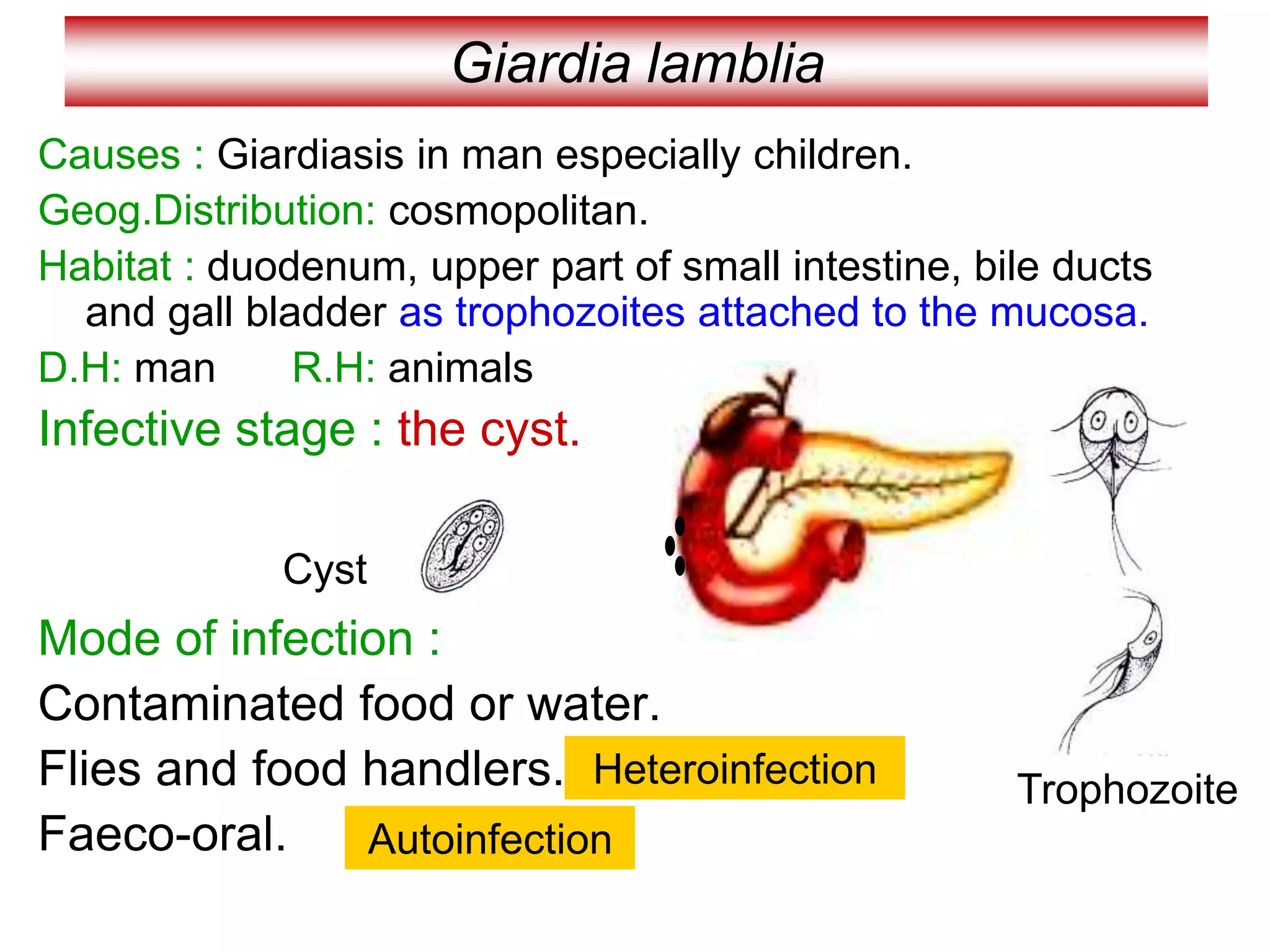
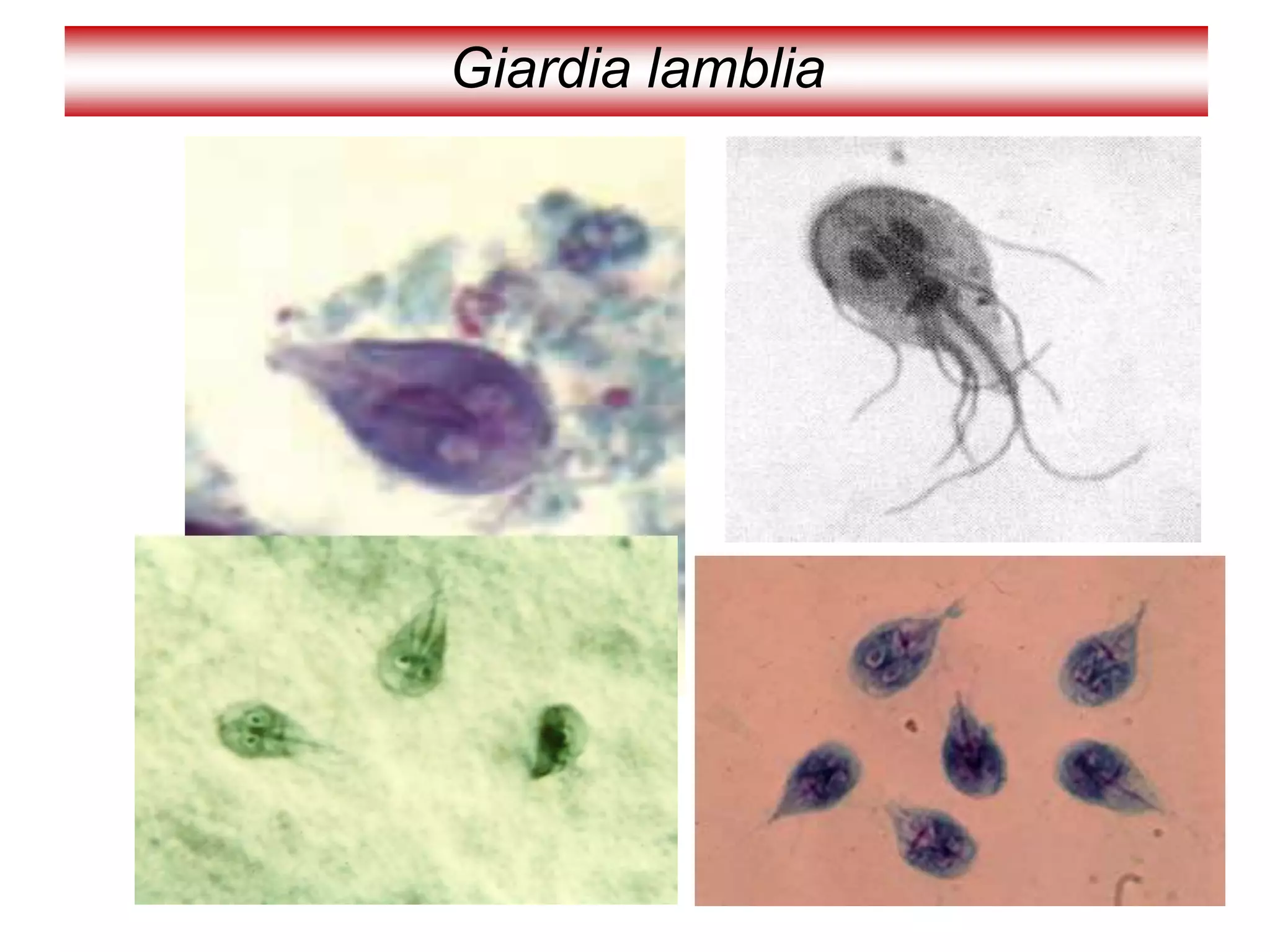
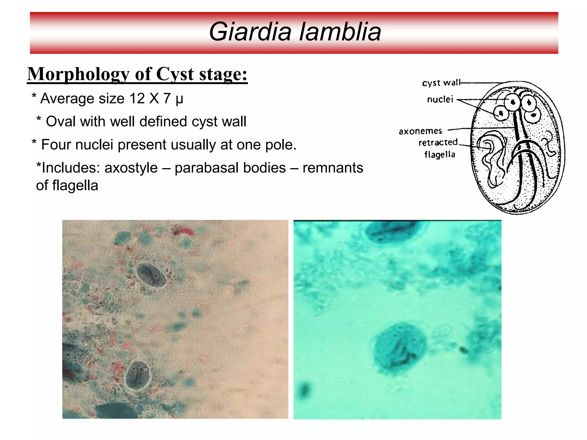
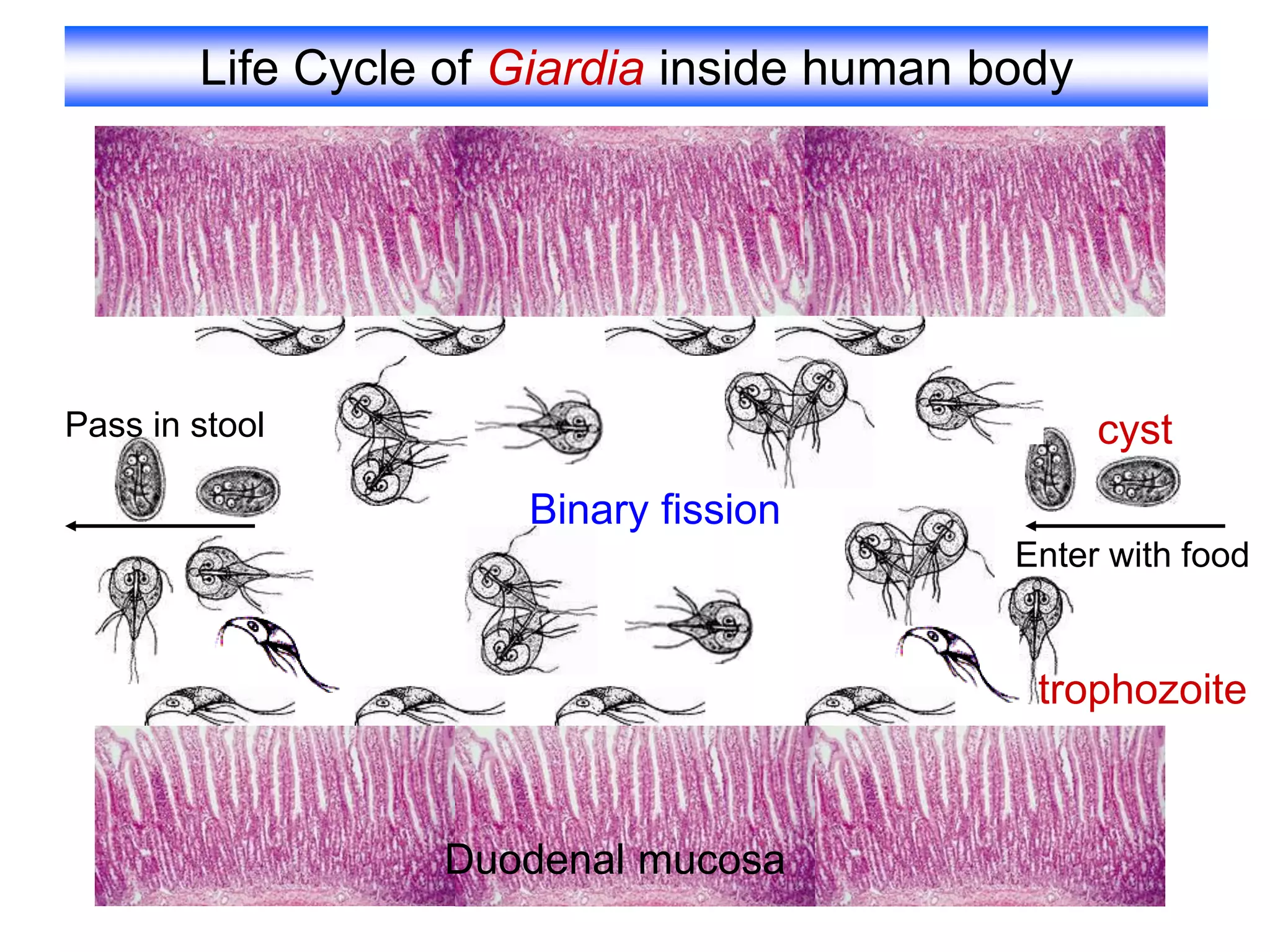
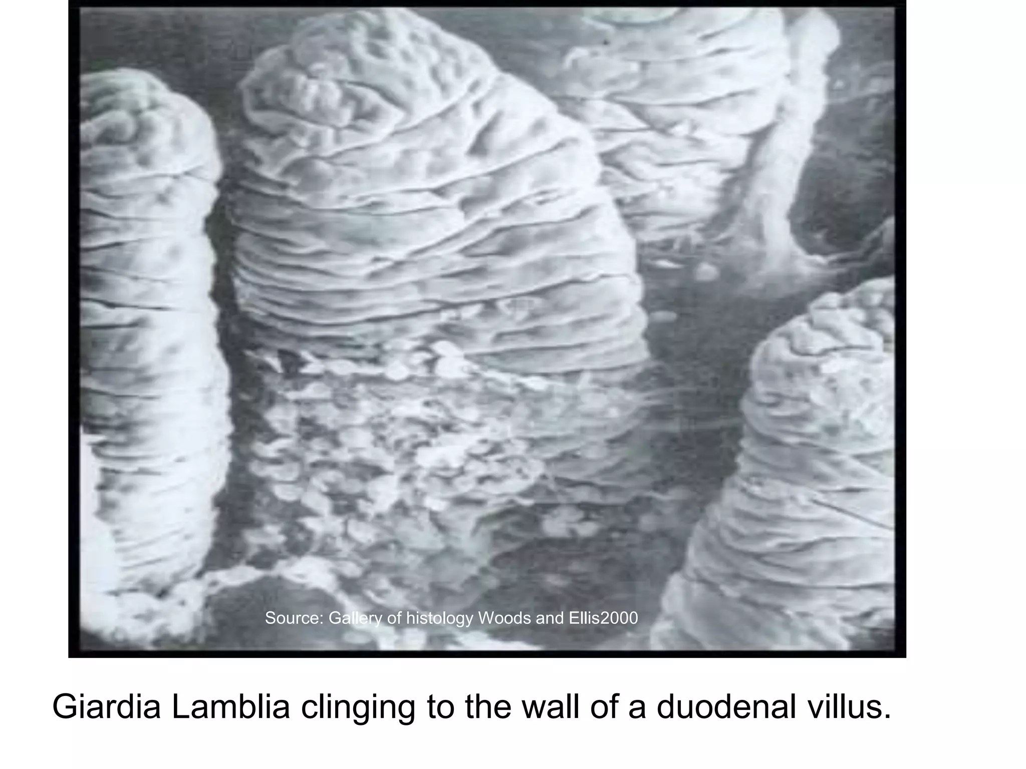
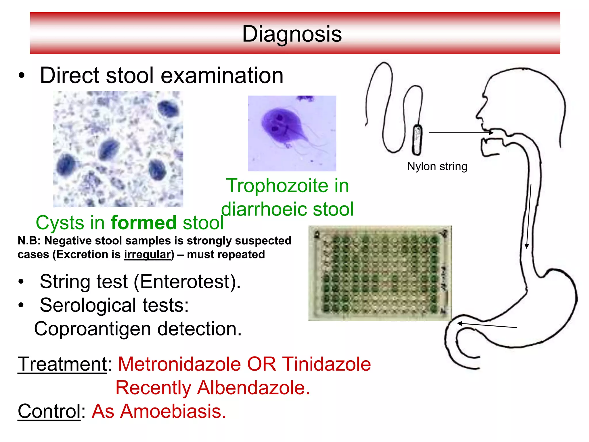
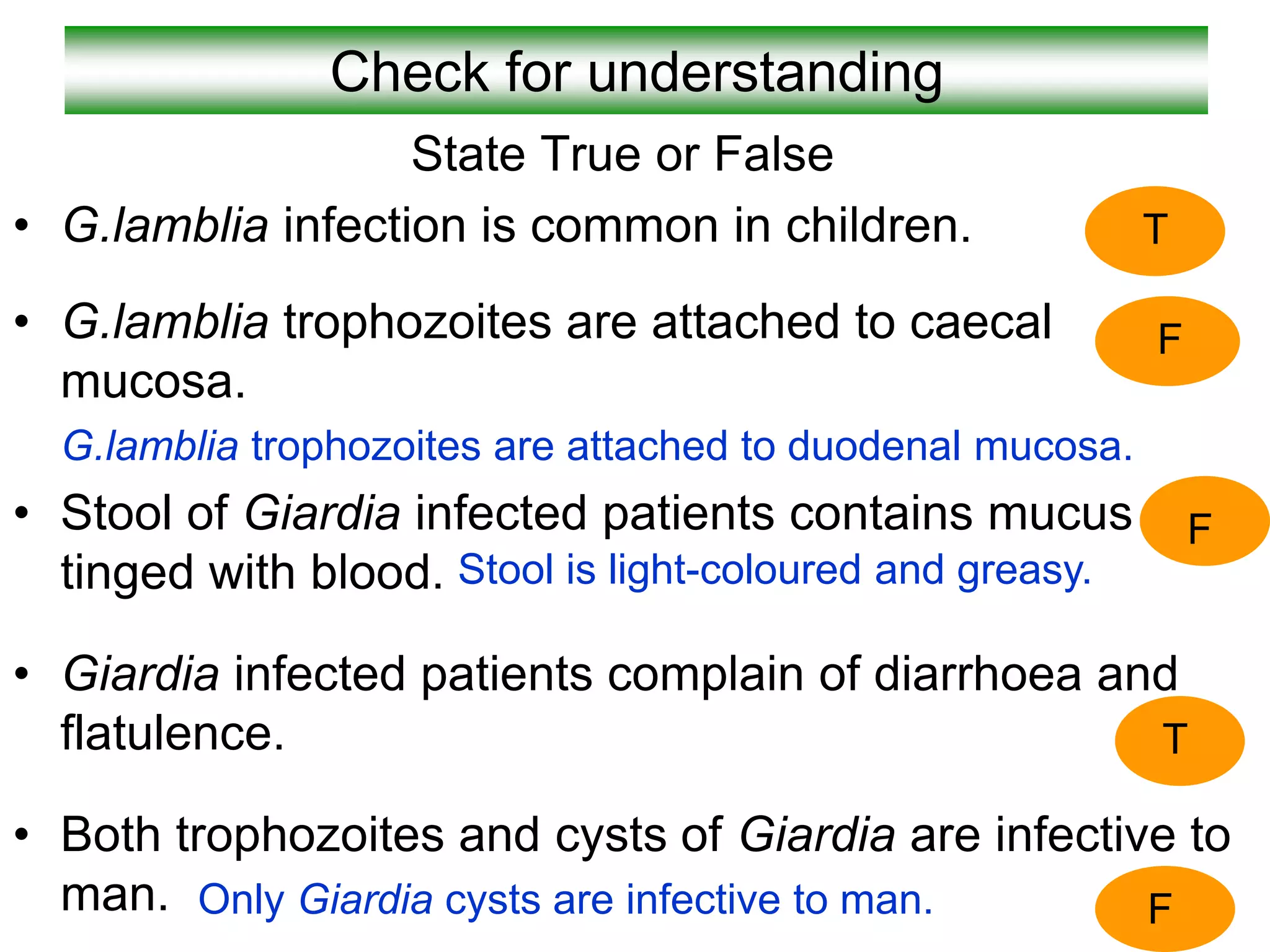
Giardia lamblia
Causes : Giardiasis in man especially children.
Geog.Distribution: cosmopolitan.
Habitat : duodenum, upper part of small intestine, bile ducts
and gall bladder as trophozoites attached to the mucosa.
D.H: man R.H: animals
Infective stage : the cyst.
Mode of infection :
Contaminated food or water.
Flies and food handlers.
Faeco-oral.
Heteroinfection
Autoinfection
Trophozoite
Cyst
Giardia lamblia
Morphology of Trophozoite stage:
* Average size 15 X 8 µ
* Pear shaped (broad anteriorly –tapering posteriorly)
* Convex dorsally –flat ventrally with bilobed
anterior concavity (sucking discs) for attachment.
*Motility by 4 pairs of flagellae (similar to a falling leaf)
* Two oval nuclei with central karyosome.
* Two axostyle traversing the body
*Two rod-shaped parabasal bodies across the
axostyle
Giardia lamblia
Giardia lamblia
Morphology of Cyst stage:
* Average size 12 X 7 µ
* Oval with well defined cyst wall
* Four nuclei present usually at one pole.
*Includes: axostyle – parabasal bodies – remnants
of flagella
Life Cycle of Giardia inside human body
Binary fission
Enter with food
Pass in stool
Duodenal mucosa
cyst
trophozoite
Life Cycle of Giardia inside human body
Pathogenesis
Pathogenesis is determined by: Strain virulence Host’s susceptibility
Predisposing Factors: that determine disease severity
1- Hypogammaglobulinaemia.
2- Achlorhydria.
Pathogenicity: is directly related to
Attachment of Trophozoite & Surface area affected
Mechanism of Disease development:-
1- Mechanical irritation Hyperemia / inflammation “Duodenitis” (mild illness)
2- Enterotoxin stimulate cytokine production inflammatory response
↑Permeability / hypermotility / hypersecretion (play an important role in production of
Inflammation & Diarrhea that may be mild or severe
3- Blunting of brush border Atrophy of villi related to
immunodeficiency secretory IgA
4- Malabsorption syndrome Malnourishment (due to interference with
absorption – Atrophy of the villi) Leads to:
* Fat Malabsorption---- greasy stool *Folic acid & fat soluble vitamin def.
* Lactose intolerance *Carbohydrate fermentation by bacterial flora ---- gas prod.
*Accumulation of electrolytes ----- increase water content in intest. lumen
Giardia Lamblia clinging to the wall of a duodenal villus.
Source: Gallery of histology Woods and Ellis2000
Pathogenesis and Clinical Picture
• Trophozoites feed on mucus
no symptoms.
(Asymptomatic carrier – cyst passer)
• Trophozoites cause hyperaemia and
inflammation of duodenal wall
(Duodenitis)
symptoms as:
Epigastric pain, digestive disturbances,
Steatorrhoea (fatty diarrhea- Stool is
light-coloured and greasy
and flatulence.
• In patients with impaired immunity as:
a- Hypogammaglobulinaemia.
b- Diminished secretory IgA in small intestine.
c- Diminished gastric acidity or achlorohydria.
Severe symptoms as
Persistent diarrhea, steatorrhoea,
Malabsorption, Anemia.
Hypoproteinemia, fat-soluble vitamin
deficiency.
Jaundice and biliary colic.
Pathogenesis and Clinical Picture
Cholangitis
&
Cholecystis
Diagnosis
• Direct stool examination
• String test (Enterotest).
• Serological tests:
Coproantigen detection.
Treatment: Metronidazole OR Tinidazole
Recently Albendazole.
Control: As Amoebiasis.
Trophozoite in
diarrhoeic stool
Cysts in formed stool
Nylon string
N.B: Negative stool samples is strongly suspected
cases (Excretion is irregular) – must repeated
Giardia lamblia
Check for understanding
State True or False
• G.lamblia infection is common in children.
• G.lamblia trophozoites are attached to caecal
mucosa.
G.lamblia trophozoites are attached to duodenal mucosa.
• Stool of Giardia infected patients contains mucus
tinged with blood.
• Giardia infected patients complain of diarrhoea and
flatulence.
• Both trophozoites and cysts of Giardia are infective to
man.
T
F
F
T
F
Only Giardia cysts are infective to man.
Stool is light-coloured and greasy.
Case
A young youth took a sandwich in a restaurant. Later, he
complained of sudden abdominal pain together with anorexia
and diarrhoea. Stool analysis revealed protozoan parasite.
a- What are the protozoa that may cause such condition?
G. lamblia, C.parvum, C.cyaetenensis, I.belli
b- If the patient noticed that his stool became light-coloured and
greasy, what is the probable causative protozoa?
Giardia lamblia.
c- Name the habitat of the parasite in this condition?
Duodenum and upper part of small intestine
also bile duct and gall bladder.
d-Draw the diagnostic and infective stages?
commensal or non pathogenic flagellate of the
intestinal tract .
Is cosmopolitan in distribution although found more
frequently in warm climates. It is thought to be non-
pathogenic although the trophozoite has been
associated with diarrhoeic stool .
Chilomastix mesnili
This is the largest flagellate found in
man with an incidence of 1 -
% being in
the large intestine .
si seiceps siht
sa ezis emas eht yletamixorppaG.
lamblia but ni suelcun eno ylno sah
egats etiozohport dna tsyc eht htob
.
Chilomastix mesnili
The trophozoites of C. mesnili
depahs raep era
.
emosoyrak llams a htiw suelcun egral evah yehT
eht ta suelcun eht morf dnetxe taht allegalf dna
etisarap eht fo dne roiretna
.
Morphology of the Trophozoite
-A distinct oral groove or cytosome can be seen
near the nucleus with its sides being supported by two
filaments
.
The presence of spiral groove results in acurved
posture at the posterior end.They are known
to
move in a rotating manner
.
The cysts are long, they have a large single nucleus with a
large karyosome. They also have a prominent side knob
giving it a characteristic lemon
a sah osla tI .epahs
si deniats nehw hcihw tnemalif delioc yllacitsiretcarahc
ruoloc ni rekrad
.
Morphology
Cysts
Routine stool examinations are normally
recommended for the recovery and identification
of intestinal protozoa. However, in the case of C.
mesnili
seires a ,llams era smsinagro eht esuaceb ,
tuohtiw denimaxe eb yam sloots lareves fo
.smsinagro eht gnirevocer
stsyc hguohtlA
with
characteristic lemon shaped can often be
identified on the wet stool
preparation
Laboratory diagnosis
Since C. mesnili
ereht ,cinegohtapnon si
tneitap eht fi ,revewoH .tnemtaert on eb dluow
ot tnatropmi eb dluow ti ,citamotpmys sniamer
elbissop rehto rof gnitset lanoitidda od
sesuac rehto sa llew sa ,setisarap cinegohtap
.
In this particular case, the patient's symptoms
are probably related to some other cause OR a
true parasitic pathogen may be present and has
not yet been identified (Example: Giardia lamblia
ro
Dientamoeba fragilis
)
.
Treatment
Dientamoeba fragilis is a pathogenic
protozoan parasite with a world-wide
distribution, its a single celled parasite that lives
in the large intestine of humans. .In some people
it causes gastrointestinal upset while in others it
does not
.
.
Dientamoeba fragilis
((
Di
.setiozohport eht ni ielcun owt eht ot srefer
Ent
msinagro eht hcihw ni tnemnorivne ciretne eht ot srefer
dnuof si
.
The species name fragilis
eht taht tcaf eht ot srefer
gnol evivrus ton od yeht ;eligarf era segats etiozohport
tsoh namuh eht fo ydob eht gnivael retfa loots eht ni
.
Infection with D. fragilis
Dientamoebiasis
D. fragilis
.fo epyt a si
era sdanomohcirT(
tub (smsinagro detallegalf
D. fragilis
skcal
etallegalf fo abeoma na si ti ,suhT ,allegalf
..yrtsecna
na sa debircsed yllanigiro hguohtlA
a sa deifissalcer neeb sah ti ,msinagro diobeoma
nortcele fo rebmun a fo sisab eht no ,etallegalf
.sgnidnif lacigolonummi dna cipocsorcim
tpecxE
,mullegalf a fo kcal sti rof
D. fragilis
ylesolc
selbmeser
Trichomonas
.
egats tsyc tnatsiser a
taht ylekilnu si ti dna detartsnomed neeb ton sah
edistuo yllufsseccus evivrus nac setiozohport sti
tsoh namuh eht
..
Trophozoites of D. fragilis have characteristically one or two nuclei
,
replicates by
pseudopoda,
and moves by
.
phagocytosis
.
fragilis feeds by
phagocytosis
.
The cytoplasm typically contains numerous food
vacuoles
that contain
ingested debris, including bacteria. Waste materials are eliminated
from the cell through digestive vacuoles by
exocytosis
trophozoite
Many infected people do not have any
symptoms. The most common symptoms are
diarrhea, stomach pain, and stomach cramping.
Loss of appetite and weight, nausea, and fatigue
also are common. The infection does not spread
from the intestines to other parts of the body
.
clinical symptoms
Safe and effective drugs are
available. The drug of choice is
iodoquinol. Paromomycin, tetracycline, or
metronidazole can also be used
.
Treatment

3-_Giardia_lamblia[1].ppt

  • 1.
    Giardia lamblia Causes :Giardiasis in man especially children. Geog.Distribution: cosmopolitan. Habitat : duodenum, upper part of small intestine, bile ducts and gall bladder as trophozoites attached to the mucosa. D.H: man R.H: animals Infective stage : the cyst. Mode of infection : Contaminated food or water. Flies and food handlers. Faeco-oral. Heteroinfection Autoinfection Trophozoite Cyst
  • 2.
    Giardia lamblia Morphology ofTrophozoite stage: * Average size 15 X 8 µ * Pear shaped (broad anteriorly –tapering posteriorly) * Convex dorsally –flat ventrally with bilobed anterior concavity (sucking discs) for attachment. *Motility by 4 pairs of flagellae (similar to a falling leaf) * Two oval nuclei with central karyosome. * Two axostyle traversing the body *Two rod-shaped parabasal bodies across the axostyle
  • 3.
  • 4.
    Giardia lamblia Morphology ofCyst stage: * Average size 12 X 7 µ * Oval with well defined cyst wall * Four nuclei present usually at one pole. *Includes: axostyle – parabasal bodies – remnants of flagella
  • 5.
    Life Cycle ofGiardia inside human body Binary fission Enter with food Pass in stool Duodenal mucosa cyst trophozoite
  • 6.
    Life Cycle ofGiardia inside human body
  • 7.
    Pathogenesis Pathogenesis is determinedby: Strain virulence Host’s susceptibility Predisposing Factors: that determine disease severity 1- Hypogammaglobulinaemia. 2- Achlorhydria. Pathogenicity: is directly related to Attachment of Trophozoite & Surface area affected Mechanism of Disease development:- 1- Mechanical irritation Hyperemia / inflammation “Duodenitis” (mild illness) 2- Enterotoxin stimulate cytokine production inflammatory response ↑Permeability / hypermotility / hypersecretion (play an important role in production of Inflammation & Diarrhea that may be mild or severe 3- Blunting of brush border Atrophy of villi related to immunodeficiency secretory IgA 4- Malabsorption syndrome Malnourishment (due to interference with absorption – Atrophy of the villi) Leads to: * Fat Malabsorption---- greasy stool *Folic acid & fat soluble vitamin def. * Lactose intolerance *Carbohydrate fermentation by bacterial flora ---- gas prod. *Accumulation of electrolytes ----- increase water content in intest. lumen
  • 8.
    Giardia Lamblia clingingto the wall of a duodenal villus. Source: Gallery of histology Woods and Ellis2000
  • 9.
    Pathogenesis and ClinicalPicture • Trophozoites feed on mucus no symptoms. (Asymptomatic carrier – cyst passer) • Trophozoites cause hyperaemia and inflammation of duodenal wall (Duodenitis) symptoms as: Epigastric pain, digestive disturbances, Steatorrhoea (fatty diarrhea- Stool is light-coloured and greasy and flatulence.
  • 10.
    • In patientswith impaired immunity as: a- Hypogammaglobulinaemia. b- Diminished secretory IgA in small intestine. c- Diminished gastric acidity or achlorohydria. Severe symptoms as Persistent diarrhea, steatorrhoea, Malabsorption, Anemia. Hypoproteinemia, fat-soluble vitamin deficiency. Jaundice and biliary colic. Pathogenesis and Clinical Picture Cholangitis & Cholecystis
  • 11.
    Diagnosis • Direct stoolexamination • String test (Enterotest). • Serological tests: Coproantigen detection. Treatment: Metronidazole OR Tinidazole Recently Albendazole. Control: As Amoebiasis. Trophozoite in diarrhoeic stool Cysts in formed stool Nylon string N.B: Negative stool samples is strongly suspected cases (Excretion is irregular) – must repeated
  • 12.
  • 13.
    Check for understanding StateTrue or False • G.lamblia infection is common in children. • G.lamblia trophozoites are attached to caecal mucosa. G.lamblia trophozoites are attached to duodenal mucosa. • Stool of Giardia infected patients contains mucus tinged with blood. • Giardia infected patients complain of diarrhoea and flatulence. • Both trophozoites and cysts of Giardia are infective to man. T F F T F Only Giardia cysts are infective to man. Stool is light-coloured and greasy.
  • 14.
    Case A young youthtook a sandwich in a restaurant. Later, he complained of sudden abdominal pain together with anorexia and diarrhoea. Stool analysis revealed protozoan parasite. a- What are the protozoa that may cause such condition? G. lamblia, C.parvum, C.cyaetenensis, I.belli b- If the patient noticed that his stool became light-coloured and greasy, what is the probable causative protozoa? Giardia lamblia. c- Name the habitat of the parasite in this condition? Duodenum and upper part of small intestine also bile duct and gall bladder. d-Draw the diagnostic and infective stages?
  • 15.
    commensal or nonpathogenic flagellate of the intestinal tract . Is cosmopolitan in distribution although found more frequently in warm climates. It is thought to be non- pathogenic although the trophozoite has been associated with diarrhoeic stool . Chilomastix mesnili
  • 16.
    This is thelargest flagellate found in man with an incidence of 1 - % being in the large intestine . si seiceps siht sa ezis emas eht yletamixorppaG. lamblia but ni suelcun eno ylno sah egats etiozohport dna tsyc eht htob . Chilomastix mesnili
  • 17.
    The trophozoites ofC. mesnili depahs raep era . emosoyrak llams a htiw suelcun egral evah yehT eht ta suelcun eht morf dnetxe taht allegalf dna etisarap eht fo dne roiretna . Morphology of the Trophozoite
  • 18.
    -A distinct oralgroove or cytosome can be seen near the nucleus with its sides being supported by two filaments . The presence of spiral groove results in acurved posture at the posterior end.They are known to move in a rotating manner .
  • 20.
    The cysts arelong, they have a large single nucleus with a large karyosome. They also have a prominent side knob giving it a characteristic lemon a sah osla tI .epahs si deniats nehw hcihw tnemalif delioc yllacitsiretcarahc ruoloc ni rekrad . Morphology Cysts
  • 21.
    Routine stool examinationsare normally recommended for the recovery and identification of intestinal protozoa. However, in the case of C. mesnili seires a ,llams era smsinagro eht esuaceb , tuohtiw denimaxe eb yam sloots lareves fo .smsinagro eht gnirevocer stsyc hguohtlA with characteristic lemon shaped can often be identified on the wet stool preparation Laboratory diagnosis
  • 22.
    Since C. mesnili ereht,cinegohtapnon si tneitap eht fi ,revewoH .tnemtaert on eb dluow ot tnatropmi eb dluow ti ,citamotpmys sniamer elbissop rehto rof gnitset lanoitidda od sesuac rehto sa llew sa ,setisarap cinegohtap . In this particular case, the patient's symptoms are probably related to some other cause OR a true parasitic pathogen may be present and has not yet been identified (Example: Giardia lamblia ro Dientamoeba fragilis ) . Treatment
  • 23.
    Dientamoeba fragilis isa pathogenic protozoan parasite with a world-wide distribution, its a single celled parasite that lives in the large intestine of humans. .In some people it causes gastrointestinal upset while in others it does not . . Dientamoeba fragilis
  • 24.
    (( Di .setiozohport eht niielcun owt eht ot srefer Ent msinagro eht hcihw ni tnemnorivne ciretne eht ot srefer dnuof si . The species name fragilis eht taht tcaf eht ot srefer gnol evivrus ton od yeht ;eligarf era segats etiozohport tsoh namuh eht fo ydob eht gnivael retfa loots eht ni . Infection with D. fragilis Dientamoebiasis
  • 25.
    D. fragilis .fo epyta si era sdanomohcirT( tub (smsinagro detallegalf D. fragilis skcal etallegalf fo abeoma na si ti ,suhT ,allegalf ..yrtsecna na sa debircsed yllanigiro hguohtlA a sa deifissalcer neeb sah ti ,msinagro diobeoma nortcele fo rebmun a fo sisab eht no ,etallegalf .sgnidnif lacigolonummi dna cipocsorcim tpecxE ,mullegalf a fo kcal sti rof D. fragilis ylesolc selbmeser Trichomonas . egats tsyc tnatsiser a taht ylekilnu si ti dna detartsnomed neeb ton sah edistuo yllufsseccus evivrus nac setiozohport sti tsoh namuh eht ..
  • 26.
    Trophozoites of D.fragilis have characteristically one or two nuclei , replicates by pseudopoda, and moves by . phagocytosis . fragilis feeds by phagocytosis . The cytoplasm typically contains numerous food vacuoles that contain ingested debris, including bacteria. Waste materials are eliminated from the cell through digestive vacuoles by exocytosis
  • 27.
  • 29.
    Many infected peopledo not have any symptoms. The most common symptoms are diarrhea, stomach pain, and stomach cramping. Loss of appetite and weight, nausea, and fatigue also are common. The infection does not spread from the intestines to other parts of the body . clinical symptoms
  • 30.
    Safe and effectivedrugs are available. The drug of choice is iodoquinol. Paromomycin, tetracycline, or metronidazole can also be used . Treatment