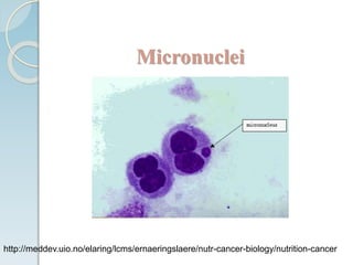
Micronucleus Assay
PRESENTED BY
G.R.HARIKA
M.S.(Pharm.) 1st semester
Dept. of Regulatory Toxicology
GUIDED BY
Dr. G.B. JENA
Associate Professor
Dept. of Pharmacology and Toxicology
National Institute of Pharmaceutical Education and Research,
S.A.S Nagar, Mohali
Flow of presentation:
• Introduction
• Mechanism
• Characteristics
• Types
• Models
• Conclusion
Introduction:
A micronucleus assay is a test used for toxicological testing of
potential genotoxic compounds
It is commonly used because of its simplicity, reliability and
reproducibility
This assay is one of the most successful assays for genotoxic
carcinogens, i.e., carcinogens that act by causing genetic
damage
 It has widespread acceptance and regulatory approval and is
the OECD guideline for the testing of chemicals
 It is performed in erythrocytes generally, but currently its
use has been extended to other tissues like liver, lung, skin
etc
 Micronucleus assay is also exploring to wide areas and
future advancements make this assay more promising in
analyzing the chromosome damage caused by the genotoxic
chemicals
Jena, et.al (2002), Indian journal of pharmacology, 34, 2 86-99
Micronuclei formation
http://meddev.uio.no/elaring/lcms/ernaeringslaere/nutr-cancer-biology/nutrition-
cancer
Mechanisms through which micronuclei
can form:
 The mitotic loss of acentric chromosome fragments (forming
structural aberrations)
 Mechanical consequences of chromosomal breakage and
exchange, such as from lagging chromosomes, an inactive
centromere or tangled chromosomes (forming structural
aberrations)
 Mitotic loss of whole chromosomes (forming numerical
aberrations)
 Apoptosis
Schmid, (1975),Mutation Research/Environmental Mutagenesis , 31,1 9-15.
Micronuclei is induced by:
A Clastogen is a mutagenic agent giving rise to or
inducing disruption or breakages of chromosomes leading to
sections of the chromosome being deleted, added, or
rearranged
An Aneugens is a substance which makes daughter cells
to have abnormal no of chromosomes
Micronuclei
http://meddev.uio.no/elaring/lcms/ernaeringslaere/nutr-cancer-biology/nutrition-cancer
Micronuclei Characteristics:
 Micronuclei are morphologically identical to the main nuclei,
but are smaller than it
 It is not linked or connected to the main nuclei
 It may touch but do not overlap the main nuclei
 It is non-retractile and they can therefore be readily
distinguished from staining particles
Fenech,(2000), Mutation Research/Fundamental and Molecular Mechanisms of Mutagenesis,
455, 1 81-95
Types of micronuclei:
Micronuclei are of different types based on 3 criteria,
Based on the presence of kinetochore
Based on the presence of centromere, they are of two types-
Centromeric micronucleus
Acentromeric micronucleus
Based on the number of nuclei present in the cell (i.e., cell
divisions that occur), they are of following types,
Mononucleate
Binucleate
Trinucleate
Quadrinucleate
Multinucleate
Fenech,(1997),Mutation Research/GeneticToxicology and Environmental Mutagenesis, 392, 1
11-18.
Models of micronucleus assay:
In-vitro: OECD guideline No. 487
In-vivo: OECD guideline No. 474
Principle : In-vitro
Cell cultures are exposed to the test substances both with and without
metabolic activation. After exposure to a test substance, and addition of
cytochalasin B for blocking cytokinesis cell cultures are grown for a
sufficient period to allow chromosomal damage to lead to the formation of
micronuclei in bi- or multinucleated interphase cells. Harvested and stained
interphase cells are then analyzed microscopically for the presence of
micronuclei. Micronuclei are scored in those cells that complete nuclear
division following exposure to the test item.
General considerations:
 Most commonly used cell lines are CHL/IU, CHO, SHE
and V79
 Mouse lymphoma L5178Y cells are also used though
they have interactions with the cytochalasinB
 Cell types with low and stable frequency of micronuclei
are mostly used because frequency of micronuclei
formation may influence the studies
Kirsch-Volders,et.al (2003),Mutation Research/Genetic Toxicology and Environmental Mutagenesis, 540, 2 153-163.
Media:
 Cells are cultured in Phytohaemaglutinin(PHA)
medium at 37°C
 Cultures should not be contaminated with mycoplasama
Ames, et.al(1975), Mutation Research/Environmental Mutagenesis and Related Subjects, 31, 6 347-363.
Metabolic activation:
 Cells should be treated with the test substance both in the
presence and absence of an appropriate metabolic activation
system
 1-10% v/v conc. of post mitochondrial fraction (S9) is the
most commonly used metabolic activating system prepared
from liver of rodents
 Initially it is treated with Aroclor1254 or in combination
with phenobarbitone and napthoflavone
Elliott, et.al (1992), Mutagenesis, 7, 3 175-177
Use of cytochalasinB:
 CytochalasinB helps in formation of binucleated cells by inhibiting
the formation of micro filament and cytokinesis
 It is used in the concentration of 3-6µg/ml to get 50% binucleated
cells; along with the test substance and it should be given at least
6hours before the first mitosis
 Evaluation of micronuclei formation can be known by reduction of
cell proliferation.
Cytokinesis-block proliferation index:
 CBPI indicates the number of cell cycles per cell during the
period of exposure to cytochalasinB.
Cytotoxicity = 100-100 {(CBPIT - 1) / (CBPI C -1)}
where; T = test ; C = control
No. mononucleate cells + 2 x No. binucleate cells + 3 x No. multinucleate cells
CBPI = ----------------------------------------------------------------------------
Total no. of cells
 CBPI = 1; indicates 100% cytotoxicity
Controls:
 Positive controls : used to identify clastogens , aneugens
and effectiveness of exogenous metabolic activation
system
 Examples: MitomycinC; Cytosine arabinoside
 Negative controls : consists of solvent alone doesn’t
requires any metabolic activators
Oecd guideline for the testing of chemicals; guideline 487
Procedure: In-vitro
Cell lines + appr.culture media acc. to test
Incubate at 37°C
cells are taken from the stock to prepare suspension
Harvested
Monolayer of cells
Stock
Step:1
Blood sample + Lithium Heparin
Separation of lymphocytes
44-48hrs
Step:2
Phytohaemaglutinin – induces cell division
This culture is added to cell lines along with test chemical
Without S9 fraction
After 3-6hrs
With S9 fraction
Aroclor 1254- induces enzymes
Cells should be exposed to test 3-6hrs
Metabolic
activation
Remove the S9 and treatment medium by
washing
Freshmedia; CytochalasinB
Harvest for 1.5- 2 cell cycle length
Cells should be exposed to test
3-6hrs
Remove the treatment
medium by washing
Fresh media; CytochalasinB
and Harvest for 1.5- 2 cell cycle
length
Step: 3
Principle : In-Vivo
Rodents are treated with the test agent by appropriate
route, bone marrow extracted at appropriate times after
treatment, smear slides are prepared either with whole
bone marrow or cellulose column-fractionated cell
suspension, stained, coded, and analyzed for the toxicity
(PCE to NCE ratio) and micro nucleated cell frequency.
Makoto Hayashi et.al(2000)Invivo Rodent Erythrocyte MicronucleusAssayII.,Environmental
and Molecular Mutagenesis 35:234–252
In-vivo micronucleus:
They are done using following
 Peripheral blood
 Bone marrow
Krishna and Hayashi, (2000),Mutation Research/Fundamental and Molecular Mechanisms of
Mutagenesis, 455, 1 155-166
Procedure: In-Vivo
5 animals should be treated with the test chemical.
Dosing done at 3levels (low, medium , high doses)
After 24-48hrs animals are sacrificed by carbon dioxide euthanasia
cut the ends of bone; flush the bonemarrow with isotonic solution
Centrifuge the marrow suspension
Preparation of smear of sample collected
Staining:
 Slides are stained using Giemsa or Fluorescent DNA specific
dyes
 Example of fluorescent stains
Acridine orange
Hoechst + pyronin Y
May - Grunwald
May - Grunwald + Giemsa
Ren, Atyah, Chen and Zhou, (2017), Journal of translational medicine, 15, 1 110
Analysis:
 Flow cytometry
 Laser scanning cytometry
 Image analysis
Applications:
 Distinguish aneugens from clastogens
 Perform long-term toxicological studies
 Examining the radioactive properties of H2 receptor
antagonists
 Evaluation of radio sensitivity of human glioma cells
Warheit and Donner, Nanotoxicology, 4, 4 409-413.
Conclusion:
 Micronucleus assay has wide spread applications in the
area of toxicology for assessing risk assessment of drugs,
pollutants, NCE etc
 Micronucleus assay results using tissues other than
hematopoietic tissue have become increasing in
toxicological studies to evaluate the multi organ toxicity
caused by different genotoxic agents
Micronucleus Assay

Micronucleus Assay

  • 1.
    Micronucleus Assay PRESENTED BY G.R.HARIKA M.S.(Pharm.)1st semester Dept. of Regulatory Toxicology GUIDED BY Dr. G.B. JENA Associate Professor Dept. of Pharmacology and Toxicology National Institute of Pharmaceutical Education and Research, S.A.S Nagar, Mohali
  • 2.
    Flow of presentation: •Introduction • Mechanism • Characteristics • Types • Models • Conclusion
  • 3.
    Introduction: A micronucleus assayis a test used for toxicological testing of potential genotoxic compounds It is commonly used because of its simplicity, reliability and reproducibility This assay is one of the most successful assays for genotoxic carcinogens, i.e., carcinogens that act by causing genetic damage
  • 4.
     It haswidespread acceptance and regulatory approval and is the OECD guideline for the testing of chemicals  It is performed in erythrocytes generally, but currently its use has been extended to other tissues like liver, lung, skin etc  Micronucleus assay is also exploring to wide areas and future advancements make this assay more promising in analyzing the chromosome damage caused by the genotoxic chemicals Jena, et.al (2002), Indian journal of pharmacology, 34, 2 86-99
  • 5.
  • 6.
    Mechanisms through whichmicronuclei can form:  The mitotic loss of acentric chromosome fragments (forming structural aberrations)  Mechanical consequences of chromosomal breakage and exchange, such as from lagging chromosomes, an inactive centromere or tangled chromosomes (forming structural aberrations)  Mitotic loss of whole chromosomes (forming numerical aberrations)  Apoptosis Schmid, (1975),Mutation Research/Environmental Mutagenesis , 31,1 9-15.
  • 7.
    Micronuclei is inducedby: A Clastogen is a mutagenic agent giving rise to or inducing disruption or breakages of chromosomes leading to sections of the chromosome being deleted, added, or rearranged An Aneugens is a substance which makes daughter cells to have abnormal no of chromosomes
  • 8.
  • 9.
    Micronuclei Characteristics:  Micronucleiare morphologically identical to the main nuclei, but are smaller than it  It is not linked or connected to the main nuclei  It may touch but do not overlap the main nuclei  It is non-retractile and they can therefore be readily distinguished from staining particles Fenech,(2000), Mutation Research/Fundamental and Molecular Mechanisms of Mutagenesis, 455, 1 81-95
  • 10.
    Types of micronuclei: Micronucleiare of different types based on 3 criteria, Based on the presence of kinetochore Based on the presence of centromere, they are of two types- Centromeric micronucleus Acentromeric micronucleus
  • 11.
    Based on thenumber of nuclei present in the cell (i.e., cell divisions that occur), they are of following types, Mononucleate Binucleate Trinucleate Quadrinucleate Multinucleate Fenech,(1997),Mutation Research/GeneticToxicology and Environmental Mutagenesis, 392, 1 11-18.
  • 12.
    Models of micronucleusassay: In-vitro: OECD guideline No. 487 In-vivo: OECD guideline No. 474
  • 13.
    Principle : In-vitro Cellcultures are exposed to the test substances both with and without metabolic activation. After exposure to a test substance, and addition of cytochalasin B for blocking cytokinesis cell cultures are grown for a sufficient period to allow chromosomal damage to lead to the formation of micronuclei in bi- or multinucleated interphase cells. Harvested and stained interphase cells are then analyzed microscopically for the presence of micronuclei. Micronuclei are scored in those cells that complete nuclear division following exposure to the test item.
  • 14.
    General considerations:  Mostcommonly used cell lines are CHL/IU, CHO, SHE and V79  Mouse lymphoma L5178Y cells are also used though they have interactions with the cytochalasinB  Cell types with low and stable frequency of micronuclei are mostly used because frequency of micronuclei formation may influence the studies Kirsch-Volders,et.al (2003),Mutation Research/Genetic Toxicology and Environmental Mutagenesis, 540, 2 153-163.
  • 15.
    Media:  Cells arecultured in Phytohaemaglutinin(PHA) medium at 37°C  Cultures should not be contaminated with mycoplasama Ames, et.al(1975), Mutation Research/Environmental Mutagenesis and Related Subjects, 31, 6 347-363.
  • 16.
    Metabolic activation:  Cellsshould be treated with the test substance both in the presence and absence of an appropriate metabolic activation system  1-10% v/v conc. of post mitochondrial fraction (S9) is the most commonly used metabolic activating system prepared from liver of rodents  Initially it is treated with Aroclor1254 or in combination with phenobarbitone and napthoflavone Elliott, et.al (1992), Mutagenesis, 7, 3 175-177
  • 17.
    Use of cytochalasinB: CytochalasinB helps in formation of binucleated cells by inhibiting the formation of micro filament and cytokinesis  It is used in the concentration of 3-6µg/ml to get 50% binucleated cells; along with the test substance and it should be given at least 6hours before the first mitosis  Evaluation of micronuclei formation can be known by reduction of cell proliferation.
  • 18.
    Cytokinesis-block proliferation index: CBPI indicates the number of cell cycles per cell during the period of exposure to cytochalasinB. Cytotoxicity = 100-100 {(CBPIT - 1) / (CBPI C -1)} where; T = test ; C = control No. mononucleate cells + 2 x No. binucleate cells + 3 x No. multinucleate cells CBPI = ---------------------------------------------------------------------------- Total no. of cells  CBPI = 1; indicates 100% cytotoxicity
  • 19.
    Controls:  Positive controls: used to identify clastogens , aneugens and effectiveness of exogenous metabolic activation system  Examples: MitomycinC; Cytosine arabinoside  Negative controls : consists of solvent alone doesn’t requires any metabolic activators Oecd guideline for the testing of chemicals; guideline 487
  • 20.
    Procedure: In-vitro Cell lines+ appr.culture media acc. to test Incubate at 37°C cells are taken from the stock to prepare suspension Harvested Monolayer of cells Stock Step:1
  • 21.
    Blood sample +Lithium Heparin Separation of lymphocytes 44-48hrs Step:2 Phytohaemaglutinin – induces cell division This culture is added to cell lines along with test chemical
  • 22.
    Without S9 fraction After3-6hrs With S9 fraction Aroclor 1254- induces enzymes Cells should be exposed to test 3-6hrs Metabolic activation Remove the S9 and treatment medium by washing Freshmedia; CytochalasinB Harvest for 1.5- 2 cell cycle length Cells should be exposed to test 3-6hrs Remove the treatment medium by washing Fresh media; CytochalasinB and Harvest for 1.5- 2 cell cycle length Step: 3
  • 23.
    Principle : In-Vivo Rodentsare treated with the test agent by appropriate route, bone marrow extracted at appropriate times after treatment, smear slides are prepared either with whole bone marrow or cellulose column-fractionated cell suspension, stained, coded, and analyzed for the toxicity (PCE to NCE ratio) and micro nucleated cell frequency. Makoto Hayashi et.al(2000)Invivo Rodent Erythrocyte MicronucleusAssayII.,Environmental and Molecular Mutagenesis 35:234–252
  • 24.
    In-vivo micronucleus: They aredone using following  Peripheral blood  Bone marrow Krishna and Hayashi, (2000),Mutation Research/Fundamental and Molecular Mechanisms of Mutagenesis, 455, 1 155-166
  • 25.
    Procedure: In-Vivo 5 animalsshould be treated with the test chemical. Dosing done at 3levels (low, medium , high doses) After 24-48hrs animals are sacrificed by carbon dioxide euthanasia cut the ends of bone; flush the bonemarrow with isotonic solution Centrifuge the marrow suspension Preparation of smear of sample collected
  • 26.
    Staining:  Slides arestained using Giemsa or Fluorescent DNA specific dyes  Example of fluorescent stains Acridine orange Hoechst + pyronin Y May - Grunwald May - Grunwald + Giemsa Ren, Atyah, Chen and Zhou, (2017), Journal of translational medicine, 15, 1 110
  • 27.
    Analysis:  Flow cytometry Laser scanning cytometry  Image analysis
  • 28.
    Applications:  Distinguish aneugensfrom clastogens  Perform long-term toxicological studies  Examining the radioactive properties of H2 receptor antagonists  Evaluation of radio sensitivity of human glioma cells Warheit and Donner, Nanotoxicology, 4, 4 409-413.
  • 29.
    Conclusion:  Micronucleus assayhas wide spread applications in the area of toxicology for assessing risk assessment of drugs, pollutants, NCE etc  Micronucleus assay results using tissues other than hematopoietic tissue have become increasing in toxicological studies to evaluate the multi organ toxicity caused by different genotoxic agents