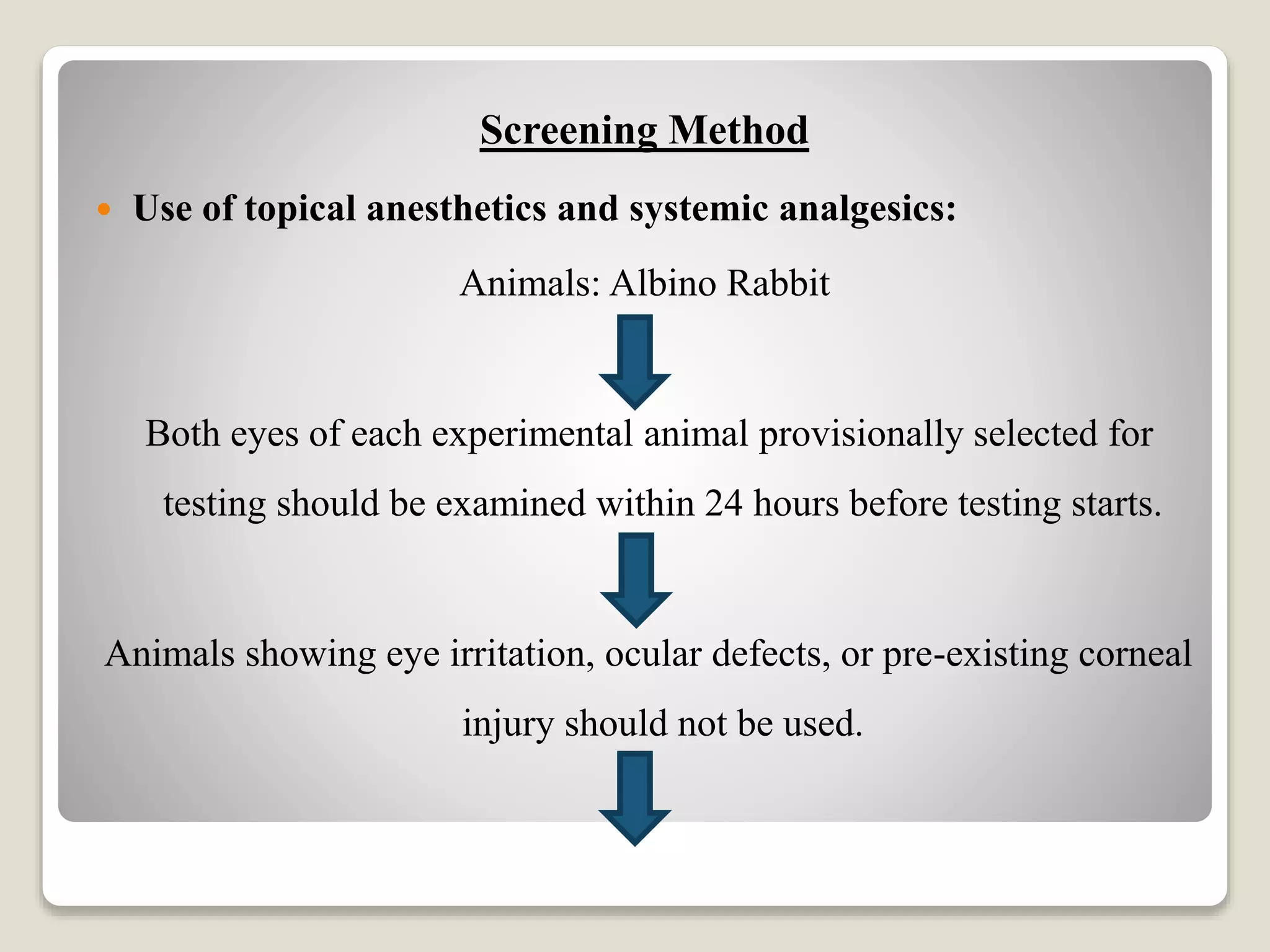
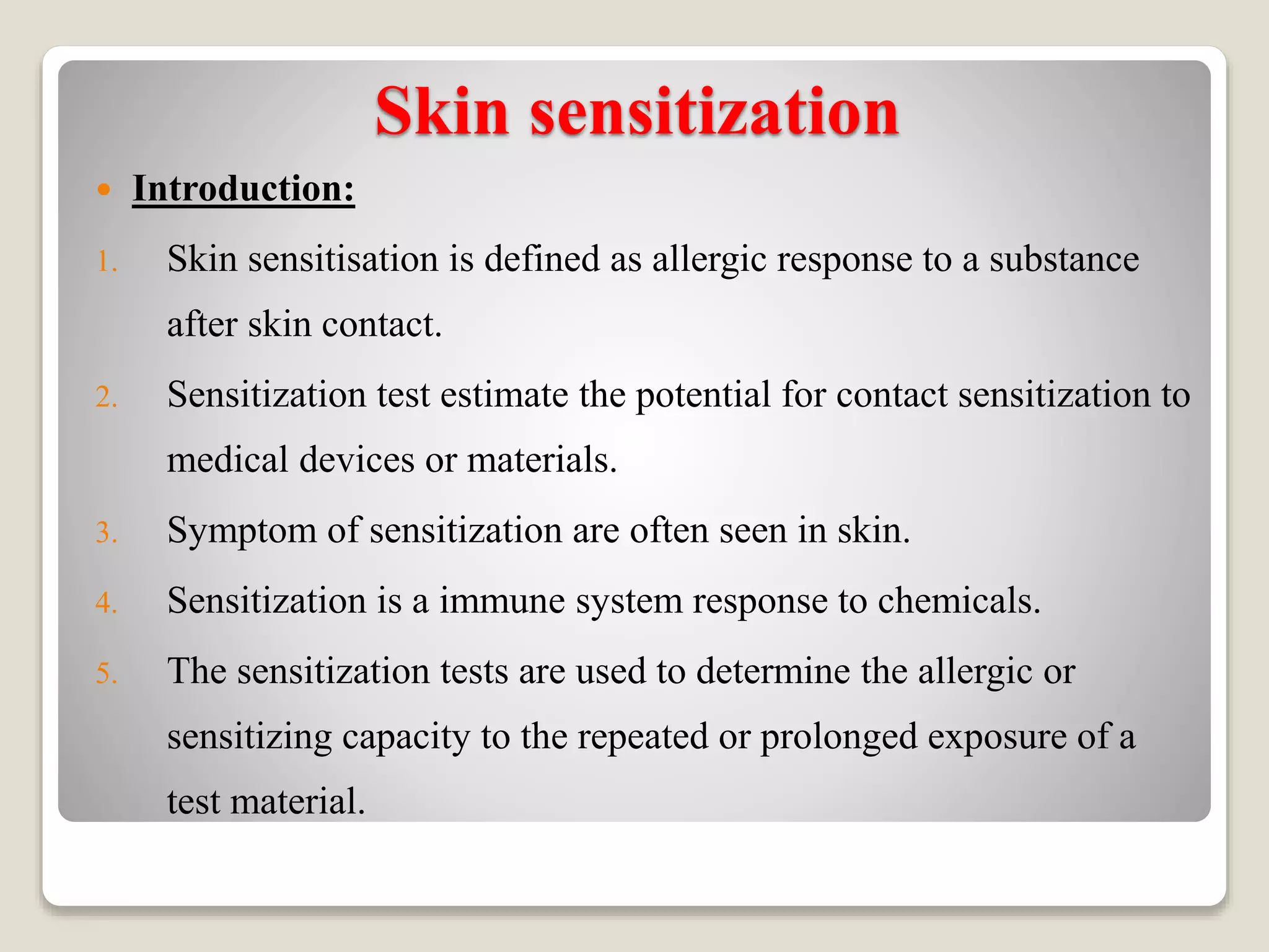
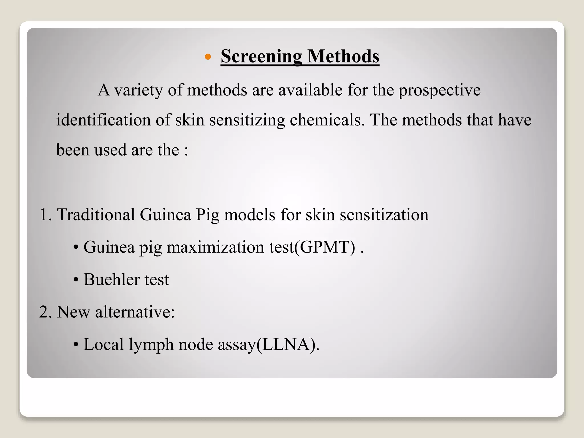
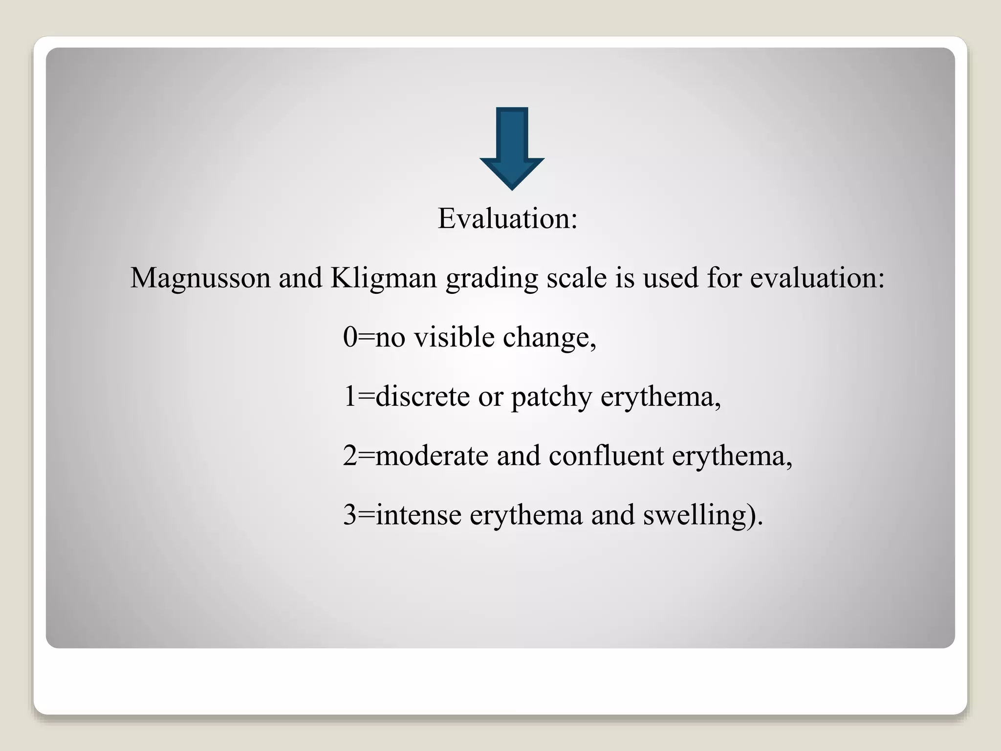
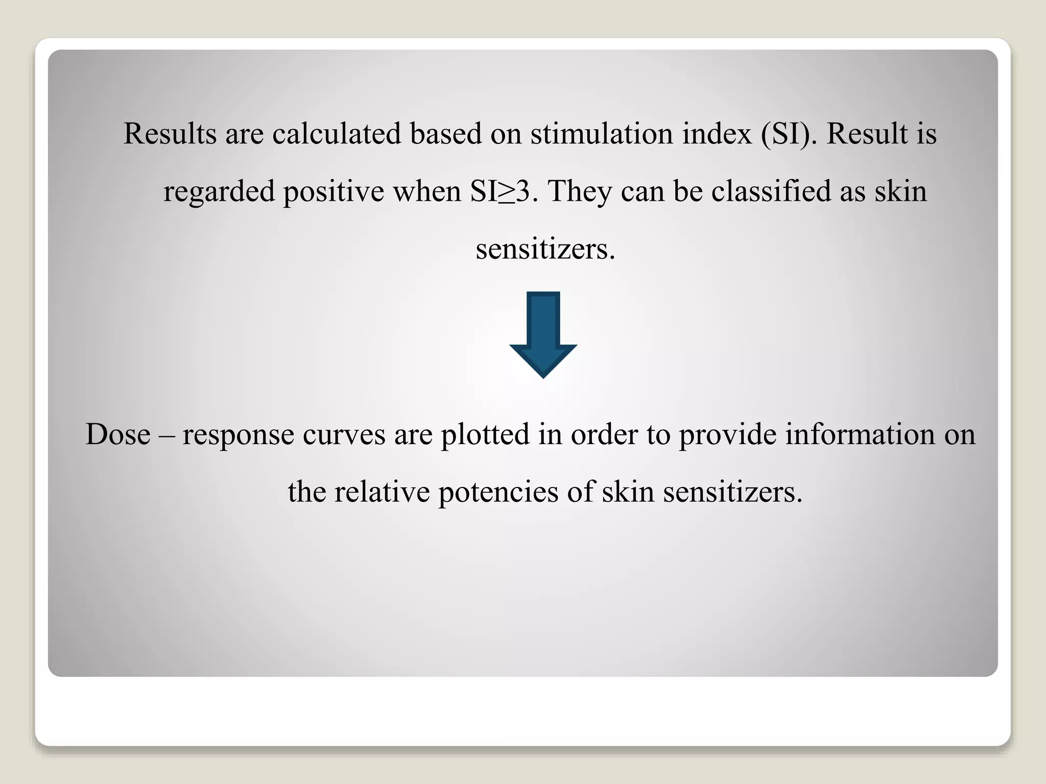
Pharmacological And
Toxicological Screening Methods
of Acute eye irritation.and Skin
sensitization
Presented By:-
Ms. Sayali S. Chavan
1ST Year M.Pharmacy
PDEA’S SGRS College of
Pharmacy.
Guided By:-
Mrs. Jagtap P.N
HOD of Pharmacology.
PDEA’S SGRS College of
Pharmacy.
1
Content :
 Screening models for:
1. Acute eye irritation.
2. Skin sensitization.
3. References.
Acute Eye Irritation
 Introduction :
1. The substance to be tested is applied in a single dose to one of the
eyes of the experimental animal; the untreated eye serves as the
control.
2. The degree of eye irritation/corrosion is evaluated by scoring
lesions of conjunctiva, cornea, and iris, at specific intervals.
3. Other effects in the eye and adverse systemic effects are also
described to provide a complete evaluation of the effects. The
duration of the study should be sufficient to evaluate the
reversibility or irreversibility of the effects.
Screening Method
 Use of topical anesthetics and systemic analgesics:
Animals: Albino Rabbit
Both eyes of each experimental animal provisionally selected for
testing should be examined within 24 hours before testing starts.
Animals showing eye irritation, ocular defects, or pre-existing corneal
injury should not be used.
Sixty minutes prior to test substance application, buprenorphine 0.01
mg/kg is administered by subcutaneous injection (SC) to provide a
therapeutic level of systemic analgesia.
Five minutes prior to TSA, one or two drops of a topical ocular
anesthetic (e.g. 0.5% proparacaine hydrochloride or 0.5% tetracaine
hydrochloride) are applied to each eye.
The eye of each animal that is not treated with a test article, but which
is treated with topical anesthetics, serves as a control.
If the test substance is anticipated to cause significant pain and
distress, it should not normally be tested in vivo.
After the initial 8-hour post-TSA treatment, buprenorphine 0.01
mg/kg SC should be administered every 12 hours, in conjunction
with meloxicam 0.5 mg/kg SC every 24 hours, until the ocular
lesions resolve and no clinical signs of pain and distress are present.
Skin sensitization
 Introduction:
1. Skin sensitisation is defined as allergic response to a substance
after skin contact.
2. Sensitization test estimate the potential for contact sensitization to
medical devices or materials.
3. Symptom of sensitization are often seen in skin.
4. Sensitization is a immune system response to chemicals.
5. The sensitization tests are used to determine the allergic or
sensitizing capacity to the repeated or prolonged exposure of a
test material.
 Screening Methods
A variety of methods are available for the prospective
identification of skin sensitizing chemicals. The methods that have
been used are the :
1. Traditional Guinea Pig models for skin sensitization
• Guinea pig maximization test(GPMT) .
• Buehler test
2. New alternative:
• Local lymph node assay(LLNA).
 Guinea pig maximization test:
Animals: Guinea pig
Sex: Male(females, should be nulliparous & non-pregnant).
Body weight: 250-500g.
Dose: An average, a guinea pig requires 10-30 mg/kg daily
Before the test, animals are randomised & assigned to the treatment
groups.
Removal of hair is by clipping , shaving or possibly by chemical
depilation , depending on the test method used.
Care should be taken to avoid abrading the skin.
The animals are weighed before the test and at the end of the test.
A minimum of 10 animals used in the treatment group and at least 5
animals in the control group are used.
Induction of saline is done initially on skin of shoulder region with
three intradermal injections on day 0.
Control animals also receive three intradermal injections, but only
vehicle is used instead of the test substance.
5–7 days later skin is painted with 0.5 mL of 10% sodium lauryl
sulfate in vaseline, to create a local irritation,
24 h later test substance is applied under occlusion for 48 h.
Challenge is done on days 20–22 with reapplication of patches for 24
h and results are assessed at 48 and 72 h after challenge.
Evaluation:
Magnusson and Kligman grading scale is used for evaluation:
0=no visible change,
1=discrete or patchy erythema,
2=moderate and confluent erythema,
3=intense erythema and swelling).
 Buehler Test Method :
A minimum of 20 animals in the treatment group and 10 animals in
the control groups is used.
Test material is applied on skin of flank under occlusion for 6 h
vehicle is applied to the control group hair on the application sites
are closely clipped.
Same process is repeated total of three times within 2 weeks.
Followed by 2 weeks rest and then challenge.
To challenge patches are again applied for 6 h under occlusion and
then evaluated at 24 and 56 h after application.
If necessary rechallenge is done by repeating the same procedure 1
week later.
Due to multiple limitations of these assays including subjective
assessment of the frequency of responses,
It is not possible to assess sensitizing potencies of a chemical using
this assay.
 Local Lymph Node Assays :
The assay is based on the fact that sensitizers induce a
primary proliferation of lymphocytes in the lymph nodes draining
the site of application.
This proliferation is proportional to the dose applied and
provides objective data on of sensitization potentials. Radioactive
labeling with (3H) thymidine is done to measure cell proliferation.
A minimum of four animals is used per dose group (female mice; 7–
12 weeks of age) with a minimum of three concentrations of the test
substance, plus a concurrent negative control group treated only
with the vehicle for the test substance, and a positive control
(concurrent or recent).
Animals are treated topically on the dorsum of both ears with 25 μL of
test material, or with an equal volume of the vehicle; once a day for
3 days.
Five days after the initial exposure, all mice will be injected with 20 μ
of tritiated (3H)-methyl thymidine via the tail vein.
Five hours later, all animals are humanely killed; draining auricular
lymph nodes are excised and processed.
Cellular proliferation is determined by measuring incorporated
radioactivity
Cellular proliferation is determined by measurement of the adenosine
triphosphate (ATP) content by bioluminescence as an indicator of
this proliferation.
Results are calculated based on stimulation index (SI). Result is
regarded positive when SI≥3. They can be classified as skin
sensitizers.
Dose – response curves are plotted in order to provide information on
the relative potencies of skin sensitizers.
References :
 Screening methods for skin sensitization, skin irritation and
dermal toxicity studies, ppt on slide share, by P. Raga
Amrutha.
 Acute eye irritation oecd guideline 405, ppt on slide share, by
Priya shukla.
Screening models for acute eye irritation & skin sentization

Screening models for acute eye irritation & skin sentization

  • 1.
    Pharmacological And Toxicological ScreeningMethods of Acute eye irritation.and Skin sensitization Presented By:- Ms. Sayali S. Chavan 1ST Year M.Pharmacy PDEA’S SGRS College of Pharmacy. Guided By:- Mrs. Jagtap P.N HOD of Pharmacology. PDEA’S SGRS College of Pharmacy. 1
  • 2.
    Content :  Screeningmodels for: 1. Acute eye irritation. 2. Skin sensitization. 3. References.
  • 3.
    Acute Eye Irritation Introduction : 1. The substance to be tested is applied in a single dose to one of the eyes of the experimental animal; the untreated eye serves as the control. 2. The degree of eye irritation/corrosion is evaluated by scoring lesions of conjunctiva, cornea, and iris, at specific intervals. 3. Other effects in the eye and adverse systemic effects are also described to provide a complete evaluation of the effects. The duration of the study should be sufficient to evaluate the reversibility or irreversibility of the effects.
  • 4.
    Screening Method  Useof topical anesthetics and systemic analgesics: Animals: Albino Rabbit Both eyes of each experimental animal provisionally selected for testing should be examined within 24 hours before testing starts. Animals showing eye irritation, ocular defects, or pre-existing corneal injury should not be used.
  • 5.
    Sixty minutes priorto test substance application, buprenorphine 0.01 mg/kg is administered by subcutaneous injection (SC) to provide a therapeutic level of systemic analgesia. Five minutes prior to TSA, one or two drops of a topical ocular anesthetic (e.g. 0.5% proparacaine hydrochloride or 0.5% tetracaine hydrochloride) are applied to each eye. The eye of each animal that is not treated with a test article, but which is treated with topical anesthetics, serves as a control.
  • 6.
    If the testsubstance is anticipated to cause significant pain and distress, it should not normally be tested in vivo. After the initial 8-hour post-TSA treatment, buprenorphine 0.01 mg/kg SC should be administered every 12 hours, in conjunction with meloxicam 0.5 mg/kg SC every 24 hours, until the ocular lesions resolve and no clinical signs of pain and distress are present.
  • 7.
    Skin sensitization  Introduction: 1.Skin sensitisation is defined as allergic response to a substance after skin contact. 2. Sensitization test estimate the potential for contact sensitization to medical devices or materials. 3. Symptom of sensitization are often seen in skin. 4. Sensitization is a immune system response to chemicals. 5. The sensitization tests are used to determine the allergic or sensitizing capacity to the repeated or prolonged exposure of a test material.
  • 8.
     Screening Methods Avariety of methods are available for the prospective identification of skin sensitizing chemicals. The methods that have been used are the : 1. Traditional Guinea Pig models for skin sensitization • Guinea pig maximization test(GPMT) . • Buehler test 2. New alternative: • Local lymph node assay(LLNA).
  • 9.
     Guinea pigmaximization test: Animals: Guinea pig Sex: Male(females, should be nulliparous & non-pregnant). Body weight: 250-500g. Dose: An average, a guinea pig requires 10-30 mg/kg daily Before the test, animals are randomised & assigned to the treatment groups.
  • 10.
    Removal of hairis by clipping , shaving or possibly by chemical depilation , depending on the test method used. Care should be taken to avoid abrading the skin. The animals are weighed before the test and at the end of the test. A minimum of 10 animals used in the treatment group and at least 5 animals in the control group are used. Induction of saline is done initially on skin of shoulder region with three intradermal injections on day 0.
  • 11.
    Control animals alsoreceive three intradermal injections, but only vehicle is used instead of the test substance. 5–7 days later skin is painted with 0.5 mL of 10% sodium lauryl sulfate in vaseline, to create a local irritation, 24 h later test substance is applied under occlusion for 48 h. Challenge is done on days 20–22 with reapplication of patches for 24 h and results are assessed at 48 and 72 h after challenge.
  • 12.
    Evaluation: Magnusson and Kligmangrading scale is used for evaluation: 0=no visible change, 1=discrete or patchy erythema, 2=moderate and confluent erythema, 3=intense erythema and swelling).
  • 13.
     Buehler TestMethod : A minimum of 20 animals in the treatment group and 10 animals in the control groups is used. Test material is applied on skin of flank under occlusion for 6 h vehicle is applied to the control group hair on the application sites are closely clipped. Same process is repeated total of three times within 2 weeks. Followed by 2 weeks rest and then challenge.
  • 14.
    To challenge patchesare again applied for 6 h under occlusion and then evaluated at 24 and 56 h after application. If necessary rechallenge is done by repeating the same procedure 1 week later. Due to multiple limitations of these assays including subjective assessment of the frequency of responses, It is not possible to assess sensitizing potencies of a chemical using this assay.
  • 15.
     Local LymphNode Assays : The assay is based on the fact that sensitizers induce a primary proliferation of lymphocytes in the lymph nodes draining the site of application. This proliferation is proportional to the dose applied and provides objective data on of sensitization potentials. Radioactive labeling with (3H) thymidine is done to measure cell proliferation.
  • 16.
    A minimum offour animals is used per dose group (female mice; 7– 12 weeks of age) with a minimum of three concentrations of the test substance, plus a concurrent negative control group treated only with the vehicle for the test substance, and a positive control (concurrent or recent). Animals are treated topically on the dorsum of both ears with 25 μL of test material, or with an equal volume of the vehicle; once a day for 3 days. Five days after the initial exposure, all mice will be injected with 20 μ of tritiated (3H)-methyl thymidine via the tail vein.
  • 17.
    Five hours later,all animals are humanely killed; draining auricular lymph nodes are excised and processed. Cellular proliferation is determined by measuring incorporated radioactivity Cellular proliferation is determined by measurement of the adenosine triphosphate (ATP) content by bioluminescence as an indicator of this proliferation.
  • 18.
    Results are calculatedbased on stimulation index (SI). Result is regarded positive when SI≥3. They can be classified as skin sensitizers. Dose – response curves are plotted in order to provide information on the relative potencies of skin sensitizers.
  • 19.
    References :  Screeningmethods for skin sensitization, skin irritation and dermal toxicity studies, ppt on slide share, by P. Raga Amrutha.  Acute eye irritation oecd guideline 405, ppt on slide share, by Priya shukla.