Kuldeep Sharma
B.Tech Biotechnology
IV SEM
Amity Institute of Biotechnology
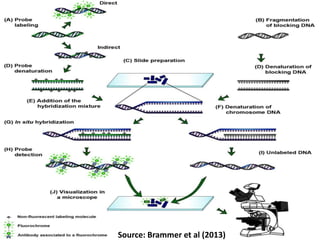
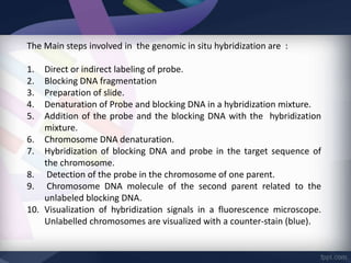
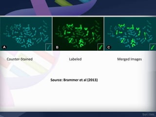
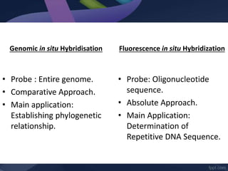
•“Genomic in situ hybridization (GISH) is a cytogenetic technique
that allows one to radiolabel parts of genome within the cells.”
•GISH was mainly developed for the animal hybrid cell lines (1986)
and later used for the plants at Plant Breeding Institute, Cambridge
(1987), where this technique got its name.
•The GISH is quick, sensitive, accurate, informative and a
comparative approach rather than absolute one.
• GISH technique is an advancement in the fluorescence in situ
hybridization (FISH) technique.
-
•The technique involves the extraction and sub-
sequentially radio labeling of whole DNA of one organism
and to use as a probe to target the genome of another
organism.
•The parts of genome that are sufficiently similar to the
probe hybridize to form a probe-target complex which is
now labeled.
•The remaining unhybridized parts of genome can be
further stained to view them.
Source: Brammer et al (2013)
The Main steps involved in the genomic in situ hybridization are :
1. Direct or indirect labeling of probe.
2. Blocking DNA fragmentation
3. Preparation of slide.
4. Denaturation of Probe and blocking DNA in a hybridization mixture.
5. Addition of the probe and the blocking DNA with the hybridization
mixture.
6. Chromosome DNA denaturation.
7. Hybridization of blocking DNA and probe in the target sequence of
the chromosome.
8. Detection of the probe in the chromosome of one parent.
9. Chromosome DNA molecule of the second parent related to the
unlabeled blocking DNA.
10. Visualization of hybridization signals in a fluorescence microscope.
Unlabelled chromosomes are visualized with a counter-stain (blue).
Counter-Stained Labeled Merged Images
Source: Brammer et al (2013)
Figure: Observed under Fluorescence Microscope
Source: Brammer et al (2013)
Counter-stained (Blue) Labeled Merged Image (A+B)
Genomic in situ Hybridisation
• Radio labeling of genome
• Quick results
• Fluorescence microscope is
used
• Site Specific
3D Genome Sequencing
• Restriction cleavage of
genome
• Time consuming technique
• Hi-C matrix and models are
used
• Multiple step technique
Genomic in situ Hybridisation
• Probe : Entire genome.
• Comparative Approach.
• Main application:
Establishing phylogenetic
relationship.
Fluorescence in situ Hybridization
• Probe: Oligonucleotide
sequence.
• Absolute Approach.
• Main Application:
Determination of
Repetitive DNA Sequence.
The major application of GISH technique are as
follows:
1. Meiotic studies
2. Determination of phylogenetic relationship.
3. Determine the positions of translocation
breakpoints
4. Comparative genomic studies of malignant and
normal cells of an individual
5. Unknown genome identification
6. To identify the hybridized genome of crop varieties

Genomic in situ Hybridization

  • 1.
    Kuldeep Sharma B.Tech Biotechnology IVSEM Amity Institute of Biotechnology
  • 3.
    •“Genomic in situhybridization (GISH) is a cytogenetic technique that allows one to radiolabel parts of genome within the cells.” •GISH was mainly developed for the animal hybrid cell lines (1986) and later used for the plants at Plant Breeding Institute, Cambridge (1987), where this technique got its name. •The GISH is quick, sensitive, accurate, informative and a comparative approach rather than absolute one. • GISH technique is an advancement in the fluorescence in situ hybridization (FISH) technique.
  • 5.
    - •The technique involvesthe extraction and sub- sequentially radio labeling of whole DNA of one organism and to use as a probe to target the genome of another organism. •The parts of genome that are sufficiently similar to the probe hybridize to form a probe-target complex which is now labeled. •The remaining unhybridized parts of genome can be further stained to view them.
  • 7.
  • 8.
    The Main stepsinvolved in the genomic in situ hybridization are : 1. Direct or indirect labeling of probe. 2. Blocking DNA fragmentation 3. Preparation of slide. 4. Denaturation of Probe and blocking DNA in a hybridization mixture. 5. Addition of the probe and the blocking DNA with the hybridization mixture. 6. Chromosome DNA denaturation. 7. Hybridization of blocking DNA and probe in the target sequence of the chromosome. 8. Detection of the probe in the chromosome of one parent. 9. Chromosome DNA molecule of the second parent related to the unlabeled blocking DNA. 10. Visualization of hybridization signals in a fluorescence microscope. Unlabelled chromosomes are visualized with a counter-stain (blue).
  • 10.
    Counter-Stained Labeled MergedImages Source: Brammer et al (2013)
  • 11.
    Figure: Observed underFluorescence Microscope Source: Brammer et al (2013) Counter-stained (Blue) Labeled Merged Image (A+B)
  • 12.
    Genomic in situHybridisation • Radio labeling of genome • Quick results • Fluorescence microscope is used • Site Specific 3D Genome Sequencing • Restriction cleavage of genome • Time consuming technique • Hi-C matrix and models are used • Multiple step technique
  • 14.
    Genomic in situHybridisation • Probe : Entire genome. • Comparative Approach. • Main application: Establishing phylogenetic relationship. Fluorescence in situ Hybridization • Probe: Oligonucleotide sequence. • Absolute Approach. • Main Application: Determination of Repetitive DNA Sequence.
  • 16.
    The major applicationof GISH technique are as follows: 1. Meiotic studies 2. Determination of phylogenetic relationship. 3. Determine the positions of translocation breakpoints 4. Comparative genomic studies of malignant and normal cells of an individual 5. Unknown genome identification 6. To identify the hybridized genome of crop varieties