BEFORE INTERPRETATING THE
RADIOGRAPH
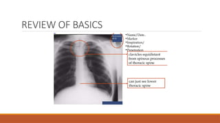
1)Patient identification details.
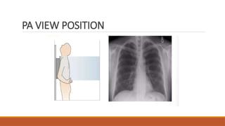
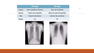
2) X-ray view PA or AP….
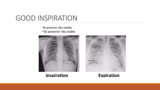
3) Breath : inspiration or expiration.
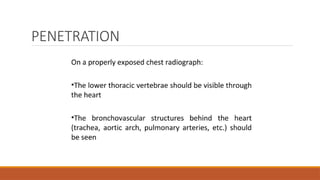
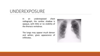
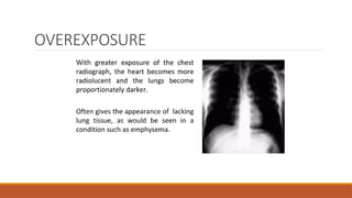
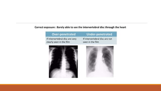
4) X-ray penetration : under or over penetrated
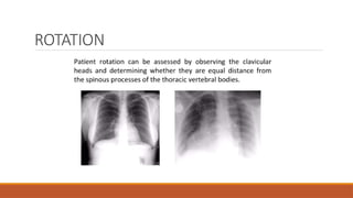
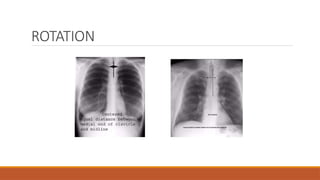
5) Rotation
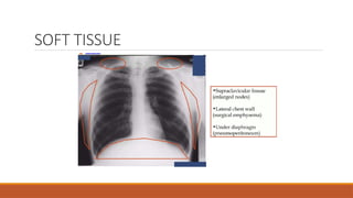
6) Extras
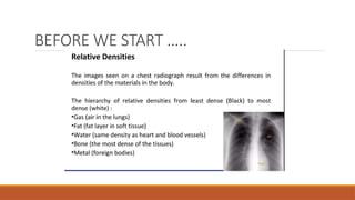
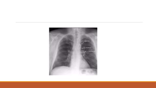
PLEURAL EFFUSION
patient ID:XYZ.
Projection: PA
Penetration: Adequate – vertebral bodies just visible behind heart
Inspiration: Limited - 5 anterior ribs visible Rotation:
The patient is rotated to the right
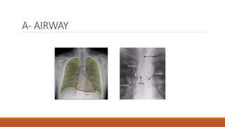
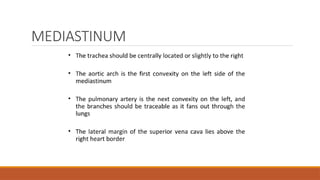
AIRWAY
The trachea is displaced slightly to the right –
this may be due to patient rotation or a mass effect.
BREATHING
There is complete, homogeneous opacification of the left hemithorax,
in keeping with a large pleural effusion.
The right lung is clear. The lungs are not hyperinflated.
51.
PLEURAL EFFUSION
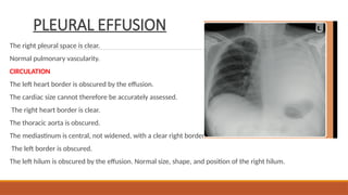
The rightpleural space is clear.
Normal pulmonary vascularity.
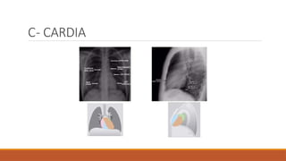
CIRCULATION
The left heart border is obscured by the effusion.
The cardiac size cannot therefore be accurately assessed.
The right heart border is clear.
The thoracic aorta is obscured.
The mediastinum is central, not widened, with a clear right border.
The left border is obscured.
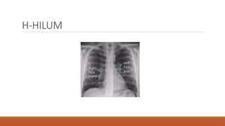
The left hilum is obscured by the effusion. Normal size, shape, and position of the right hilum.
52.
PLEURAL EFFUSION
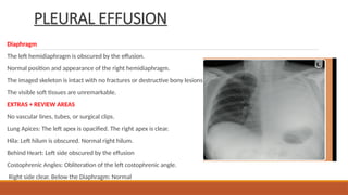
Diaphragm
The lefthemidiaphragm is obscured by the effusion.
Normal position and appearance of the right hemidiaphragm.
The imaged skeleton is intact with no fractures or destructive bony lesions visible.
The visible soft tissues are unremarkable.
EXTRAS + REVIEW AREAS
No vascular lines, tubes, or surgical clips.
Lung Apices: The left apex is opacified. The right apex is clear.
Hila: Left hilum is obscured. Normal right hilum.
Behind Heart: Left side obscured by the effusion
Costophrenic Angles: Obliteration of the left costophrenic angle.
Right side clear. Below the Diaphragm: Normal
53.
PLEURAL EFFUSION
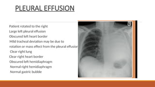
Patient rotatedto the right
Large left pleural effusion
Obscured left heart border
Mild tracheal deviation may be due to
rotation or mass effect from the pleural effusion
Clear right lung
Clear right heart border
Obscured left hemidiaphragm
Normal right hemidiaphragm
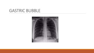
Normal gastric bubble