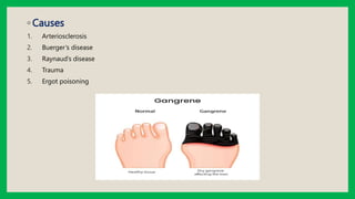
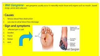
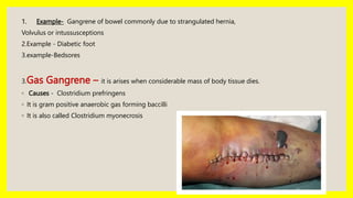
3. ◦INTRODUCTION
. Gangrene is also called as Edematous myonecrosis
It is the death of tissue caused by lack of blood Flow or necrosis
. The basic type of necrosis in GANGRENE is coagulative necrosis due to ischemia onto which
bacterial infection is superimposed
. Ischemia-it is the lack of blood supply to body part
◦ Definition- Gangrene is nothing but the death of tissue caused by infection or lack of blood
supply to body part.
4. ◦TYPES
1. Dry Gangrene
2. Wet Gangrene
3. Gas Gangrene
1. Dry Gangrene – it is usually seen in the distal part of limbs due to ischemia
. E.g - Dry gangrene in the toes and feet of old patient due to arteriosclerosis.
6. ◦ One the most important thing in dry gangrene is the affected part falls continuously there is no
need to remove it and no any surgery require
◦ Affected part in dry gangrene is dark black because liberation of hemoglobin which react with
hydrogen disulfide produced by bacteria resulting in formation of black iron sulfied.
◦ Sign and symptoms
1. Affected part is dry
2. Shrunken
3. Dark black
7. ◦ Wet Gangrene- wet gangrene usually occur in naturally moist tissue and organs such as mouth , bowel
,lungs ,cervix and vulva ect.
◦ Causes
1. Venous blood flow obstruction
2. Less due to atrial blood flow blockage
◦ Sign and symptoms
1. affected part is soft
2. Swollen
3. Putrid
4. Rotten
5. dark
8. 1. Example- Gangrene of bowel commonly due to strangulated hernia,
Volvulus or intussusceptions
2.Example - Diabetic foot
3.example-Bedsores
3.Gas Gangrene – it is arises when considerable mass of body tissue dies.
◦ Causes - Clostridium prefringens
◦ It is gram positive anaerobic gas forming baccilli
◦ It is also called Clostridium myonecrosis
9. ◦ It is usually occur in skeletal muscle and colon
◦Sign and symptoms
1. Grossly affected organ swollen
2. Edematous
3. Crepitus
4. Blisters with foul smelling discharges
5. Microscopically, bacterial colonies seen
10. ◦ Diagnostic evaluation
1.X-ray of blood vessels after injection of
dye Ateriogram
2.Blood test to rule out infection
3.CT- scan and MRI
4.Culture
5.Biopsy
6.Surgery
11. ◦ MANAGEMENT
◦ Pharmacological therapy
1. Penicillin Antibiotic
◦ Other broad spectrum antibiotics
1. Ampicillin plus sulbactum
2. Piperacillin plus tazobactum
◦ SUPPORTIVE CARE
1. i.v fluid
2. Anticoagulant
3. High protein diet
4. Oxygen therapy if needed
◦ SUGICAL MANAGEMENT
◦ Amputation
◦ Angioplasty