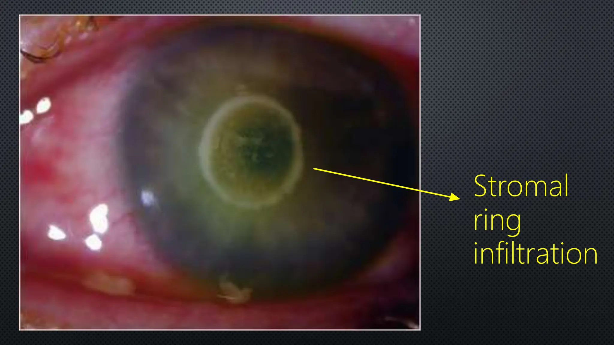
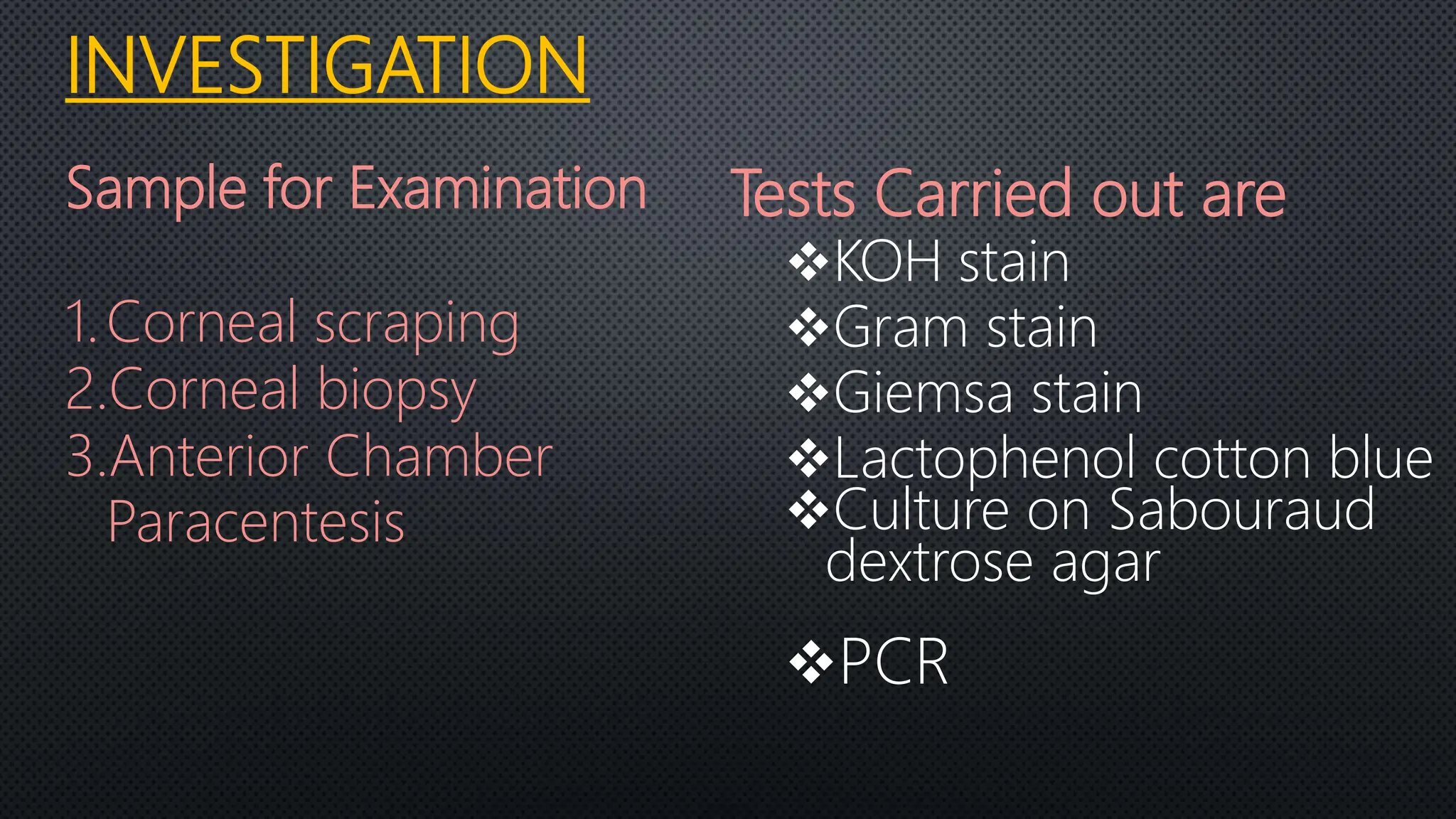
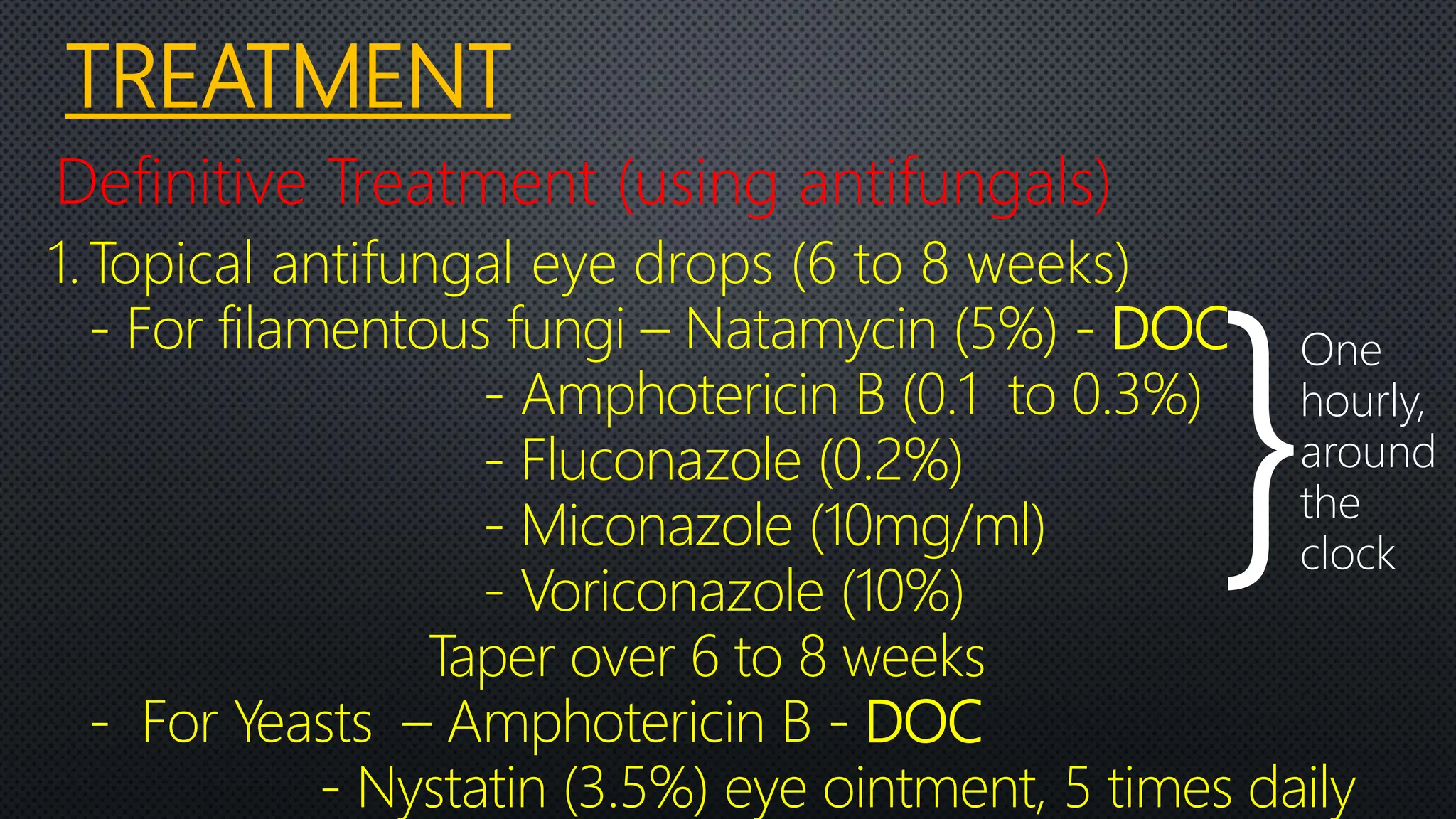
MYCOTIC / FUNGAL
CORNEAL ULCER
CORNEAL ULCER
“It is defined as discontinuation of corneal
epithelium associated necrosis of
surrounding tissue”
FUNGAL CORNEAL ULCER:
A fungal keratitis is an inflammation of
the cornea (called keratitis) that result
from infection by a fungal organism.
Keratomycosis is the Greek terminology
equivalent of fungal keratitis. It is the
fungal infection of the cornea.
PATHOLOGY OF CORNEAL ULCER
•
•
•
CAUSATIVE ORGANISM
Yeasts
 Candida albicans
 Cryptococcus
Filamentous
Aseptate
Rhizopus
Septate
I. Aspergilus
II. Curvularia
III. Fusarium
IV. Cladosporium
“FUNGI”
Dimprphic
 Histoplasma
 Coccidioides
 Blastomyces
ASEPTATE FUNGAL
HYPHAE (GRAM STAIN)
FUNGAL HYPHAE
(LPCB)
ASPERGILUS(SDA) FUSARIUM(SDA)
FUNGAL HYPHAE
(CALCOFLORITE)
MODE OF INFECTION
• Injury by vegetative material
- Seen in Field Workers
• Injury by Animal Tail
• Secondary fungal Ulcer
- Dry eyes,
- Herpetic keratopathy,
- Bullous Keratopathy,
- Post operative cases.
•
CLINICAL FEATURES
Hypopyon
Infiltration
Conjunctival
Hyperaemia
Lid swelling
• SYMPTOMS
*Pain, foreign body sensation – due to mechanical
effects of lids and chemical effects of toxins on exposed
nerve ending
*Watering of eyes – due to reflex lacrimation
*Photophobia – intolerance to light due to stimulation
of nerve ending
*Blurred vision – due to corneal haze
*Redness – congession of circumcorneal vessels
SIGNS
Corneal ulcer - Greyish white
- Dry looking
- Elevated rolled out margin
Delicate Feathery finger like extentions
present into the stroma under intact
epithelium
Sterile immune ring (Stromal ring
infiltration)
Satellite lesion
Endothelial plaque (Fibrin and Leucocytes)
Hypopyon - Non sterile
- Large
Perforation (Rare)
Corneal vascularisation
Feathery
margin
Elevated
surface
Stromal
ring
infiltration
DIAGNOSIS
Clinical diagnosis
• Typical Clinical Manifestation
• Chronic Ulcers
• Confocal Microscopic Examination
Fungal
hyphae in
confocal
microscope
INVESTIGATION
Sample for Examination
1.Corneal scraping
2.Corneal biopsy
3.Anterior Chamber
Paracentesis
Tests Carried out are
KOH stain
Gram stain
Giemsa stain
Lactophenol cotton blue
Culture on Sabouraud
dextrose agar
PCR
TREATMENT
Definitive Treatment (using antifungals)
1.Topical antifungal eye drops (6 to 8 weeks)
- For filamentous fungi – Natamycin (5%) - DOC
- Amphotericin B (0.1 to 0.3%)
- Fluconazole (0.2%)
- Miconazole (10mg/ml)
- Voriconazole (10%)
Taper over 6 to 8 weeks
- For Yeasts – Amphotericin B - DOC
- Nystatin (3.5%) eye ointment, 5 times daily
}
One
hourly,
around
the
clock
2. Intracameral and Intracorneal/Intrastromal
- Voriconazole (in case of anterior
chamber involvement.)
3. Systemic antifungal drugs
(in severe cases of deeper fungal keratitis)
- Tab. Fluconazole
- Tab. Ketoconazole
- Tab. Voriconazole
For 3 weeks
B. Adjunctive/concurrent therapy
(Non specific treatment)
- Cycloplegic drugs
- Systemic analgesics and anti-inflamatory
- Vitamins (A,B-complex and C)
C. Therapeutic Penetrating Keratoplasty
For non responsive cases
Fungal Corneal Ulcer Ophthalmology PPT.pptx

Fungal Corneal Ulcer Ophthalmology PPT.pptx