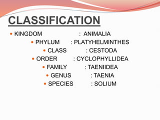
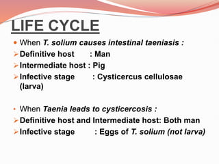
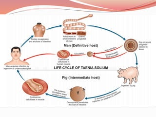
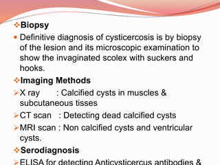
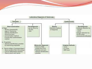
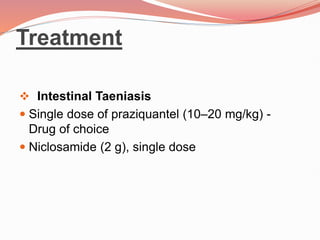
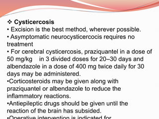
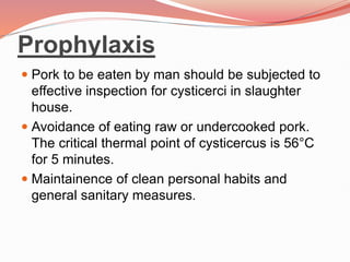
💕SONAM KUMAR💕Premium Call Girls Jaipur ↘️9257276172 ↙️One Night Stand With Lo...
TAENIA SOLIUM.pptx
1.
2. CLASSIFICATION
KINGDOM : ANIMALIA
PHYLUM : PLATYHELMINTHES
CLASS : CESTODA
ORDER : CYCLOPHYLLIDEA
FAMILY : TAENIIDEA
GENUS : TAENIA
SPECIES : SOLIUM
3. HISTORY
T. saginata has been known as an intestinal
parasite of man from very ancient times.
But it was only in 1782 when Goeze
differentiated it from the pork tapeworm,T.
solium.
Its life cycle was elucidated when Leuckart,
in1861, first experimentally demonstrated that
cattle serve as the intermediate host for the
worm
The name Taenia is derived from the Greek word
meaning tape or band. It was originally used to
4. DISTRIBUTION
T. solium is also worldwide in distribution except
in the countries and communities, which
proscribe pork as taboo.
HABITAT
The adult worms of T. solium live in the human
small intestine, commonly in the jejunum
5. MORPHOLOGY
Length : 2–3 m
Scolex : Small and globular.
Rostellum and hooks are
present.
. Suckers not pigmented.
Neck : Short
Proglottids : Below 1,000
Gravid segment : 12 mm × 6 mm
Expulsion : Expelled passively in
chains of 5 or 6
Uterus : Lateral branches 5–10 on
6. Vagina : Absent
Accessary lobe of ovary : Present
Testes : 150–200 follicles
Genital pore : present, alternating
between the . . right
and left sides of adjacent .
segments.
7. EGGS
Spherical, measuring 30–40 μm in diameter.
It has a thin hyaline embryonic membrane
around it which soon disappears after release.
The inner embryophore is radially striated and is
yellow-brown due to bile staining.
In the center is a fully-developed embryo
(oncosphere) with 3 pairs of hooklets
(hexacanth embryo).
Do not float in saturated salt solution.
The eggs of T. solium are infective to pigs and
humans.
8. LARVA : Cysticercus
cellulosae
The larval stage of Taenia is called as
cysticercus
Cysticercus cellulosae is the larva of T. solium.
The infective form of the parasite.
It can develop in various organs of pig as well as
in man.
The cysticercus cellulosae or ‘bladder worm’ is
ovoid opalescent milky-white, measuring 8–10
mm in breadth and 5 mm in length.
The scolex of the larva, with its suckers, lies
invaginated within the bladder and can be seen
9. LIFE CYCLE
When T. solium causes intestinal taeniasis :
Definitive host : Man
Intermediate host : Pig
Infective stage : Cysticercus cellulosae
(larva)
• When Taenia leads to cysticercosis :
Definitive host and Intermediate host: Both man
Infective stage : Eggs of T. solium (not larva)
10.
11. Pathogenicity and Clinical
Features
Intestinal Taeniasis
Caused by both T. saginata and T. solium.
When the infection is symptomatic, vague
abdominal discomfort, indigestion, nausea,
diarrhea, and weightloss may be present.
Occasional cases of acute intestinal
obstruction, acute appendicitis, and
pancreatitis have also been reported.
12. Cysticercosis
• It is caused by larval stage (cysticecus
cellulosae) of T. solium.
Cysticercus cellulosae may be solitary or more
often multiple.
Any organ or tissue may be involved, the most
common being subcutaneous tissues and
muscles. It may also affect the eyes, brain, and
less often other organs.
The cysticercus is surrounded by a fibrous
capsule except in the eye and ventricles of the
brain.
The larvae evoke a cellular reaction starting with
13. The clinical features depend on the site affected
Subcutaneous nodules are mostly
asymptomatic
€
Muscular cysticerosis may cause acute
myositis
Neurocysticerosis (cysticercosis of brain) is
the most common and most serious form of
cysticercosis.
Clinical features of neurocysticercosis are
epilepsy, increased intracranial
tension,hydrocephalus, psychiatric disturbances,
meningoencephalitis, behavioral disorders and
visual disturbances.
14. Laboratory Diagnosis
Stool Examination
Eggs
• Microscopic examination of feces
Formol ether sedimentation method of stool
concentration is useful.
cellophane swab method (NIH Swab)
Species identification cannot be made from the
eggs.
Proglottids
• Species identification can be done
When the gravid proglotlid pressed between 2
slides, branching can be made out (under 13
15. Scolex
Definitive diagnosis can also be established by
demonstration of armed scolex in case of T.
solium.
Detection of Taenia Antigen in feces
Antigen capture ELISA using polyclonal antisera
against Taenia is employed to detect
coproantigen in feces and is more sensitive than
microscopy.
The drawback of the test is that it cannot
differentiate between T. saginata and T. solium
16. Serodiagnosis
Specific antibodies in serum can be
demonstrated by ELISA, indirect
immunofluroscence test and indirect
hemagglutination (IHA) test.
Molecular Diagnosis
Both DNA probes and polymerase chain reaction
(PCR) technique are used to detect and
differentiate between eggs and proglottids of T.
saginata and T. solium. It can also differentiate
between their subspecies.
17. Biopsy
Definitive diagnosis of cysticercosis is by biopsy
of the lesion and its microscopic examination to
show the invaginated scolex with suckers and
hooks.
Imaging Methods
X ray : Calcified cysts in muscles &
subcutaneous tisses
CT scan : Detecting dead calcified cysts
MRI scan : Non calcified cysts and ventricular
cysts.
Serodiagnosis
20. Cysticercosis
• Excision is the best method, wherever possible.
• Asymptomatic neurocysticercocis requires no
treatment
• For cerebral cysticercosis, praziquantel in a dose of
50 mg/kg in 3 divided doses for 20–30 days and
albendazole in a dose of 400 mg twice daily for 30
days may be administered.
•Corticosteroids may be given along with
praziquantel or albendazole to reduce the
inflammatory reactions.
•Antiepileptic drugs should be given until the
reaction of the brain has subsided.
21. Prophylaxis
Pork to be eaten by man should be subjected to
effective inspection for cysticerci in slaughter
house.
Avoidance of eating raw or undercooked pork.
The critical thermal point of cysticercus is 56°C
for 5 minutes.
Maintainence of clean personal habits and
general sanitary measures.