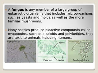
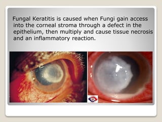
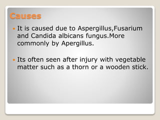
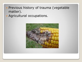
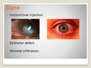
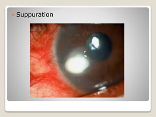
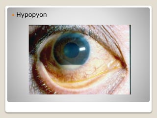
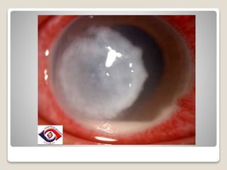
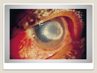
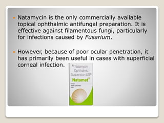
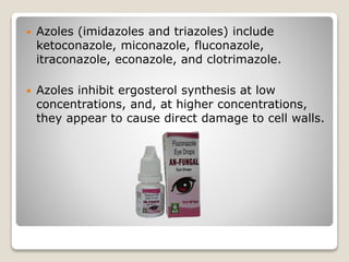
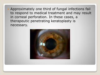
FUNGAL KERATITIS
By:Sina Motallebi
M.Optom(2nd Sem)
Amity Medical School
 A fungus is any member of a large group of
eukaryotic organisms that includes microorganisms
such as yeasts and molds,as well as the more
familiar mushrooms.
 Many species produce bioactive compounds called
mycotoxins, such as alkaloids and polyketides, that
are toxic to animals including humans.
Fusarium Candida albicans
Fungal Keratitis is caused when Fungi gain access
into the corneal stroma through a defect in the
epithelium, then multiply and cause tissue necrosis
and an inflammatory reaction.
Causes
 It is caused due to Aspergillus,Fusarium
and Candida albicans fungus.More
commonly by Apergillus.
 Its often seen after injury with vegetable
matter such as a thorn or a wooden stick.
Risk factors
 Trauma (eg, contact lenses, foreign
body).
 Topical corticosteroid use.
 Corneal surgery such as penetrating
keratoplasty, clear cornea (sutureless)
cataract surgery, photorefractive
keratectomy, or laser in situ
keratomileusis (LASIK)
 Previous history of trauma (vegetable
matter).
 Agricultural occupations.
Workup
Laboratory:
 Corneal scrapings are obtained using a
platinum spatula, surgical blade, or
calcium alginate swab inoculated on
Sabouraud agar plates, and then
maintained at 25°C to enhance fungal
growth.
Symptoms
 Foreign body sensation.
 Increasing eye pain or discomfort.
 Sudden blurry vision.
 Unusual redness of the eye.
 Excessive tearing and discharge from the
eye.
 Increased light sensitivity.
Signs
 Conjunctival injection.
 Epithelial defect.
 Stromal infiltration.
 Suppuration
 Hypopyon
Presenting clinical features:
 Fine or coarse granular infiltrate within the epithelium and
anterior stroma.
 Gray-white color, dry, and rough corneal surface that may
appear elevated.
 Typical irregular feathery-edged infiltrate.
 White ring in the cornea and satellite lesions near the edge
of the primary focus of the infection.
Ophthalmic imaging:
 If clinical evidence or suspicion of
posterior segment involvement exists,
ophthalmic B-scan ultrasound may be
necessary to rule out concurrent fungal
endophthalmitis.
Other tests:
 Immunofluorescence staining.
 Electron microscopy.
 Confocal microscopy- It may help in correctly
diagnosing early stages of fungal keratitis and in
monitoring disease progress at the edges and
depth.
Treatment
Medical care:
Antifungal agents are classified into the following groups:
 Polyenes include natamycin, nystatin, and
amphotericin B. Polyenes disrupt the cell by
binding to fungal cell wall.
 Amphotericin B is the drug of choice for
treatment of fungal keratitis caused by Candida.
 Natamycin is the only commercially available
topical ophthalmic antifungal preparation. It is
effective against filamentous fungi, particularly
for infections caused by Fusarium.
 However, because of poor ocular penetration, it
has primarily been useful in cases with superficial
corneal infection.
 Azoles (imidazoles and triazoles) include
ketoconazole, miconazole, fluconazole,
itraconazole, econazole, and clotrimazole.
 Azoles inhibit ergosterol synthesis at low
concentrations, and, at higher concentrations,
they appear to cause direct damage to cell walls.
Biomicroscopic signs to assess he
efficacy of the medications being
used:
 Blunting of the perimeters of the infiltrate.
 Reduction of the density of the
suppuration.
 Reduction in cellular infiltrate and edema
in the surrounding stroma.
 Reduction in anterior chamber
inflammation.
 Progressive reepithelization.
 Loss of the feathery perimeter of the
stromal inflammation.
Surgical care:
 Patients who do not respond to medical
treatment of topical and oral antifungal
medications usually require surgical intervention,
including corneal transplantation.
 Frequent corneal debridement with a spatula is
helpful; it debulks fungal organisms and
epithelium and enhances penetration of the
topical antifungal agent.
 Approximately one third of fungal infections fail
to respond to medical treatment and may result
in corneal perforation. In these cases, a
therapeutic penetrating keratoplasty is
necessary.
 The main goals of surgery are to control the
infection and to maintain the integrity of the
globe.
 Topical antifungal therapy, in addition to systemic
fluconazole or ketoconazole, should be continued
following penetrating keratoplasty.
References
 Vaddavalli PK, Garg P, Sharma S, Sangwan VS, Rao GN,
Thomas R. Role of confocal microscopy in the diagnosis of
fungal and acanthamoeba keratitis. Ophthalmology. Jan
2011;118(1):29-35.
 Dunlop AA, Wright ED, Howlader SA, Nazrul I, Husain R,
McClellan K. Suppurative corneal ulceration in Bangladesh.
A study of 142 cases examining the microbiological
diagnosis, clinical and epidemiological features of bacterial
and fungal keratitis. Aust N Z J Ophthalmol. May
1994;22(2):105-10.
 Kanski J Jack;Clinical ophthalmology a systemic
approach;6th edition;butterworth and heinnmen.
 Ramanjit Sihota,Radhika Tandon(eds);Parson’s Diseases of
the Eye;15;199-200.
THANK YOU

Fungal keratitis

  • 1.
  • 2.
     A fungusis any member of a large group of eukaryotic organisms that includes microorganisms such as yeasts and molds,as well as the more familiar mushrooms.  Many species produce bioactive compounds called mycotoxins, such as alkaloids and polyketides, that are toxic to animals including humans. Fusarium Candida albicans
  • 3.
    Fungal Keratitis iscaused when Fungi gain access into the corneal stroma through a defect in the epithelium, then multiply and cause tissue necrosis and an inflammatory reaction.
  • 4.
    Causes  It iscaused due to Aspergillus,Fusarium and Candida albicans fungus.More commonly by Apergillus.  Its often seen after injury with vegetable matter such as a thorn or a wooden stick.
  • 5.
    Risk factors  Trauma(eg, contact lenses, foreign body).  Topical corticosteroid use.  Corneal surgery such as penetrating keratoplasty, clear cornea (sutureless) cataract surgery, photorefractive keratectomy, or laser in situ keratomileusis (LASIK)
  • 6.
     Previous historyof trauma (vegetable matter).  Agricultural occupations.
  • 7.
    Workup Laboratory:  Corneal scrapingsare obtained using a platinum spatula, surgical blade, or calcium alginate swab inoculated on Sabouraud agar plates, and then maintained at 25°C to enhance fungal growth.
  • 8.
    Symptoms  Foreign bodysensation.  Increasing eye pain or discomfort.  Sudden blurry vision.  Unusual redness of the eye.
  • 9.
     Excessive tearingand discharge from the eye.  Increased light sensitivity.
  • 10.
    Signs  Conjunctival injection. Epithelial defect.  Stromal infiltration.
  • 11.
  • 12.
  • 13.
    Presenting clinical features: Fine or coarse granular infiltrate within the epithelium and anterior stroma.  Gray-white color, dry, and rough corneal surface that may appear elevated.  Typical irregular feathery-edged infiltrate.  White ring in the cornea and satellite lesions near the edge of the primary focus of the infection.
  • 16.
    Ophthalmic imaging:  Ifclinical evidence or suspicion of posterior segment involvement exists, ophthalmic B-scan ultrasound may be necessary to rule out concurrent fungal endophthalmitis.
  • 17.
    Other tests:  Immunofluorescencestaining.  Electron microscopy.  Confocal microscopy- It may help in correctly diagnosing early stages of fungal keratitis and in monitoring disease progress at the edges and depth.
  • 18.
    Treatment Medical care: Antifungal agentsare classified into the following groups:  Polyenes include natamycin, nystatin, and amphotericin B. Polyenes disrupt the cell by binding to fungal cell wall.  Amphotericin B is the drug of choice for treatment of fungal keratitis caused by Candida.
  • 19.
     Natamycin isthe only commercially available topical ophthalmic antifungal preparation. It is effective against filamentous fungi, particularly for infections caused by Fusarium.  However, because of poor ocular penetration, it has primarily been useful in cases with superficial corneal infection.
  • 20.
     Azoles (imidazolesand triazoles) include ketoconazole, miconazole, fluconazole, itraconazole, econazole, and clotrimazole.  Azoles inhibit ergosterol synthesis at low concentrations, and, at higher concentrations, they appear to cause direct damage to cell walls.
  • 21.
    Biomicroscopic signs toassess he efficacy of the medications being used:  Blunting of the perimeters of the infiltrate.  Reduction of the density of the suppuration.  Reduction in cellular infiltrate and edema in the surrounding stroma.
  • 22.
     Reduction inanterior chamber inflammation.  Progressive reepithelization.  Loss of the feathery perimeter of the stromal inflammation.
  • 23.
    Surgical care:  Patientswho do not respond to medical treatment of topical and oral antifungal medications usually require surgical intervention, including corneal transplantation.  Frequent corneal debridement with a spatula is helpful; it debulks fungal organisms and epithelium and enhances penetration of the topical antifungal agent.
  • 24.
     Approximately onethird of fungal infections fail to respond to medical treatment and may result in corneal perforation. In these cases, a therapeutic penetrating keratoplasty is necessary.
  • 25.
     The maingoals of surgery are to control the infection and to maintain the integrity of the globe.  Topical antifungal therapy, in addition to systemic fluconazole or ketoconazole, should be continued following penetrating keratoplasty.
  • 26.
    References  Vaddavalli PK,Garg P, Sharma S, Sangwan VS, Rao GN, Thomas R. Role of confocal microscopy in the diagnosis of fungal and acanthamoeba keratitis. Ophthalmology. Jan 2011;118(1):29-35.  Dunlop AA, Wright ED, Howlader SA, Nazrul I, Husain R, McClellan K. Suppurative corneal ulceration in Bangladesh. A study of 142 cases examining the microbiological diagnosis, clinical and epidemiological features of bacterial and fungal keratitis. Aust N Z J Ophthalmol. May 1994;22(2):105-10.  Kanski J Jack;Clinical ophthalmology a systemic approach;6th edition;butterworth and heinnmen.
  • 27.
     Ramanjit Sihota,RadhikaTandon(eds);Parson’s Diseases of the Eye;15;199-200.
  • 28.