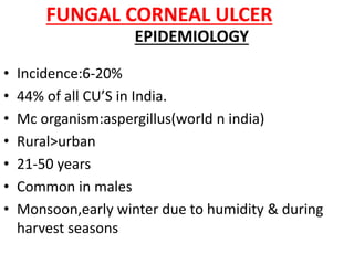
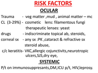
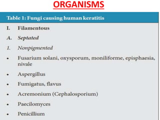
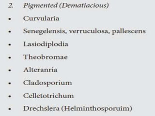
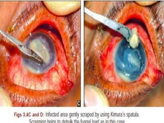
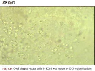
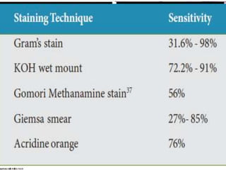
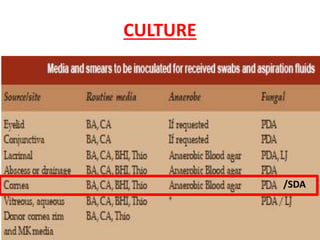
FUNGAL
CORNEAL ULCER
FUNGAL CORNEAL ULCER
• Incidence:6-20%
• 44% of all CU’S in India.
• Mc organism:aspergillus(world n india)
• Rural>urban
• 21-50 years
• Common in males
• Monsoon,early winter due to humidity & during
harvest seasons
EPIDEMIOLOGY
RISK FACTORS
OCULAR
Trauma - veg matter ,mud , animal matter – mc
CL (3-29%) - cosmetic lens: filamentous fungi
therapeutic lenses: yeast
drugs - indiscriminate topical ab, steroids,
corneal sx - any sx :PK ,cataract & refractive sx
steroid abuse,
c/c keratitis- VKC,allergic cojunctivits,neurotropic
ulcers,SJS,dry eye,
SYSTEMIC
P/t on immunosuppresants,DM,ICU p/t, HIV,leprosy.
ORGANISMS
Fusarium : S.India
aspergillus : N.India
PATHOGENESIS
• Fungi are saprophytic pathogenic organisms.
• Saprophytic :obtain their nutrients from decaying
organic matter,
• pathogenic :feed on living cells.
• Rarely cause infection in human cornea.
• gain access into the corneal stroma through epithelial
defectproliferate tissue necrosis host
inflammatory reaction. penetrate intact
Descemet’s membrane.
• when entered into AC/iris, eradication is difficult as
Blood-borne growth inhibiting factors may not reach
the avasculr cornea, AC & sclera, fungi continue to
multiply and persist despite treatment.
• basis for the vasculoplasty procedure
C/F
• SYMPTOMS (RELATIVELY LESS)
• insidious onset
• nonspecific symptoms
• duration : 5-10 days
• Fb sensation & Slow onset of pain
• decreased vision if on visual axis.
• SIGNS
conjunctival injection
Epithelial defect
AC reaction
General signs
• hyphate/branching ulcer
• Irregular/feathery margins(70%)with base filled with soft creamy
raised exudate
• margins: elevated
• Dry/ rough texture.
• Satellite lesions (10%)
• Fixed, unsterile,thick hypopyon.(66%)
• Grey white /yellow white infiltrate
• Weesley immune ring(rare):due to immune complex deposition
• Posterior corneal abscess
• Endothelial plaque
SPECIFIC FEATURES:
• demetiaceous fungi:
brown pigments in curvularia due to melanin
indicate superficial infection with low virulence &
inflammation.
• Fusarium:solani sp:
severe case with deep extension & perforation in 1
week
• aspergillus:
less severe,no rapid progression
• Candida:
keratitis resembling bacteria(discrete margins,epithelial
defect,slow progression,pre existing corneal d/s seen at
junction of superior & inferior corneal junction)
• Yeast
A “collar button” configuration is typical of the
keratitis associated with a small ulceration and an
expanding discreet stromal infiltrate
INVESTIGATIONS
1. Scraping
• aim :to debulk the organisms,
to diagnose the organism
to decrease the barrier for entry of antifungal
procedure
– aseptic precautions
– topical anesthesia
– leading edge & base debrided with spatula,sx blade,
diamond tipped motorized burr.
2.PARACENTESIS
• deep stromal lesions with outpouchings into AC.
• done with 2 cc 26 G tuberculin syringe.
3. KERATECTOMY /BIOPSY
• If corneal scrapings for smears and cultures are negative
• procedure
• performed in the minor OT / slit lamp
• under topical /eyelid and retrobulbar anesthesia.
• a round 2 to 3mm sterile disposable dermatologic trephine is
used 4 partial thickness trephination to take both clinically
infected area & the adjacent clear cornea.
• Care is taken to avoid the visual axis, if possible.
• The base is then undermined with a surgical blade to
complete the lamellar keratectomy.
• superior to corneal scraping for the isolation of the fungal
organisms.
LABORATORY DIAGNOSIS
• direct microscopy
• fungal cultures
• Polymerase chain reaction (PCR)
• Confocal microscopy
• DIRECT MICROSCOPY
• uses KOH wet mount preparation and smears,
• stained by Gram and Giemsa stain.
NOCARDIA : SLENDER THIN FILAMENTS
STAINING
• INTERPRETATION
• filamentous fungi:variable response
• co existing bacterial infection identified.
– GRAM+ve :
– thick peptidoglycan cell wall more 1 dye penetration.
– appears violet.
– GRAM –VE:
– appears pink.
• yeast:gram + ve oval/round shape
• Nocardia:thin gram +ve branched beaded filament.
CULTURE
/SDA
• SDA :should contain 50 micrograms /ml gentamicin
without cycloheximide as it inhibits saprophytic
fungi.
• incubated at 25-30 in regular incubator
• Thioglycollate broth
• inoculated for growth of anaerobic bacteria at 35⁰ -
37 ⁰ C.
• A definitive diagnosis of fungal keratitis is made if
1. Corneal scrapings reveal fungal elements in smears,
2. Fungus grows in more than one medium in the
absence of fungus in smears,
3. Fungus grows on a single medium in the presence
of fungus in smears,
4. Confluent growth of fungus appears at the
inoculated site on a single solid medium.
• incubated for 10 days to facilitate sporulation.
• +ve test yielded : 3 days (83%) ,>1 wk(97%).
• no growth :in 1 wk & reported in 3 wks
• identification based on its macroscopic and
microscopic features.
• MICROSCOPIC EXAMINATION METHODS
• direct examination of a portion of slide
• adhesive tape technique
• slide culture technique
POLYMERASE CHAIN REACTION
• newer method
• equivalent to culture
• only needs 4 hrs ,but cannot replace culture as false
+ve results are possible.
• indications:
• signs & symptoms of infection but no def diagnosis can
b made.
• not responding to Rx.
• h/o doesot match with clinical presentation.
• procedure;
• DNA extraction
• DNA amplification
• examination using agarose gel electrophoresis.
CONFOCAL MICROSCOPY
• imaging technique which allows the optical section of
any material with lateral n axial spatial
resolution,better contrast.
• early identification of hyphal elements & yeast is
possible.
• invivo scanning slit :establishes the diagnosis
:demonstrating non-responsiveness
to Rx( load of fungal filaments).
disadv
• low reproducibility,lack of distinct morphology to
pathogens,limited resolution,microscope or tissue may
move.
TREATMENT
1. ANTIFUNGALS
classification
• POLYENES:binds cell wall & alters cell permeability
natamycin
amphotericin b
• AZOLES: inhibit ergosterol
econazole,clotrimazole,imidazole,voriconazole,ketoconaz
ole
• ECHINOCANDINS:acts on 1,3-b D glucan synthase
cuspofungin,micafungin,anidulafungin
• Indicated only when scraping /culture report : +ve.
• Empirical treatment shud not b given.
• Debridement of epithelium helps in penetration of drug.
• Topical :initially -5% natamycin hrly :day time n 2 hrly: nite
:+ FQ 4 2⁰ bacterial.
• Bd eye checkups under s/l
• resolving  give natamycin 2 hrly ->2 wks t
• worsening topical ampho B 0.15%/fluconazole 2% is
given.
• candida I ampho B 0.15%/ fluconazole 0.3% 1st
choice.
• Patomycetes miconazole 1st choice
• echinocandins,ampho:not effective against fusarium.
•Nata 5% = econazole 1% = voriconazole 0.5 µg/ml.
• VORICONAZOLE
• derived from Fluconazole
• wider spectrum of activity against Candida, Aspergillus and
Fusarium.
• exerts its effect from inhibition of cytochrome P450-
dependant 14 alpha sterol demethylase, an enzyme involved
in the ergosterol biosynthetic pathway.
• INTRACAMERAL THERAPY
• In severe keratomycosis not responding to topical natamycin.
• It ensures adequate drug delivery into AC and avoid surgical
intervention in the acute stage of the disease.
• performed under strict aseptic conditions.
• If the infection involves the anterior capsule of the lens, care
should be taken to avoid injury to the lens.
• 5 μg ampho B in 0.1 mL 5% dextrose through a paracentesis.
• Injections repeated in case of inadequate response
INTRACORNEAL THERAPY
• for non healing fungal corneal ulcers
• Amphotericin B injection 5-7.5 μg , given near to stromal site of growth.
• raise the local concentration of the antifungal agent enough to be effective
in the eradication of the deep corneal infection  total elimination
• repeated after 48 to 72 hours
SUB CONJUNCTIVAL THERAPY
• not routinely used .s/ctoxicity and the intense pain.
• Miconazole : least toxic & best tolerated Rx(5 to 10 mg of 10mg/ml
suspension)
SIGNS OF IMPROVEMENT
• Decreased pain
• Decreased size of infiltrate.
• Disappeared satellite lesions
• Rounding of feathery margins
DURATION OF TREATMENT :
• 4-6 WKS only due to toxicity
DRUG INTERACTIONS
• Synergism :
• amphotericin B +flucytosine, (Candida )
• natamycin + ketoconazole (Aspergillus ).
• antagonism
• amphotericin B and imidazoles
• RESISTANCE
• rare and generally occurs in systemic mycoses.
Competition for volume in the pre corneal tear film
when using two topical anti-fungals.
SYSTEMIC Rx
• very large ulcers, severe deep keratitis, scleritis , endophthalmitis ,as
prophylactic treatment after PK.
• ketoconazole (oral), miconazole (intravenous) itraconazole (orally
200mg/day) and fluconazole.
• ketoconazole:600 mg per day.
• assess LFT every 2Wks after starting Ketoconazole.
• TOPICAL CORTICOSTEROIDS
• controversial.
• worsen the disease when given alone and adversely influence the
efficacy of natamycin, flucytosine and miconazole when given in
combination.
• Indication
• to decrease corneal inflammation and scarring .
• current regimen is to no steroids until at least 2 weeks of anti-fungal
treatment and clear clinical evidence of control of the infection
• used in conjunction with the topical anti-fungal and never without it.
• SURGICAL THERAPY
• DEBRIDEMENT
• Daily debridement with a spatula or blade is the
simplest form of surgical intervention .
• done at the slit lamp under topical anesthesia.
• performed every 24 to 48 hours
• debulks organisms and necrotic material and
enhance the penetration of the topical antifungal.
• BIOPSY
• for the diagnosis but also as a therapeutic
intervention.
• THERAPEUTIC KERATOPLASTY
• one third of fungal infections corneal perforations.
• goal:control the infection & maintain globe integrity.
• indications:
• When progression of the keratitis is noted
• If it p rogress to involve limbus or sclerascleritis,
endophthalmitis,recurrence .
• impending perforations, frank perforations > 2mm
• if there is no response to therapy.
• procedure :
• should leave a 1 to 1.5mm clear zone of clinically
uninvolved cornea to reduce the possibility of residual
fungal organisms peripheral to the trephination
• Interrupted sutures with slightly longer bites should be
used to avoid cheese wiring of the suture if the edge of
the recipient becomes involved .
• Irrigation of the AS to eliminate any organisms.
• the lens should be left untouched to prevent the spread
of infection in the posterior segment.
• if affected the intraocular structures including the iris,
lens, and vitreous may be excised.
• The specimens mcirobiology and pathology for
culture and fixed section examination.
• If involvement of intraocular structures or
endophthalmitis is suspected, an
• antifungal agents :amphotericin B (5μg/0.1ml) or
miconazole (25μg/0.1ml).
• Fungal hyphae usually lie parallel to the corneal surface
and lamellae.
• A vertical or perpendicular arrangement of fungal
hyphae in the corneal stroma: increased virulence and
in patients on topical corticosteroid therapy.
• PENETRATING KERATOPLASTY,
• topical antifungals to prevent recurrence of infection.
• + Postoperatively, systemic keatoconazole/ fluconazole.
• pathology :
• no organisms at the edge of the corneal specimen,
antifungals stopped after 2 weeks followup for
recurrences.
• microbiology +ve :more prolonged topical and systemic
Rx for 6 to 8 weeks
Fungal corneal ulcer

Fungal corneal ulcer

  • 1.
  • 2.
    FUNGAL CORNEAL ULCER •Incidence:6-20% • 44% of all CU’S in India. • Mc organism:aspergillus(world n india) • Rural>urban • 21-50 years • Common in males • Monsoon,early winter due to humidity & during harvest seasons EPIDEMIOLOGY
  • 3.
    RISK FACTORS OCULAR Trauma -veg matter ,mud , animal matter – mc CL (3-29%) - cosmetic lens: filamentous fungi therapeutic lenses: yeast drugs - indiscriminate topical ab, steroids, corneal sx - any sx :PK ,cataract & refractive sx steroid abuse, c/c keratitis- VKC,allergic cojunctivits,neurotropic ulcers,SJS,dry eye, SYSTEMIC P/t on immunosuppresants,DM,ICU p/t, HIV,leprosy.
  • 4.
  • 6.
  • 7.
    PATHOGENESIS • Fungi aresaprophytic pathogenic organisms. • Saprophytic :obtain their nutrients from decaying organic matter, • pathogenic :feed on living cells. • Rarely cause infection in human cornea. • gain access into the corneal stroma through epithelial defectproliferate tissue necrosis host inflammatory reaction. penetrate intact Descemet’s membrane. • when entered into AC/iris, eradication is difficult as Blood-borne growth inhibiting factors may not reach the avasculr cornea, AC & sclera, fungi continue to multiply and persist despite treatment. • basis for the vasculoplasty procedure
  • 8.
    C/F • SYMPTOMS (RELATIVELYLESS) • insidious onset • nonspecific symptoms • duration : 5-10 days • Fb sensation & Slow onset of pain • decreased vision if on visual axis.
  • 9.
    • SIGNS conjunctival injection Epithelialdefect AC reaction General signs • hyphate/branching ulcer • Irregular/feathery margins(70%)with base filled with soft creamy raised exudate • margins: elevated • Dry/ rough texture. • Satellite lesions (10%) • Fixed, unsterile,thick hypopyon.(66%) • Grey white /yellow white infiltrate • Weesley immune ring(rare):due to immune complex deposition • Posterior corneal abscess • Endothelial plaque
  • 10.
    SPECIFIC FEATURES: • demetiaceousfungi: brown pigments in curvularia due to melanin indicate superficial infection with low virulence & inflammation. • Fusarium:solani sp: severe case with deep extension & perforation in 1 week • aspergillus: less severe,no rapid progression • Candida: keratitis resembling bacteria(discrete margins,epithelial defect,slow progression,pre existing corneal d/s seen at junction of superior & inferior corneal junction)
  • 11.
    • Yeast A “collarbutton” configuration is typical of the keratitis associated with a small ulceration and an expanding discreet stromal infiltrate
  • 12.
    INVESTIGATIONS 1. Scraping • aim:to debulk the organisms, to diagnose the organism to decrease the barrier for entry of antifungal procedure – aseptic precautions – topical anesthesia – leading edge & base debrided with spatula,sx blade, diamond tipped motorized burr.
  • 13.
    2.PARACENTESIS • deep stromallesions with outpouchings into AC. • done with 2 cc 26 G tuberculin syringe. 3. KERATECTOMY /BIOPSY • If corneal scrapings for smears and cultures are negative • procedure • performed in the minor OT / slit lamp • under topical /eyelid and retrobulbar anesthesia. • a round 2 to 3mm sterile disposable dermatologic trephine is used 4 partial thickness trephination to take both clinically infected area & the adjacent clear cornea. • Care is taken to avoid the visual axis, if possible. • The base is then undermined with a surgical blade to complete the lamellar keratectomy. • superior to corneal scraping for the isolation of the fungal organisms.
  • 14.
    LABORATORY DIAGNOSIS • directmicroscopy • fungal cultures • Polymerase chain reaction (PCR) • Confocal microscopy • DIRECT MICROSCOPY • uses KOH wet mount preparation and smears, • stained by Gram and Giemsa stain. NOCARDIA : SLENDER THIN FILAMENTS
  • 15.
  • 16.
    • INTERPRETATION • filamentousfungi:variable response • co existing bacterial infection identified. – GRAM+ve : – thick peptidoglycan cell wall more 1 dye penetration. – appears violet. – GRAM –VE: – appears pink. • yeast:gram + ve oval/round shape • Nocardia:thin gram +ve branched beaded filament.
  • 17.
  • 18.
    • SDA :shouldcontain 50 micrograms /ml gentamicin without cycloheximide as it inhibits saprophytic fungi. • incubated at 25-30 in regular incubator • Thioglycollate broth • inoculated for growth of anaerobic bacteria at 35⁰ - 37 ⁰ C.
  • 19.
    • A definitivediagnosis of fungal keratitis is made if 1. Corneal scrapings reveal fungal elements in smears, 2. Fungus grows in more than one medium in the absence of fungus in smears, 3. Fungus grows on a single medium in the presence of fungus in smears, 4. Confluent growth of fungus appears at the inoculated site on a single solid medium. • incubated for 10 days to facilitate sporulation. • +ve test yielded : 3 days (83%) ,>1 wk(97%). • no growth :in 1 wk & reported in 3 wks • identification based on its macroscopic and microscopic features.
  • 20.
    • MICROSCOPIC EXAMINATIONMETHODS • direct examination of a portion of slide • adhesive tape technique • slide culture technique
  • 21.
    POLYMERASE CHAIN REACTION •newer method • equivalent to culture • only needs 4 hrs ,but cannot replace culture as false +ve results are possible. • indications: • signs & symptoms of infection but no def diagnosis can b made. • not responding to Rx. • h/o doesot match with clinical presentation. • procedure; • DNA extraction • DNA amplification • examination using agarose gel electrophoresis.
  • 22.
    CONFOCAL MICROSCOPY • imagingtechnique which allows the optical section of any material with lateral n axial spatial resolution,better contrast. • early identification of hyphal elements & yeast is possible. • invivo scanning slit :establishes the diagnosis :demonstrating non-responsiveness to Rx( load of fungal filaments). disadv • low reproducibility,lack of distinct morphology to pathogens,limited resolution,microscope or tissue may move.
  • 23.
    TREATMENT 1. ANTIFUNGALS classification • POLYENES:bindscell wall & alters cell permeability natamycin amphotericin b • AZOLES: inhibit ergosterol econazole,clotrimazole,imidazole,voriconazole,ketoconaz ole • ECHINOCANDINS:acts on 1,3-b D glucan synthase cuspofungin,micafungin,anidulafungin
  • 24.
    • Indicated onlywhen scraping /culture report : +ve. • Empirical treatment shud not b given. • Debridement of epithelium helps in penetration of drug. • Topical :initially -5% natamycin hrly :day time n 2 hrly: nite :+ FQ 4 2⁰ bacterial. • Bd eye checkups under s/l • resolving  give natamycin 2 hrly ->2 wks t • worsening topical ampho B 0.15%/fluconazole 2% is given. • candida I ampho B 0.15%/ fluconazole 0.3% 1st choice. • Patomycetes miconazole 1st choice • echinocandins,ampho:not effective against fusarium. •Nata 5% = econazole 1% = voriconazole 0.5 µg/ml.
  • 25.
    • VORICONAZOLE • derivedfrom Fluconazole • wider spectrum of activity against Candida, Aspergillus and Fusarium. • exerts its effect from inhibition of cytochrome P450- dependant 14 alpha sterol demethylase, an enzyme involved in the ergosterol biosynthetic pathway. • INTRACAMERAL THERAPY • In severe keratomycosis not responding to topical natamycin. • It ensures adequate drug delivery into AC and avoid surgical intervention in the acute stage of the disease. • performed under strict aseptic conditions. • If the infection involves the anterior capsule of the lens, care should be taken to avoid injury to the lens. • 5 μg ampho B in 0.1 mL 5% dextrose through a paracentesis. • Injections repeated in case of inadequate response
  • 26.
    INTRACORNEAL THERAPY • fornon healing fungal corneal ulcers • Amphotericin B injection 5-7.5 μg , given near to stromal site of growth. • raise the local concentration of the antifungal agent enough to be effective in the eradication of the deep corneal infection  total elimination • repeated after 48 to 72 hours SUB CONJUNCTIVAL THERAPY • not routinely used .s/ctoxicity and the intense pain. • Miconazole : least toxic & best tolerated Rx(5 to 10 mg of 10mg/ml suspension) SIGNS OF IMPROVEMENT • Decreased pain • Decreased size of infiltrate. • Disappeared satellite lesions • Rounding of feathery margins DURATION OF TREATMENT : • 4-6 WKS only due to toxicity
  • 27.
    DRUG INTERACTIONS • Synergism: • amphotericin B +flucytosine, (Candida ) • natamycin + ketoconazole (Aspergillus ). • antagonism • amphotericin B and imidazoles • RESISTANCE • rare and generally occurs in systemic mycoses. Competition for volume in the pre corneal tear film when using two topical anti-fungals.
  • 28.
    SYSTEMIC Rx • verylarge ulcers, severe deep keratitis, scleritis , endophthalmitis ,as prophylactic treatment after PK. • ketoconazole (oral), miconazole (intravenous) itraconazole (orally 200mg/day) and fluconazole. • ketoconazole:600 mg per day. • assess LFT every 2Wks after starting Ketoconazole. • TOPICAL CORTICOSTEROIDS • controversial. • worsen the disease when given alone and adversely influence the efficacy of natamycin, flucytosine and miconazole when given in combination. • Indication • to decrease corneal inflammation and scarring . • current regimen is to no steroids until at least 2 weeks of anti-fungal treatment and clear clinical evidence of control of the infection • used in conjunction with the topical anti-fungal and never without it.
  • 29.
    • SURGICAL THERAPY •DEBRIDEMENT • Daily debridement with a spatula or blade is the simplest form of surgical intervention . • done at the slit lamp under topical anesthesia. • performed every 24 to 48 hours • debulks organisms and necrotic material and enhance the penetration of the topical antifungal. • BIOPSY • for the diagnosis but also as a therapeutic intervention.
  • 30.
    • THERAPEUTIC KERATOPLASTY •one third of fungal infections corneal perforations. • goal:control the infection & maintain globe integrity. • indications: • When progression of the keratitis is noted • If it p rogress to involve limbus or sclerascleritis, endophthalmitis,recurrence . • impending perforations, frank perforations > 2mm • if there is no response to therapy. • procedure : • should leave a 1 to 1.5mm clear zone of clinically uninvolved cornea to reduce the possibility of residual fungal organisms peripheral to the trephination
  • 31.
    • Interrupted sutureswith slightly longer bites should be used to avoid cheese wiring of the suture if the edge of the recipient becomes involved . • Irrigation of the AS to eliminate any organisms. • the lens should be left untouched to prevent the spread of infection in the posterior segment. • if affected the intraocular structures including the iris, lens, and vitreous may be excised. • The specimens mcirobiology and pathology for culture and fixed section examination. • If involvement of intraocular structures or endophthalmitis is suspected, an • antifungal agents :amphotericin B (5μg/0.1ml) or miconazole (25μg/0.1ml).
  • 32.
    • Fungal hyphaeusually lie parallel to the corneal surface and lamellae. • A vertical or perpendicular arrangement of fungal hyphae in the corneal stroma: increased virulence and in patients on topical corticosteroid therapy. • PENETRATING KERATOPLASTY, • topical antifungals to prevent recurrence of infection. • + Postoperatively, systemic keatoconazole/ fluconazole. • pathology : • no organisms at the edge of the corneal specimen, antifungals stopped after 2 weeks followup for recurrences. • microbiology +ve :more prolonged topical and systemic Rx for 6 to 8 weeks