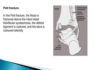
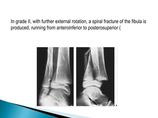
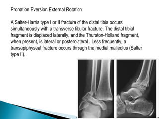
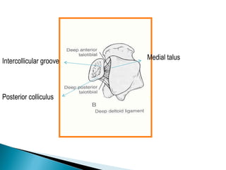
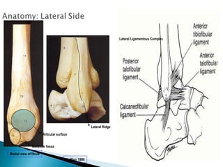
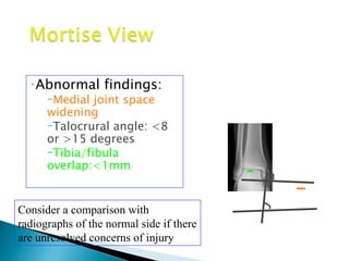
The document discusses the anatomy and biomechanics of the ankle joint. It describes the ankle joint as a three bone joint composed of the tibia, fibula, and talus. It notes that the talus articulates superiorly with the tibial plafond and posteriorly with the posterior malleolus of the tibia. The lateral articulation is with the malleolus of the fibula. The joint is saddle-shaped and wider anteriorly to accommodate dorsiflexion. Disruption of the ankle mortise can decrease contact area by 42%.
ANKLE FRACTURES
Pott’s fracture
A Pott’s fracture is a type of ankle fracture that is characterized by a break in one or more bony prominences on the sides of the ankle known as the malleoli.
Also known as Broken Ankle, Ankle Fracture and malleolar fracture.
Pott’s fracture often occurs in combination with other injuries such as a sprained ankle or other fractures of the foot, ankle or lower leg.
We all have good and bad thoughts from time to time and situation to situation. We are bombarded daily with spiraling thoughts(both negative and positive) creating all-consuming feel , making us difficult to manage with associated suffering. Good thoughts are like our Mob Signal (Positive thought) amidst noise(negative thought) in the atmosphere. Negative thoughts like noise outweigh positive thoughts. These thoughts often create unwanted confusion, trouble, stress and frustration in our mind as well as chaos in our physical world. Negative thoughts are also known as “distorted thinking”.
2024.06.01 Introducing a competency framework for languag learning materials ...Sandy Millin
http://sandymillin.wordpress.com/iateflwebinar2024
Published classroom materials form the basis of syllabuses, drive teacher professional development, and have a potentially huge influence on learners, teachers and education systems. All teachers also create their own materials, whether a few sentences on a blackboard, a highly-structured fully-realised online course, or anything in between. Despite this, the knowledge and skills needed to create effective language learning materials are rarely part of teacher training, and are mostly learnt by trial and error.
Knowledge and skills frameworks, generally called competency frameworks, for ELT teachers, trainers and managers have existed for a few years now. However, until I created one for my MA dissertation, there wasn’t one drawing together what we need to know and do to be able to effectively produce language learning materials.
This webinar will introduce you to my framework, highlighting the key competencies I identified from my research. It will also show how anybody involved in language teaching (any language, not just English!), teacher training, managing schools or developing language learning materials can benefit from using the framework.
The Roman Empire A Historical Colossus.pdfkaushalkr1407
The Roman Empire, a vast and enduring power, stands as one of history's most remarkable civilizations, leaving an indelible imprint on the world. It emerged from the Roman Republic, transitioning into an imperial powerhouse under the leadership of Augustus Caesar in 27 BCE. This transformation marked the beginning of an era defined by unprecedented territorial expansion, architectural marvels, and profound cultural influence.
The empire's roots lie in the city of Rome, founded, according to legend, by Romulus in 753 BCE. Over centuries, Rome evolved from a small settlement to a formidable republic, characterized by a complex political system with elected officials and checks on power. However, internal strife, class conflicts, and military ambitions paved the way for the end of the Republic. Julius Caesar’s dictatorship and subsequent assassination in 44 BCE created a power vacuum, leading to a civil war. Octavian, later Augustus, emerged victorious, heralding the Roman Empire’s birth.
Under Augustus, the empire experienced the Pax Romana, a 200-year period of relative peace and stability. Augustus reformed the military, established efficient administrative systems, and initiated grand construction projects. The empire's borders expanded, encompassing territories from Britain to Egypt and from Spain to the Euphrates. Roman legions, renowned for their discipline and engineering prowess, secured and maintained these vast territories, building roads, fortifications, and cities that facilitated control and integration.
The Roman Empire’s society was hierarchical, with a rigid class system. At the top were the patricians, wealthy elites who held significant political power. Below them were the plebeians, free citizens with limited political influence, and the vast numbers of slaves who formed the backbone of the economy. The family unit was central, governed by the paterfamilias, the male head who held absolute authority.
Culturally, the Romans were eclectic, absorbing and adapting elements from the civilizations they encountered, particularly the Greeks. Roman art, literature, and philosophy reflected this synthesis, creating a rich cultural tapestry. Latin, the Roman language, became the lingua franca of the Western world, influencing numerous modern languages.
Roman architecture and engineering achievements were monumental. They perfected the arch, vault, and dome, constructing enduring structures like the Colosseum, Pantheon, and aqueducts. These engineering marvels not only showcased Roman ingenuity but also served practical purposes, from public entertainment to water supply.
Students, digital devices and success - Andreas Schleicher - 27 May 2024..pptxEduSkills OECD
Andreas Schleicher presents at the OECD webinar ‘Digital devices in schools: detrimental distraction or secret to success?’ on 27 May 2024. The presentation was based on findings from PISA 2022 results and the webinar helped launch the PISA in Focus ‘Managing screen time: How to protect and equip students against distraction’ https://www.oecd-ilibrary.org/education/managing-screen-time_7c225af4-en and the OECD Education Policy Perspective ‘Students, digital devices and success’ can be found here - https://oe.cd/il/5yV
Operation “Blue Star” is the only event in the history of Independent India where the state went into war with its own people. Even after about 40 years it is not clear if it was culmination of states anger over people of the region, a political game of power or start of dictatorial chapter in the democratic setup.
The people of Punjab felt alienated from main stream due to denial of their just demands during a long democratic struggle since independence. As it happen all over the word, it led to militant struggle with great loss of lives of military, police and civilian personnel. Killing of Indira Gandhi and massacre of innocent Sikhs in Delhi and other India cities was also associated with this movement.
Synthetic Fiber Construction in lab .pptxPavel ( NSTU)
Synthetic fiber production is a fascinating and complex field that blends chemistry, engineering, and environmental science. By understanding these aspects, students can gain a comprehensive view of synthetic fiber production, its impact on society and the environment, and the potential for future innovations. Synthetic fibers play a crucial role in modern society, impacting various aspects of daily life, industry, and the environment. ynthetic fibers are integral to modern life, offering a range of benefits from cost-effectiveness and versatility to innovative applications and performance characteristics. While they pose environmental challenges, ongoing research and development aim to create more sustainable and eco-friendly alternatives. Understanding the importance of synthetic fibers helps in appreciating their role in the economy, industry, and daily life, while also emphasizing the need for sustainable practices and innovation.
The Indian economy is classified into different sectors to simplify the analysis and understanding of economic activities. For Class 10, it's essential to grasp the sectors of the Indian economy, understand their characteristics, and recognize their importance. This guide will provide detailed notes on the Sectors of the Indian Economy Class 10, using specific long-tail keywords to enhance comprehension.
For more information, visit-www.vavaclasses.com
Read| The latest issue of The Challenger is here! We are thrilled to announce that our school paper has qualified for the NATIONAL SCHOOLS PRESS CONFERENCE (NSPC) 2024. Thank you for your unwavering support and trust. Dive into the stories that made us stand out!
2. Ankle is a three bone joint
composed of the tibia ,
fibula an talus
Talus articulates with the
tibial plafond superiorly ,
posterior malleolus of the
tibia posteriorly and
medial malleolus medially
Lateral articulation is with
malleolus of fibula
3. The joint is considered saddle-shaped with the dome itself is wider
anteriorly than posteriorly, and as the ankle dorsiflexes, the fibula rotates
externally through the tibiofibular syndesmosis, to accommodate this
widened anterior surface of the talar dome
The tibiotalar articulation is considered to be highly congruent such that 1
mm talar shift within the mortise decreases the contact area by 42 %
11. INTRODUCTION
Ankle fractures are among the most common injuries and
management of these fractures depends upon careful
identification of the extent of bony injury as well as soft tissue
and ligamentous damage.
Once defined, the key to successful outcome following
rotational ankle fractures is anatomic restoration and healing of
ankle mortise.
12. IMAGING AND DIAGNOSTIC MODALITIES
OTTAWA ANKLE RULES
To manage the large volume of ankle injuries of patients who
presented to emergency certain criteria has been established for
requiring ankle radiographs.
Pain exists near one or both of the malleoli PLUS one or more of the
following:
•Age > 55 yrs old
•Inability to bear weight
•Bone tenderness over the posterior edge or tip of either malleolus .
13. Although the OTTAWA RULES have been validated and found to be both cost
effective and reliable (up to 100% sensitivity their implementation has been
inconsistent in general clinical practice
•Plain Films
–AP, Mortise, Lateral
views of the ankle
–Image the entire
tibia to knee joint
–Foot films when
tender to palpation
– Common
associated fractures
are:
•5th metatarsal
base fracture
•Calcaneal
fracture
14. An initial evaluation of the radiograph should 1st focus on
•Tibiotalar articulation and access for fibular shortening
•Widening of joint space
•Malrotation of fibula
•Talar tilt
15. Identifies fractures
of
◦ malleoli
◦ distal tibia/fibula
◦ plafond
◦ talar dome
◦ body and lateral
process of talus
◦ calcaneous
16.
17.
18. On the anteroposterior view,
the distal tibia and fibula, including the
medial and lateral malleoli, are well
demonstrated .
important note is that the fibular
(lateral) malleolus is longer than the tibial
(medial) malleolus.
This anatomic feature, important for maintaining ankle stability, is crucial
for reconstruction of the fractured ankle joint. Even minimal displacement
or shortening of the lateral malleolus allows lateral talar shift to occur and
may cause incongruity in the ankle joint, possibly leading to posttraumatic
arthritis.
19. Quantitative analysis
◦Tibiofibular overlap
◦<10mm is abnormal - implies
syndesmotic injury
◦Tibiofibular clear space
◦>5mm is abnormal - implies
syndesmotic injury
◦Talar tilt
◦>2mm is considered abnormal
Consider a comparison with
radiographs of the normal side if there
are unresolved concerns of injury
20. Lateral malleolar fracture
Tib/fib clear space
<5mm
Tib/fib overlap >10 mm
No evidence of
syndesmotic injury
21. Taken with ankle in
15-25 degrees of
internal rotation
Useful in
evaluation of
articular surface
between talar
dome and mortise
24. Medial clear space
◦ Between lateral border of
medial malleous and
medial talus
◦ <4mm is normal
◦ >4mm suggests lateral
shift of talus
25. •Abnormal findings:
–Medial joint space
widening
–Talocrural angle: <8
or >15 degrees
–Tibia/fibula
overlap:<1mm
Consider a comparison with
radiographs of the normal side if there
are unresolved concerns of injury
26. FIBULAR LENGTH: 1. Shenton’s Line of the ankle
2. The dime test
27. •Posterior mallelolar
fractures
•AP talar subluxation
•Distal fibular translation
&/or angulation
•Syndesmotic relationship
•Associated or occult
injuries
–Lateral process talus
–Posterior process talus
–Anterior process calcaneus
28. The ankle is a ring
◦ Tibial plafond
◦ Medial malleolus
◦ Deltoid ligaments
◦ calcaneous
◦ Lateral collateral ligaments
◦ Lateral malleolus
◦ Syndesmosis
Fracture of single part
usually stable
Fracture > 1 part =
unstable
Source: Rosen
30. Some ligament injuries may be diagnosed on the basis of disruption of the ankle
mortise and displacement of the talus; others can be deduced from the
appearance of fractured bones.
For example,
fibular fracture above the level of the ankle joint indicates that the distal anterior
tibiofibular ligament is torn.
Fracture of the fibula above its anterior tubercle strongly suggests that the
tibiofibular syndesmosis is completely disrupted.
Fracture of the fibula above the level of the ankle joint without accompanying
fracture of the medial malleolus indicates rupture of the deltoid ligament.
31. Transverse fracture of the medial malleolus indicates that the deltoid
ligament is intact.
High fracture of the fibula associated with a fracture of the medial
malleolus or tear of the tibiofibular ligament, the so-called Maisonneuve
fracture (see later), indicates rupture of the interosseous membrane up to
the level of the fibular fracture
32. When radiographs of the ankle are normal,
however, stress views are extremely important in
evaluating ligament injuries .
Inversion (adduction) and anterior-draw stress
films are most frequently obtained; only rarely is
an eversion (abduction)-stress examination
required.
33. Inversion stress view. (A) For inversion
(adduction)-stress examination of the ankle, the
foot is fixed in the device while the patient is
supine. The pressure plate, positioned
approximately 2 cm above the ankle joint, applies
varus stress adducting the heel. (If the
examination is painful, 5 to 10 mL of 1%
Xylocaine or a similar local anesthetic is injected
at the site of maximum pain.) (B) On the
anteroposterior film, the degree of talar tilt is
measured by the angle formed by lines drawn
along the tibial plafond and the dome of the talus.
The contralateral ankle is subjected to the same
procedure for comparison.
This angle helps diagnose tears of the
lateral collateral ligament
34. The anterior-draw stress film, obtained in the lateral projection, provides a
useful measurement for determining injury to the anterior talofibular ligament
Values of up to 5 mm of
separation between the
talus and the distal tibia
are considered normal;
values between 5 and 10
mm may be normal or
abnormal, and the opposite
ankle should be stressed
for comparison. Values
above 10 mm always
indicate abnormality.
35. Radiography after reduction should be studied with
following requirements in mind:
•Normal relationship of ankle mortise must be restored.
•Weight bearing alignment of ankle must be at right angle to the
longitudinal axis of leg
•Counters of the articular surface must be as smooth as possible
36. • Classification systems
– Lauge-Hansen
– Weber
– OTA
• Additional Anatomic Evaluation
– Posterior Malleolar Fractures
– Syndesmotic Injuries
– Common Eponyms
37. Based on cadaveric study
• First word: position of foot at time of injury
• Second word: force applied to foot relative to
tibia at time of injury
Types:
Supination External Rotation
Supination Adduction
Pronation External Rotation
Pronation Abduction
38. • In each type there are several stages of injury
• Imperfect system:
– Not every fracture fits exactly into one category
– Even mechanismspecific pattern has been
questioned
– Inter and intraobserver variation not ideal
– Still useful and widely used
Remember the injury starts on the tight side of the ankle!
The lateral side is tight in supination, while the medial
side is tight in pronation.
39. Primary advantage :
Characteristic fibular # pattern
useful for reconstructing the mechanism of injury
a guide for the closed reduction
Sequential pattern – inference of ligament injuries
Disadvantages:
complicated, variable inter observer reliability
doesn’t signify prognosis
internal rotation injuries (Weber A3) missed
doesn’t indicate stability
40.
41. Stage 1 Anterior
tibio- fibular
ligament
Stage 2 Fibula fx
Stage 3 Posterior
malleolus fx or
posterior tibio-
fibular ligament
4 1 Stage 4 Deltoid
ligament tear or
3 2
medial malleolus
fx
52. • Must x-ray knee to ankle to assess
injury
• Syndesmosis is disrupted in most cases
– Eponym: Maissoneuve Fracture
• Restore:
– Fibular length and rotation
– Ankle mortise
– Syndesmotic stability
53. Stage 1 Transverse
medial malleolus fx
distal to mortise
Stage 2 Posterior
malleolus fx or
posterior tibio-fibular
ligament
Stage 3 Fibula fracture,
1 typically proximal to
mortise, often with a
2 3 butterfly fragment
54. Medial injury: tranverse to short oblique medial malleolar fracture
Lateral Injury: comminuted impaction type distal lateral malleolar fracture
55. • Classification systems
– Lauge-Hansen
– Weber
– OTA
• Additional Anatomic Evaluation
– Posterior Malleolar Fractures
– Syndesmotic Injuries
– Common Eponyms
56. Based on location of fibula
fracture relative to mortise
and appearance
Weber A fibula distal to
mortise
Weber B fibula at level
of mortise
Weber C fibula
proximal to mortise
Concept - the higher the
fibula the more severe the
injury
69. • Classification systems
– Lauge-Hansen
– Weber
– OTA
• Additional Anatomic Evaluation
– Posterior Malleolar Fractures
– Syndesmotic Injuries
– Common Eponyms
70. • Maisonneuve Fracture
– Fracture of proximal fibula with
syndesmotic disruption
• Volkmann Fracture
– Fracture of tibial attachment of
PITFL
– Posterior malleolar fracture type
• Tillaux-Chaput Fracture
– Fracture of tibial attachment of
AITFL
71. Pott fracture.
In the Pott fracture, the fibula is
fractured above the intact distal
tibiofibular syndesmosis, the deltoid
ligament is ruptured, and the talus is
subluxed laterally
72. Dupuytren fracture.
(A) This fracture usually
occurs 2 to 7 cm above
the distal tibiofibular
syndesmosis, with
disruption of the medial
collateral ligament and,
typically, tear of the
syndesmosis leading to
ankle instability. (B) In
the low variant, the
fracture occurs more
distally and the
tibiofibular ligament
remains intact.
73. Wagstaffe-LeFort fracture.
In the Wagstaffe-LeFort
fracture, seen here
schematically on the
anteroposterior view, the
medial portion of the fibula is
avulsed at the insertion of the
anterior tibiofibular ligament.
The ligament, however,
remains intact.
74. •Collicular Fractures INTERCOLLICULAR GROOVE
–Avulsion fracture of distal
portion of medial malleolus
–Injury may continue and
rupture the deep deltoid
ligament
•Bosworth fracture POSTERIOR COLLICULUS ANTERIOR COLLICULUS
dislocation
–Fibular fracture with posterior
dislocation of proximal fibular
segment behind tibia
75. Tibial Pilon Fractures
The terms tibial plafond fracture, pilon fracture, and distal tibial
explosion fracture all have been used to describe intraarticular fractures
of the distal tibia.
These terms encompass a spectrum of skeletal injury ranging from
fractures caused by low-energy rotational forces to fractures caused by
high-energy axial compression forces arising from motor vehicle
accidents or falls from a height.
Rotational variants typically have a more favorable prognosis, whereas
high-energy fractures frequently are associated with open wounds or
severe, closed, soft-tissue trauma.
77. Rotational fracture of the ankle can be viewed as a continuum,
progressing from single malleolar fractures to bimalleolar fractures to
fractures involving the distal tibial articular surface.
Lauge-Hansen described a pronation-dorsiflexion injury that produces
an oblique medial malleolar fracture, a large anterior lip fracture, a
supraarticular fibular fracture, and a posterior tibial fracture.
Giachino and Hammond described a fracture caused by a combination
of external rotation, dorsiflexion, and abduction that consisted of an
oblique fracture of the medial malleolus and an anterolateral tibial
plafond fracture..
78. These fractures generally have little comminution, no significant
metaphyseal involvement, and minimal soft-tissue injury. They can be
treated similarly to other ankle fractures with internal fixation of the
fibula and lag screw fixation of the distal tibial articular surface through
limited surgical approaches
79. CLASSIFICATION OF ANKLE FRACTURES IN CHILDREN
Salter-Harris anatomic classification as applied to injuries of the distal
tibial epiphysis.
81. Supination Inversion
grade I adduction or inversion force avulses the distal fibular epiphysis
(Salter-Harris type I or II fracture). Occasionally, the fracture is
transepiphyseal; rarely, the lateral ligaments fail.
grade II further inversion produces a tibial fracture, usually a Salter-Harris
type III or IV and, rarely, a Salter-Harris type I or II injury, or the fracture
passes through the medial malleolus below the physis
82. Variants of grade II supination inversion injuries (Dias-Tachdjian
classification).
B.Salter-Harris I fracture of the distal tibia
and fibula.
D. B. Salter-Harris I fracture of the fibula,
Salter-Harris II tibial fracture.
F.C. Salter-Harris I fibular fracture, Salter-
Harris III tibial fracture.
H.D. Salter-Harris I fibular fracture, Salter-
Harris IV tibial fracture.
83. Supination Plantarflexion
The plantarflexion force displaces the epiphysis directly posteriorly,
resulting in a Salter-Harris type I or II fracture. Fibular fractures were not
reported with this mechanism. The tibial fracture usually is difficult to see
on anteroposterior x-rays
84. Supination External Rotation
In grade I the external rotation force results in a Salter-Harris type II
fracture of the distal tibia The distal fragment is displaced posteriorly, as in
a supination plantarflexion injury, but the Thurston-Holland fragment is
visible on an anteroposterior x-ray, with the fracture line extending
proximally and medially. Occasionally, the distal tibial epiphysis is rotated
but not displaced.
85. In grade II, with further external rotation, a spiral fracture of the fibula is
produced, running from anteroinferior to posterosuperior (
86. Pronation Eversion External Rotation
A Salter-Harris type I or II fracture of the distal tibia occurs
simultaneously with a transverse fibular fracture. The distal tibial
fragment is displaced laterally, and the Thurston-Holland fragment,
when present, is lateral or posterolateral . Less frequently, a
transepiphyseal fracture occurs through the medial malleolus (Salter
type II).
Editor's Notes
How do you tell AP from mortise?
AP defined as long axis of foot in true vertical position. Tib fib overlap defined by Pettrone in classic article [JBJS 1983] Tibiofibular clear space defined in the same article. It has subsequently been reevaluated multiple times [Harper Foot Ankle 1993; Park et al JOT 2006…] Talar tilt originated ??? One early reference is Joy et al JBJS 1974. In this it was defined by measuring the distance between the articular surfaces of the tibia and talus in the medial and lateral parts of the joint as seen on the AP.
“ In the adult, the coronal plane of the ankle is oriented in about 15 – 20 degrees of ER with reference to the coronal plane of the knee, and therefore the lateral malleolus is slightly posterior to the medial malleolus. To obtain a true AP of the tibiotalar articulation [i.e. a mortise view], the ankle must be positioned with the medial and lateral malleoli parallel to the tabletop; that is, in about 15-20 degrees of internal rotation.” This was best achieved by internally rotating the foot so that the lateral border of the fifth metatarsal was 10 degrees internally rotated with respect to a vertical line.
The medial clear space has been defined as the distance between the lateral border of the medial malleolus and the medial border of the talus at the level of the talar dome [Joy et al JBJS 1974]. The idea dates back at least to the 1940s [Burns 1943]. It is considered to be representative of the status of the deep deltoid ligament. It varies depending on the position of the radiograph, the stress on the ankle, and the injury to the ankle. Historically a space wider than 4mm was considered to be abnormal. More recently, a medial clear space of greater than or equal to 5mm on radiographs taken in dorsiflexion with an external rotation stress was found to be most predictive of deep deltoid ligament transection after distal fibular fracture [Park et al. JOT 2006]. The talocrural angle is the superomedial angle formed by the intersection of a line joining the tips of both malleoli and of a line perpendicular to the distal tibial articular surface. This originated in 1976 [Sarkisian , Cody, J Trauma]. Note tib fib overlap is measured on both the AP and the mortise view. [Pettrone et al. JBJS 1983]. The number revealing likely instability is different by a factor of ten.
Fibular length can be defined by: Shenton’s line of the ankle The dime test Other measurements [eg bimalleolar angular measurements [Rolfe et al Foot and Ankle 1989] Comparison radiographs always useful
Widened anterior joint space on true lateral radiograph should increase suspicion for external rotation/posterior translation of talus which can occur with syndesmotic widening
Recently even this has been questioned [Koval Presentation OTA 2006]. It is plausible that the degree of instability makes a difference in functional outcome. That is, incomplete deep deltoid injuries could lead to a widened medial joint space with stress…but still heal with nonoperative treatment in a stable position, with no apparent functional problems in the short term [average 18 months].
A medial injury is thought to be required for a syndesmotic injury to alter loading [Boden JBJS 1989]