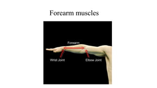
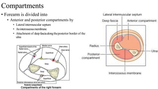
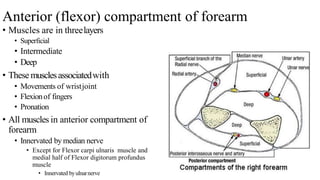
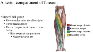
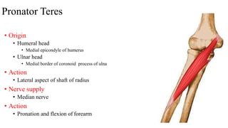
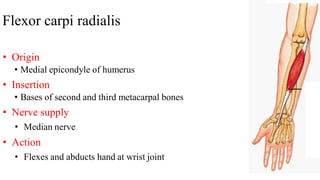
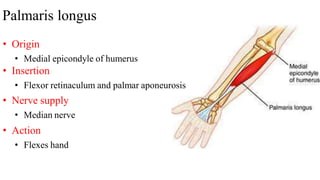
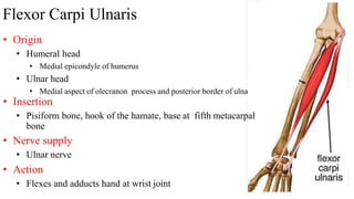
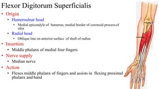
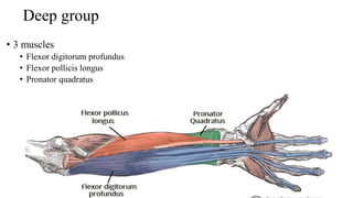
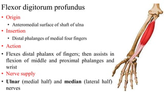
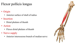
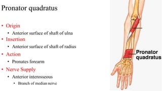
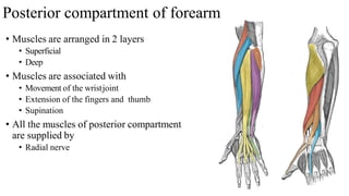
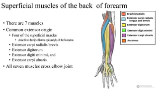
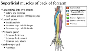
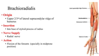
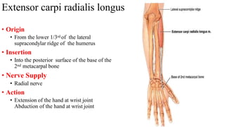
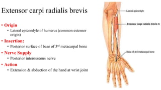
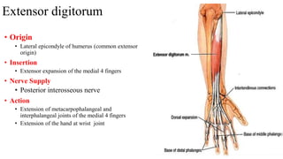
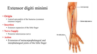
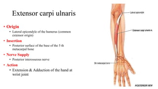
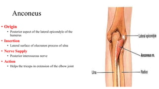
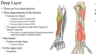
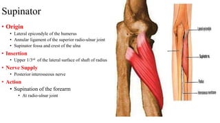
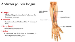
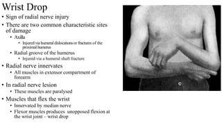
The forearm is divided into anterior and posterior compartments by septa. The anterior compartment contains flexor muscles in superficial, intermediate, and deep layers innervated by the median and ulnar nerves. Key muscles are pronator teres, flexor carpi radialis, palmaris longus, flexor carpi ulnaris, and flexor digitorum superficialis. The posterior compartment contains extensor muscles in superficial and deep layers innervated by the radial nerve. Key muscles are brachioradialis, extensor carpi radialis longus/brevis, extensor digitorum, and abductor pollicis longus. Radial nerve injury can cause wrist drop due to
femoral triangle, boundaries and contents of femoral triangle. femoral shetah. compartments of femoral sheath. ddfemoral canal. contets of femoral canal. femoral hernia. definition. causes. sign and symptoms. and treatment of femoral hernia.
The scapula, also known as the shoulder blade, is a flat triangular bone located at the back of the trunk and resides over the posterior surface of ribs two to seven. ... It also articulates with the humerus and clavicle, forming the glenohumeral (shoulder) joint and acromioclavicular joint respectively.
brachial plexus, branches of brachial plexus, main nerves of brachial plexus and their innervations, disorders of brachial plexus injury, Erb's palsy, Klumpke's palsy, compression of brachial plexus
Forearm bones and the work of right sided mid shaft femur and tibia and distal end of radius of right sided intertrochanteric femur and tibia and distal femur
USMLE MSK L006 Lower 04 Muscles of leg anatomy medical .pdfAHMED ASHOUR
The muscles of the leg are responsible for various movements, including flexion, extension, inversion, and eversion, as well as providing support during activities such as walking and running.
The muscles of the leg can be categorized into several groups based on their functions.
Understanding the actions and functions of these leg muscles is crucial for assessing and treating conditions affecting the lower extremity, such as injuries, imbalances, or musculoskeletal disorders.
Health Education on prevention of hypertensionRadhika kulvi
Hypertension is a chronic condition of concern due to its role in the causation of coronary heart diseases. Hypertension is a worldwide epidemic and important risk factor for coronary artery disease, stroke and renal diseases. Blood pressure is the force exerted by the blood against the walls of the blood vessels and is sufficient to maintain tissue perfusion during activity and rest. Hypertension is sustained elevation of BP. In adults, HTN exists when systolic blood pressure is equal to or greater than 140mmHg or diastolic BP is equal to or greater than 90mmHg. The
The Importance of Community Nursing Care.pdfAD Healthcare
NDIS and Community 24/7 Nursing Care is a specific type of support that may be provided under the NDIS for individuals with complex medical needs who require ongoing nursing care in a community setting, such as their home or a supported accommodation facility.
Empowering ACOs: Leveraging Quality Management Tools for MIPS and BeyondHealth Catalyst
Join us as we delve into the crucial realm of quality reporting for MSSP (Medicare Shared Savings Program) Accountable Care Organizations (ACOs).
In this session, we will explore how a robust quality management solution can empower your organization to meet regulatory requirements and improve processes for MIPS reporting and internal quality programs. Learn how our MeasureAble application enables compliance and fosters continuous improvement.
Navigating Challenges: Mental Health, Legislation, and the Prison System in B...Guillermo Rivera
This conference will delve into the intricate intersections between mental health, legal frameworks, and the prison system in Bolivia. It aims to provide a comprehensive overview of the current challenges faced by mental health professionals working within the legislative and correctional landscapes. Topics of discussion will include the prevalence and impact of mental health issues among the incarcerated population, the effectiveness of existing mental health policies and legislation, and potential reforms to enhance the mental health support system within prisons.
LGBTQ+ Adults: Unique Opportunities and Inclusive Approaches to CareVITASAuthor
This webinar helps clinicians understand the unique healthcare needs of the LGBTQ+ community, primarily in relation to end-of-life care. Topics include social and cultural background and challenges, healthcare disparities, advanced care planning, and strategies for reaching the community and improving quality of care.
The dimensions of healthcare quality refer to various attributes or aspects that define the standard of healthcare services. These dimensions are used to evaluate, measure, and improve the quality of care provided to patients. A comprehensive understanding of these dimensions ensures that healthcare systems can address various aspects of patient care effectively and holistically. Dimensions of Healthcare Quality and Performance of care include the following; Appropriateness, Availability, Competence, Continuity, Effectiveness, Efficiency, Efficacy, Prevention, Respect and Care, Safety as well as Timeliness.
TOP AND BEST GLUTE BUILDER A 606 | Fitking FitnessFitking Fitness
"Feature:
• Intelligent Ergonomically Design Glute Builder Is A Must Have For Those Looking To Target Their Gluteal Muscles And Hamstrings With Precision.
• The Ability To Adjust The Starting Position, This Machine Allows For A More Targeted Workout That Is Tailored To Your Specific Needs.
• Spacious And Supportive Cushioned Seat Provide Added Comfort And Stability During Your Workout."
Get more information visit on:- www.fitking.in
Our mail I.D:-care@fitking.in, fitking.in@gmail.com
Call us at :- 9958880790, 9870336406, 8800695917
How many patients does case series should have In comparison to case reports.pdfpubrica101
Pubrica’s team of researchers and writers create scientific and medical research articles, which may be important resources for authors and practitioners. Pubrica medical writers assist you in creating and revising the introduction by alerting the reader to gaps in the chosen study subject. Our professionals understand the order in which the hypothesis topic is followed by the broad subject, the issue, and the backdrop.
https://pubrica.com/academy/case-study-or-series/how-many-patients-does-case-series-should-have-in-comparison-to-case-reports/
CHAPTER 1 SEMESTER V - ROLE OF PEADIATRIC NURSE.pdfSachin Sharma
Pediatric nurses play a vital role in the health and well-being of children. Their responsibilities are wide-ranging, and their objectives can be categorized into several key areas:
1. Direct Patient Care:
Objective: Provide comprehensive and compassionate care to infants, children, and adolescents in various healthcare settings (hospitals, clinics, etc.).
This includes tasks like:
Monitoring vital signs and physical condition.
Administering medications and treatments.
Performing procedures as directed by doctors.
Assisting with daily living activities (bathing, feeding).
Providing emotional support and pain management.
2. Health Promotion and Education:
Objective: Promote healthy behaviors and educate children, families, and communities about preventive healthcare.
This includes tasks like:
Administering vaccinations.
Providing education on nutrition, hygiene, and development.
Offering breastfeeding and childbirth support.
Counseling families on safety and injury prevention.
3. Collaboration and Advocacy:
Objective: Collaborate effectively with doctors, social workers, therapists, and other healthcare professionals to ensure coordinated care for children.
Objective: Advocate for the rights and best interests of their patients, especially when children cannot speak for themselves.
This includes tasks like:
Communicating effectively with healthcare teams.
Identifying and addressing potential risks to child welfare.
Educating families about their child's condition and treatment options.
4. Professional Development and Research:
Objective: Stay up-to-date on the latest advancements in pediatric healthcare through continuing education and research.
Objective: Contribute to improving the quality of care for children by participating in research initiatives.
This includes tasks like:
Attending workshops and conferences on pediatric nursing.
Participating in clinical trials related to child health.
Implementing evidence-based practices into their daily routines.
By fulfilling these objectives, pediatric nurses play a crucial role in ensuring the optimal health and well-being of children throughout all stages of their development.
Deep Leg Vein Thrombosis (DVT): Meaning, Causes, Symptoms, Treatment, and Mor...The Lifesciences Magazine
Deep Leg Vein Thrombosis occurs when a blood clot forms in one or more of the deep veins in the legs. These clots can impede blood flow, leading to severe complications.
This document is designed as an introductory to medical students,nursing students,midwives or other healthcare trainees to improve their understanding about how health system in Sri Lanka cares children health.
2. Compartments
• Forearm is divided into
• Anterior and posterior compartments by
• Lateral intermuscular septum
• Aninterosseousmembrane
• Attachment of deepfasciaalongtheposterior border of the
ulna
3. Anterior (flexor) compartment of forearm
• Muscles are in threelayers
• Superficial
• Intermediate
• Deep
• Thesemusclesassociatedwith
• Movements of wristjoint
• Flexionof fingers
• Pronation
• All muscles in anterior compartment of
forearm
• Innervated by median nerve
• Except for Flexor carpi ulnaris muscle and
medial half of Flexor digitorum profundus
muscle
• Innervated byulnarnerve
4. Anterior compartment of forearm
• Superficial group
• Five muscles cross the elbow joint
• Three musclesdonot
• Flexor compartment is much more
bulky
• Than extensor compartment
• Necessary power of grip
5. Pronator Teres
• Origin
• Humeral head
• Medial epicondyle of humerus
• Ulnar head
• Medial border of coronoid process of ulna
• Action
• Lateral aspect of shaft of radius
• Nerve supply
• Median nerve
• Action
• Pronation and flexion of forearm
6. Flexor carpi radialis
• Origin
• Medial epicondyle of humerus
• Insertion
• Bases of second and third metacarpal bones
• Nerve supply
• Median nerve
• Action
• Flexes and abducts hand at wrist joint
7. Palmaris longus
• Origin
• Medial epicondyle of humerus
• Insertion
• Flexor retinaculum and palmar aponeurosis
• Nerve supply
• Median nerve
• Action
• Flexes hand
8. Flexor Carpi Ulnaris
• Origin
• Humeral head
• Medial epicondyle of humerus
• Ulnar head
• Medial aspect of olecranon process and posterior border of ulna
• Insertion
• Pisiform bone, hook of the hamate, base at fifth metacarpal
bone
• Nerve supply
• Ulnar nerve
• Action
• Flexes and adducts hand at wrist joint
9. Flexor Digitorum Superficialis
• Origin
• Humeroulnar head
• Medial epicondyle of humerus; medial border of coronoid process of
ulna
• Radial head
• Oblique line on anterior surface of shaft of radius
• Insertion
• Middle phalanx of medial four fingers
• Nerve supply
• Median nerve
• Action
• Flexes middle phalanx of fingers and assists in flexing proximal
phalanx and hand
10. Deep group
• 3 muscles
• Flexor digitorum profundus
• Flexor pollicis longus
• Pronator quadratus
11. Flexor digitorum profundus
• Origin
• Anteromedial surface of shaft of ulna
• Insertion
• Distal phalanges of medial four fingers
• Action
• Flexes distal phalanx of fingers; then assists in
flexion of middle and proximal phalanges and
wrist
• Nerve supply
• Ulnar (medial half) and median (lateral half)
nerves
12. Flexor pollicis longus
• Origin
• Anterior surface of shaft of radius
• Insertion
• Distal phalanx of thumb
• Action
• Flexes distal phalanx of thumb
• Nerve supply
• Anterior interosseous branch of median nerve
13. Pronator quadratus
• Origin
• Anterior surface of shaft of ulna
• Insertion
• Anterior surface of shaft of radius
• Action
• Pronates forearm
• Nerve Supply
• Anterior interosseous
• Branch of median nerve
14. Space of Parona
• Space in front of pronator quadratus
• Deep to long flexor tendons of fingers
and their synovial sheaths
• Limited proximally by
• Oblique origin of flexor digitorum
superficialis
• Space involved in proximal extensions
of synovial sheath infections
• Can be drained through radial and
ulnar incisions to the side of the flexor
tendons
15. Posterior compartment of forearm
• Muscles are arranged in 2 layers
• Superficial
• Deep
• Muscles are associated with
• Movement of the wristjoint
• Extension of the fingers and thumb
• Supination
• All the muscles of posterior compartment
are supplied by
• Radial nerve
16. Arrangement of posterior compartment muscles
• Upper part
• Anconeus (superficial)
• Supinator (deep)
• Lateral part of humerus
• Pass along the radial side
• Brachioradialis
• Extensors carpi radialis longus
• Extensors carpi radialis brevis
• Passing along posterior surface of the
forearm
• Extensor digitorum
• Extensor digiti minimi and
• Extensor carpi ulnaris
• At lower end of forearm
• These two groups are separated by
three muscles
• They emerge from deeply in between
them and go to the thumb
• Abductor pollicis longus
• Extensors pollicis longus and
• Extensors pollicis longus brevis
• One muscle for the forefinger runs
deeply to reach the back of the hand
• Extensor indicis
17. Superficial muscles of the back of forearm
• There are 7 muscles
• Common extensor origin
• Four of the superficialmuscles
• Arisefromthetipoflateralepicondyleof thehumerus
• Extensor carpi radialis brevis
• Extensor digitorum
• Extensor digiti minimi, and
• Extensor carpi ulnaris
• All seven muscles cross elbow joint
18. Superficial muscles of back of forearm
• Categorized into two groups
• Lateral and posterior
• Each group consists of three muscles
• Lateral group
• Brachioradialis
• Extensor carpi radialis longus
• Extensor carpi radialis brevis
• Posterior group
• Extensor digitorum
• Extensor digiti minimi
• Extensor carpi ulnaris
• In the upper end
• Anconeus
19. Brachioradialis
• Origin
• Upper 2/3rd of lateral supracondylar ridge of
humerus
• Insertion
• Into base of styloid process of radius
• Nerve Supply
• Radial nerve
• Action
• Flexion of the forearm (specially in midprone
position)
20. Extensor carpi radialis longus
• Origin
• From the lower 1/3rd of the lateral
supracondylar ridge of the humerus
• Insertion
• Into the posterior surface of the base of the
2nd metacarpal bone
• Nerve Supply
• Radial nerve
• Action
• Extension of the hand at wrist joint
Abduction of the hand at wrist joint
21. Extensor carpi radialis brevis
• Origin
• Lateral epicondyle of humerus (common extensor
origin)
• Insertion:
• Posterior surface of base of 3rd metacarpal bone
• Nerve Supply
• Posterior interosseous nerve
• Action
• Extension & abduction of the hand at wrist joint
22. Extensor digitorum
• Origin
• Lateral epicondyle of humerus (common extensor
origin)
• Insertion
• Extensor expansion of the medial 4 fingers
• Nerve Supply
• Posterior interosseous nerve
• Action
• Extension of metacarpophalangeal and
interphalangeal joints of the medial 4 fingers
• Extension of the hand at wrist joint
23. Extensor digiti minimi
• Origin
• Lateral epicondyle of the humerus (common
extensor origin)
• Insertion
• Extensor expansion of the little finger
• Nerve Supply
• Posterior interosseous nerve
• Action
• Extension of metacarpophalangeal and
interphalangeal joints of the little finger
24. Extensor carpi ulnaris
• Origin
• Lateral epicondyle of the humerus (common
extensor origin)
• Insertion
• Posterior surface of the base of the 5 th
metacarpal bone
• Nerve Supply
• Posterior interosseous nerve
• Action
• Extension & Adduction of the hand at
wrist joint
25. Anconeus
• Origin
• Posterior aspect of the lateral epicondyle of the
humerus
• Insertion
• Lateral surface of olecranon process of ulna
• Nerve Supply
• Posterior interosseous nerve
• Action
• Helps the triceps in extension of the elbow joint
26. Deep Layer
• There are five deep muscles
• Three deepextensorsof the forearm
• Function on thumb
• Abductor pollicis longus(APL)
• Extensor pollicis brevis (EPB)
• Extensor pollicis longus (EPL)
• To acquire insertion on the three bones of
thumb ‘crop out’
• From furrow in lateralelementof forearmbetweenlateral
andposterior groupsof superficial extensor
• One muscle
• Function onindex finger
• Extensor indicis
• In the upper part
• Supinator
27. Supinator
• Origin
• Lateral epicondyle of the humerus
• Annular ligament of the superior radio-ulnar joint
• Supinator fossa and crest of the ulna
• Insertion
• Upper 1/3rd of the lateral surface of shaft of radius
• Nerve Supply
• Posterior interosseous nerve
• Action
• Supination of the forearm
• At radio-ulnar joint
28. Abductor pollicis longus
• Origin
• Middle of the posterior surface of radius and ulna
• Interosseus membrane
• Insertion
• Posterior surface of the base of the 1st metacarpal
bone
• Nerve Supply
• Posterior interosseous nerve
• Action
• Abduction and extension of the thumb at
the carpo-metacarpal joint
29. Extensor pollicis brevis
• Origin
• Posterior surface of the shaft of radius
• Insertion
• Posterior surface of the base of the proximal
phalanx of the thumb
• Nerve Supply
• Posterior interosseous nerve
• Action
• Extension of the metacarpophalangeal joint of
the thumb
30. Extensor pollicis longus
• Origin
• Posterior surface of ulna
• Insertion
• Posterior surface of the base of the distal
phalanx of the thumb
• Nerve Supply
• Posterior interosseous nerve
• Action
• Extension of the interphalangeal joints of
the thumb
31. Extensor indicis
• Origin
• Posterior surface of ulna (below extensor
pollicis longus)
• Insertion
• Extensor expansion of the index finger.
• Nerve Supply
• Posterior interosseous nerve
• Action
• Extension of the interphalangeal joints of the
thumb
32. Wrist Drop
• Sign of radial nerve injury
• There are two common characteristic sites
of damage
• Axilla
• Injured viahumeral dislocationsor fractures of the
proximal humerus
• Radial groove of the humerus
• Injured via a humeral shaft fracture
• Radial nerve innervates
• All muscles in extensor compartment of
forearm
• In radial nerve lesion
• These muscles are paralysed
• Muscles that flex the wrist
• Innervated by median nerve
• Flexor muscles produces unopposed flexion at
the wrist joint – wrist drop