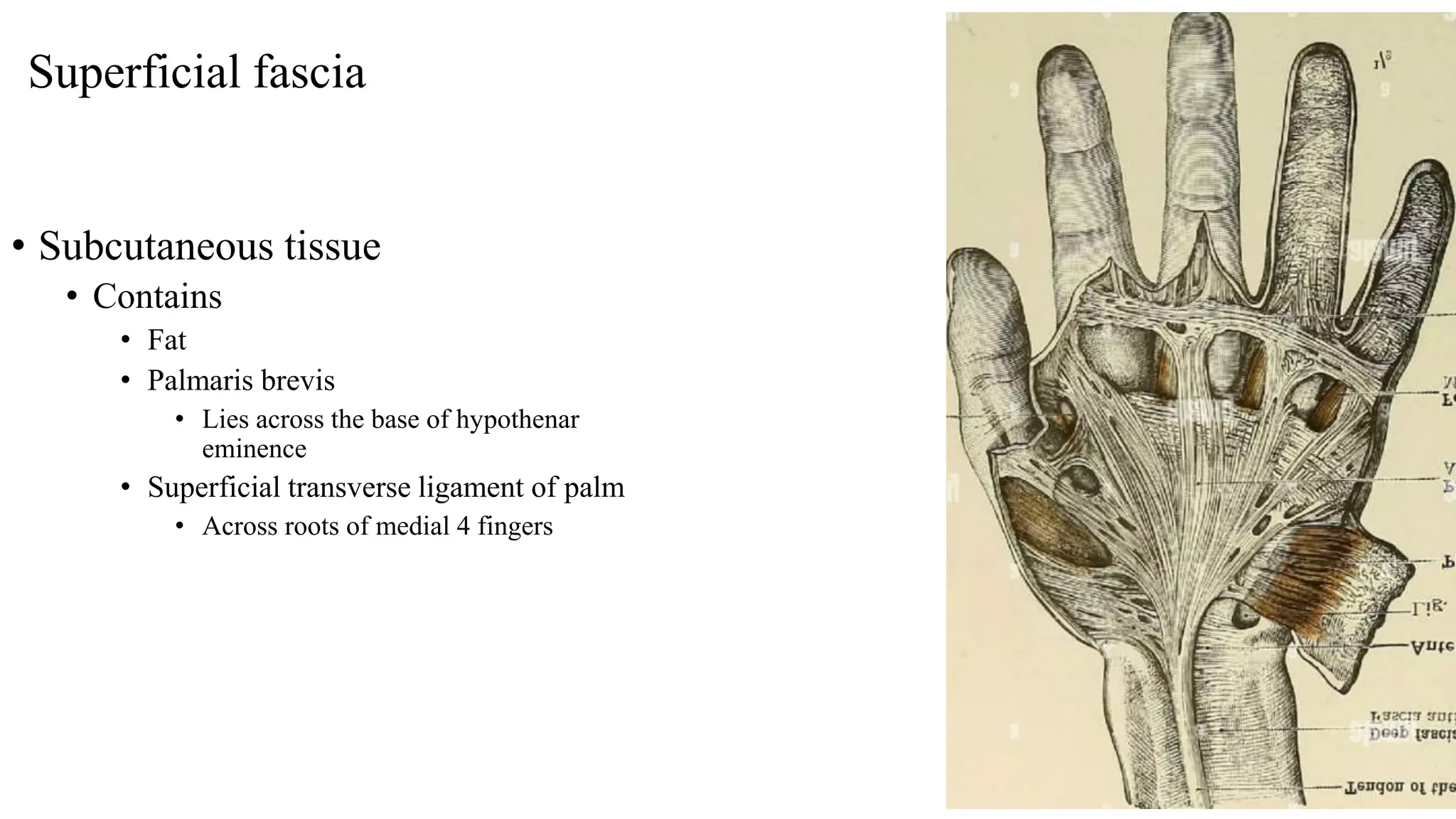
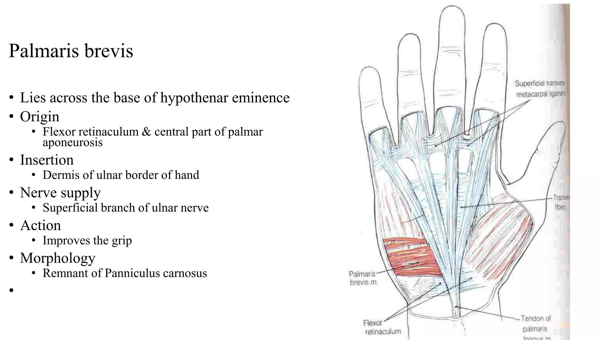
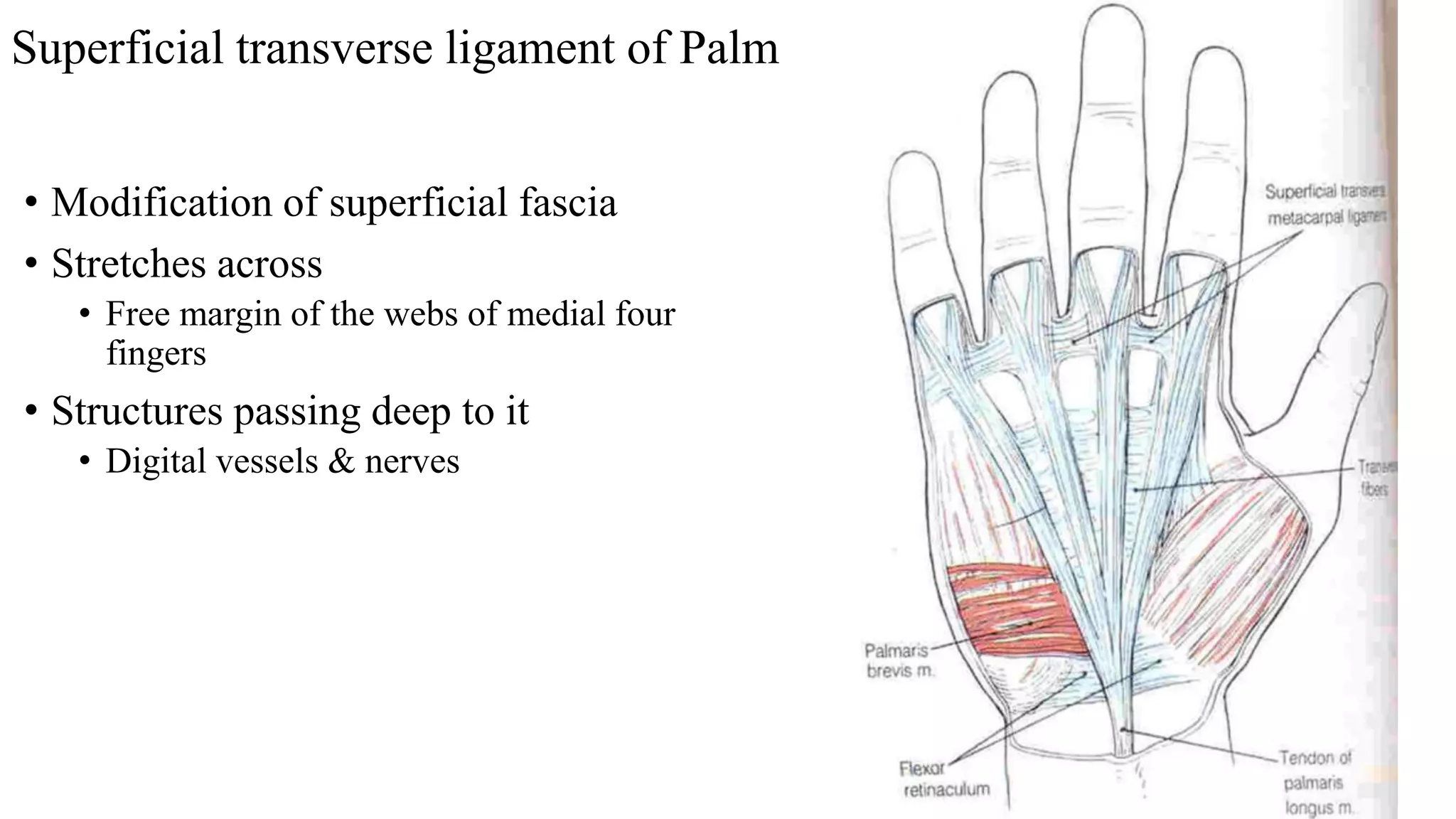
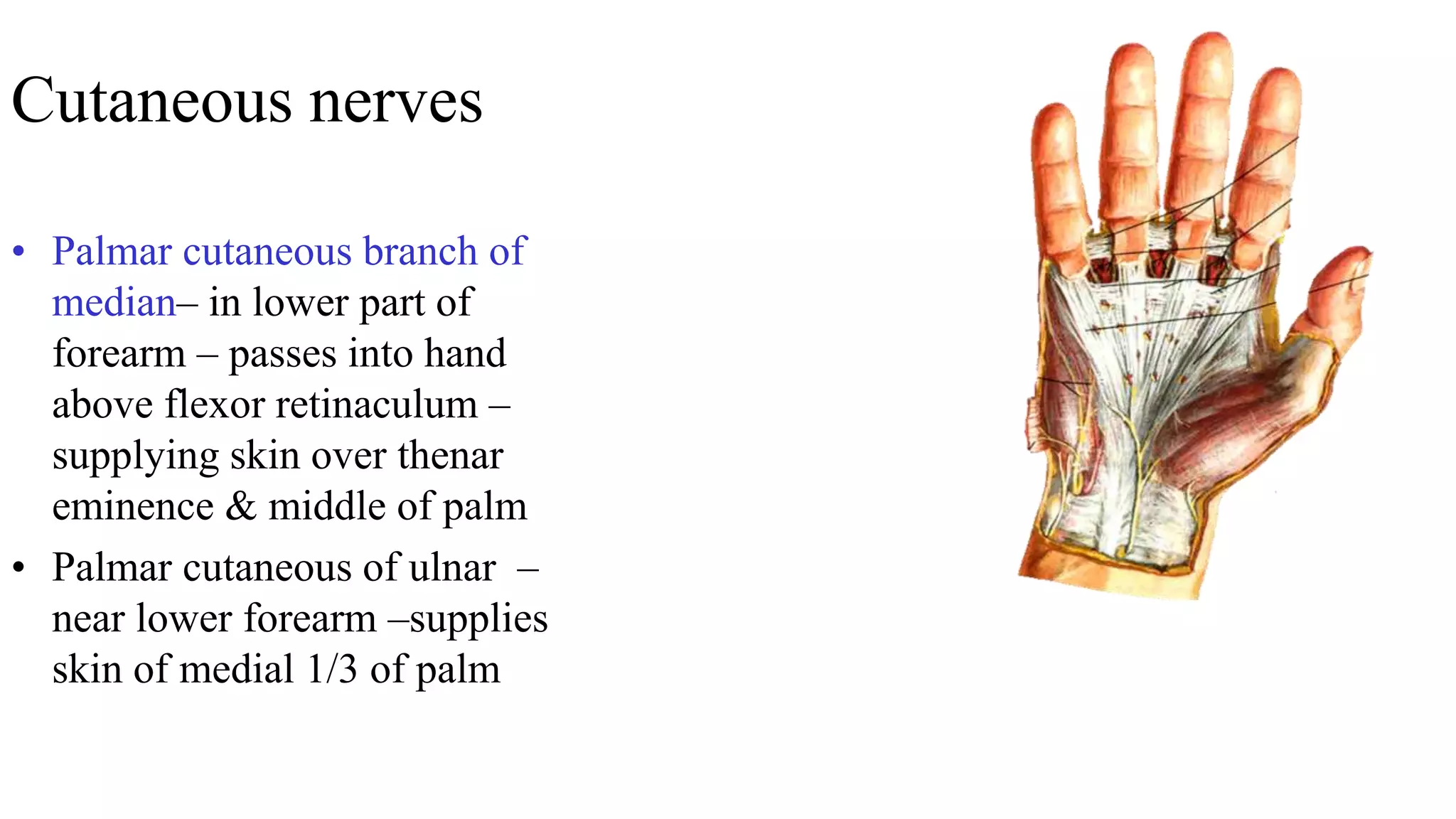
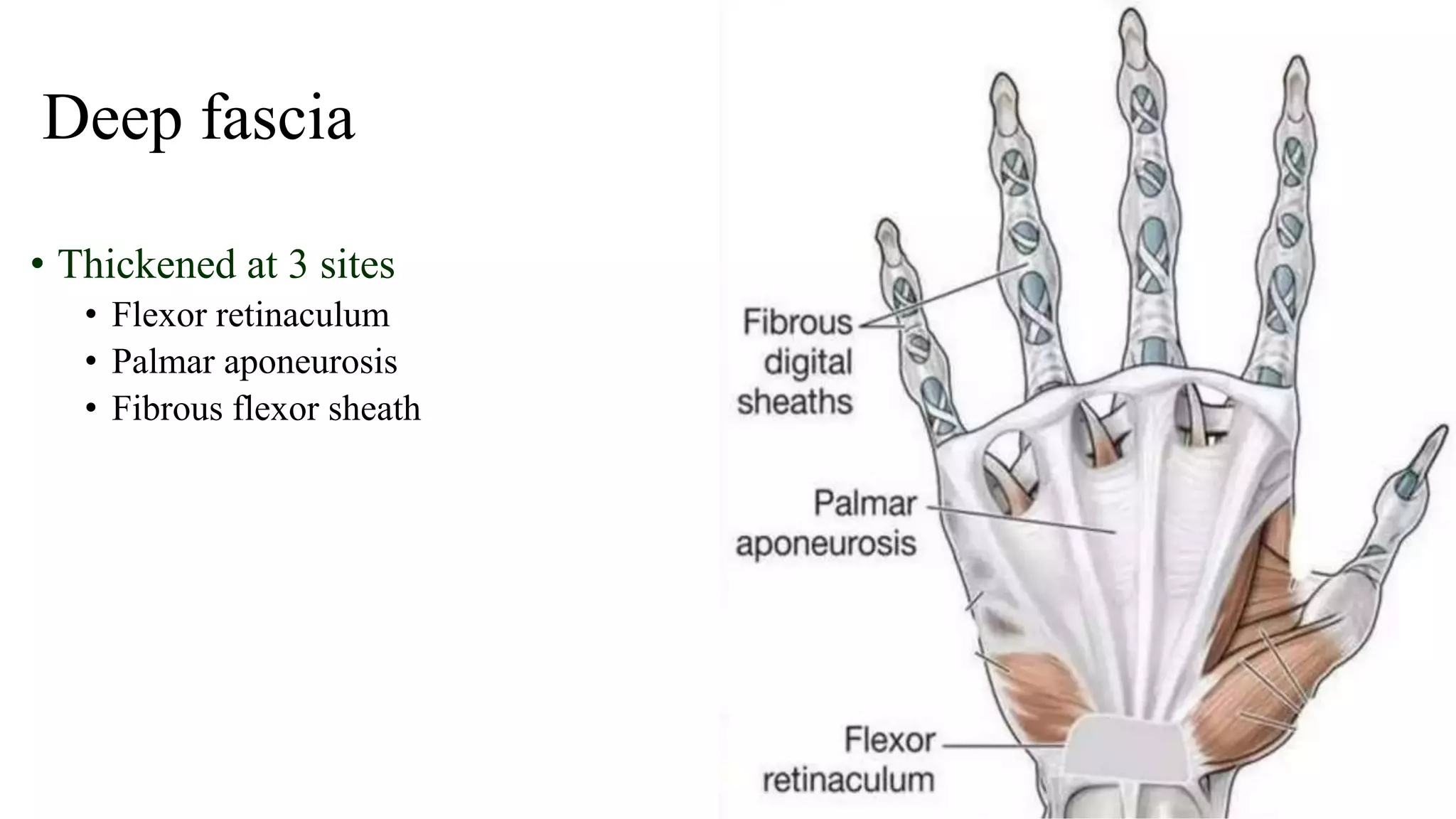
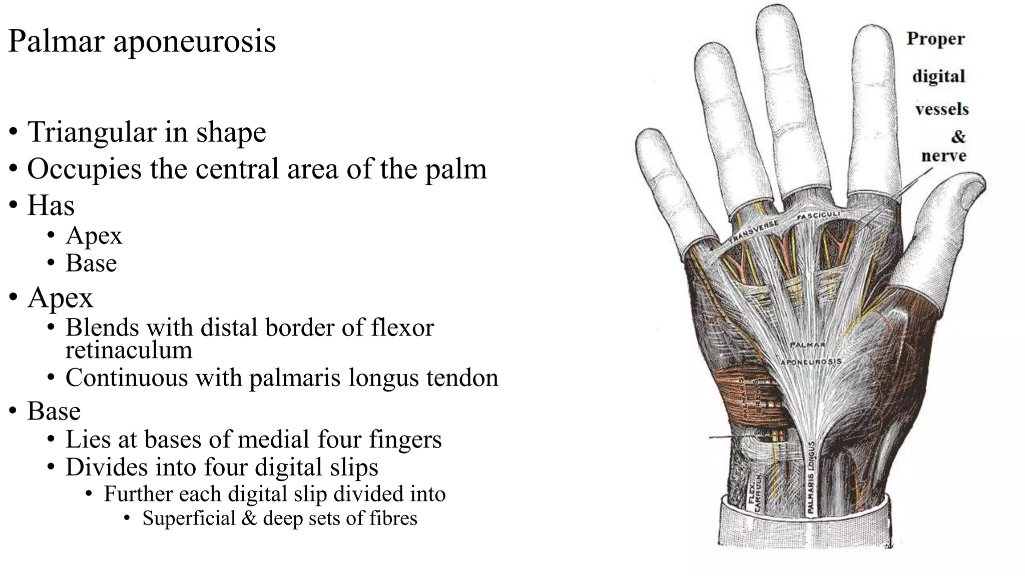
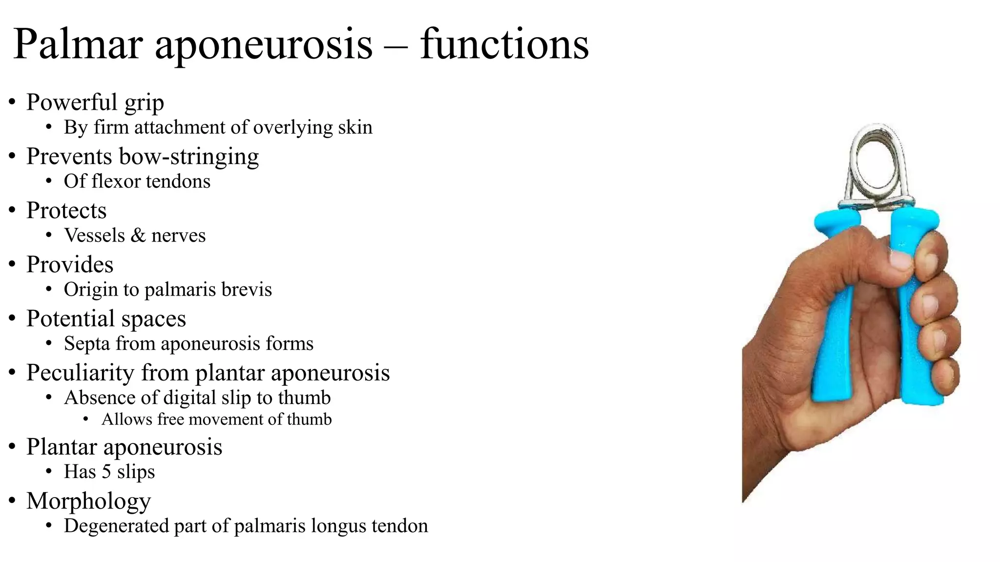
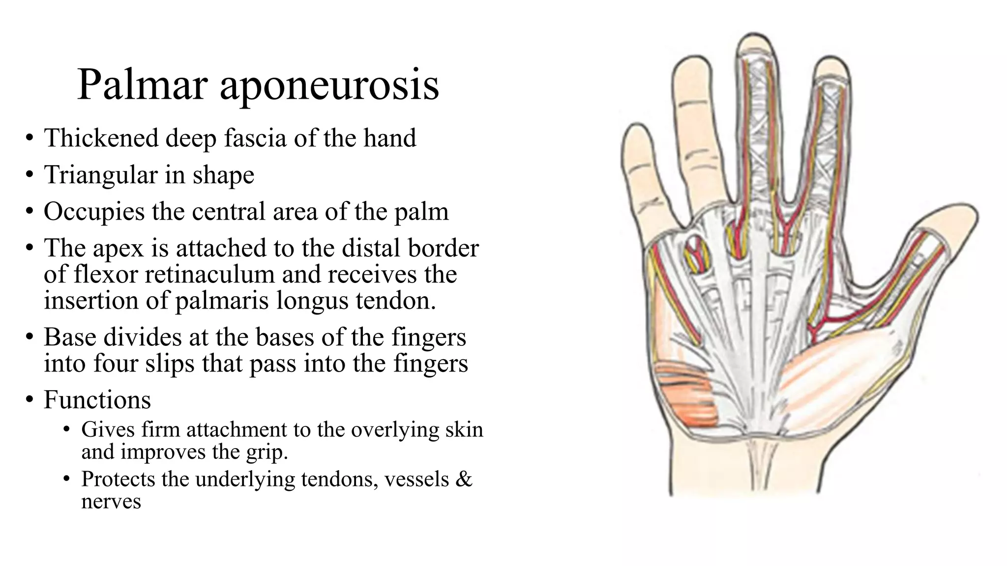
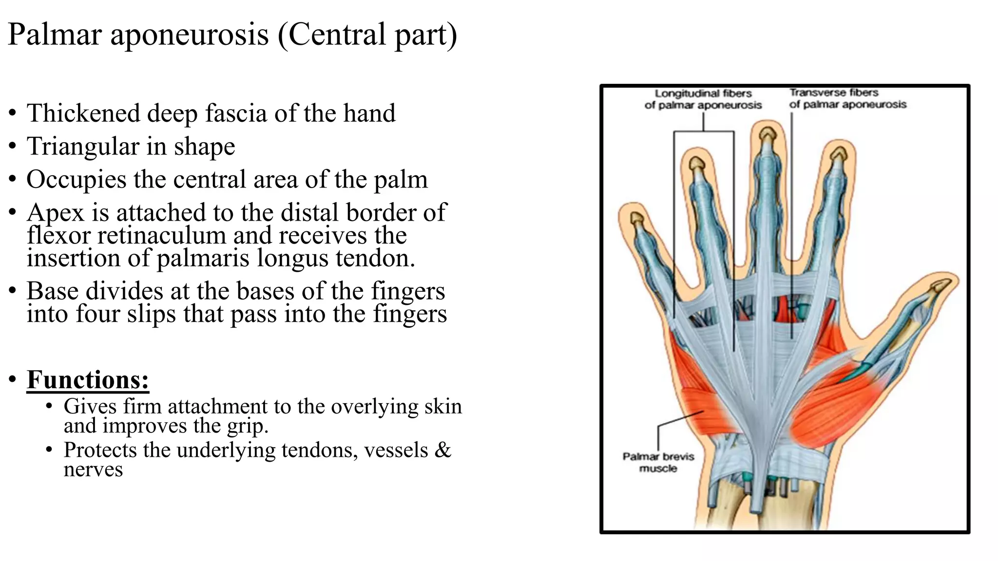
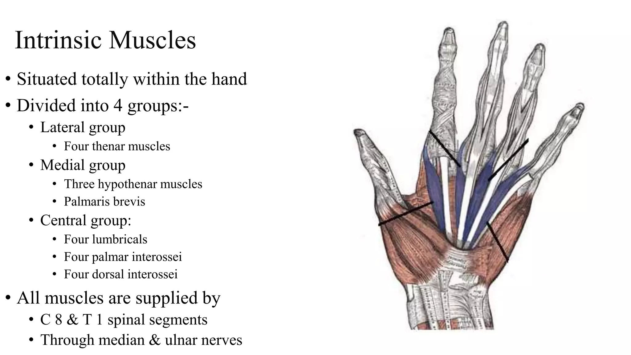
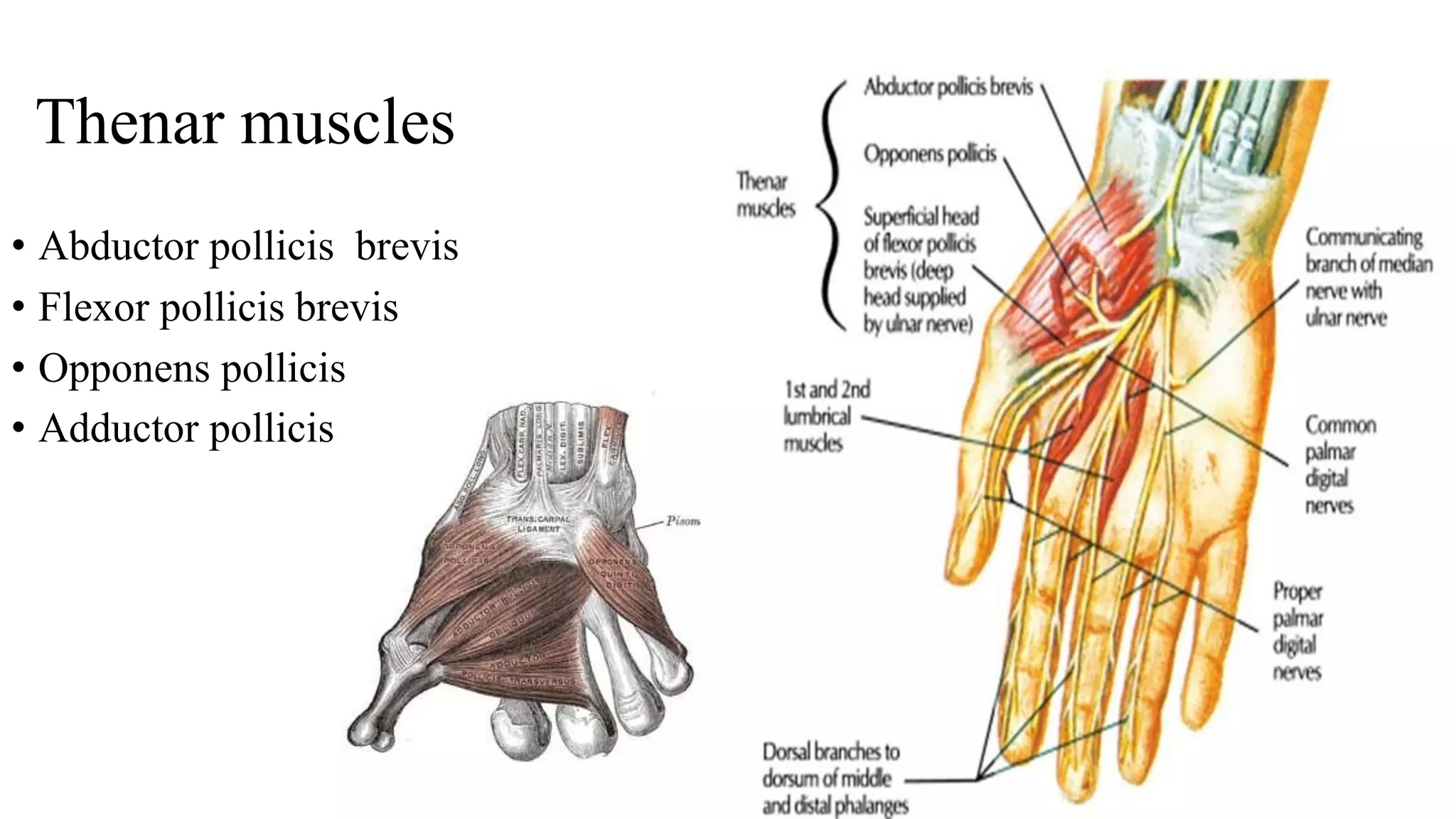
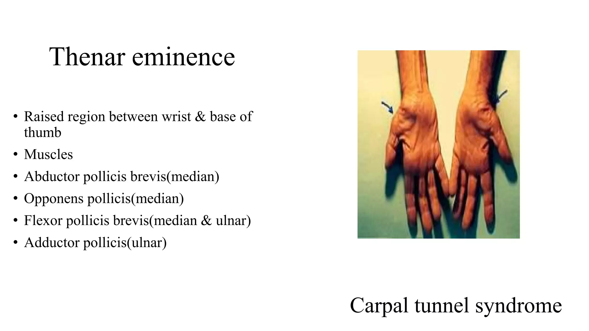
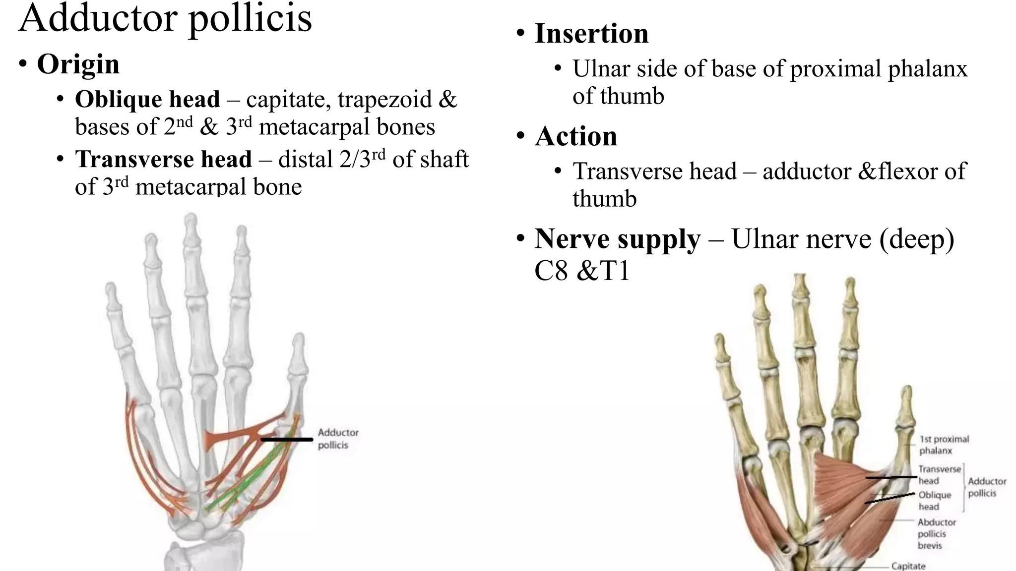
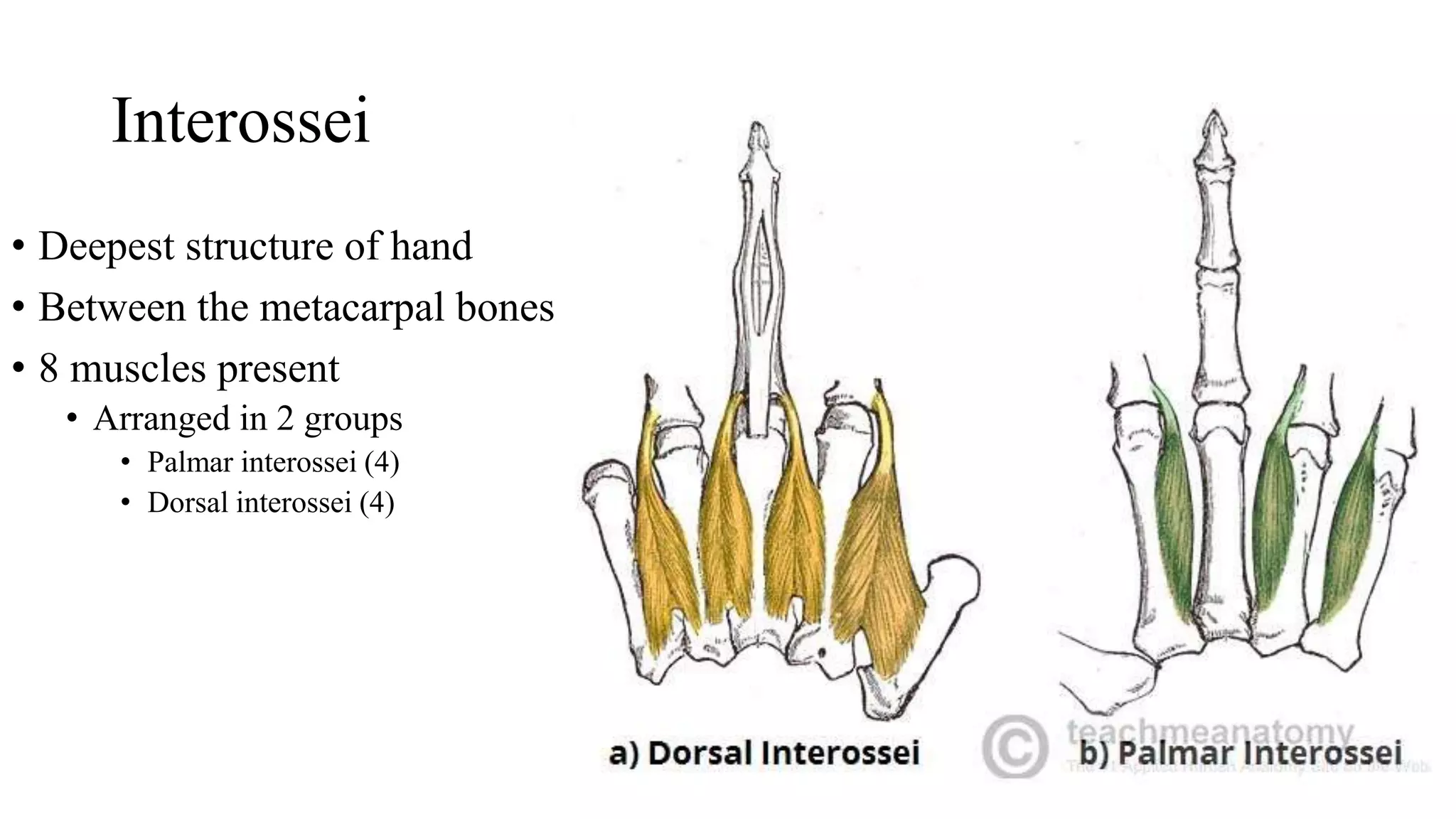
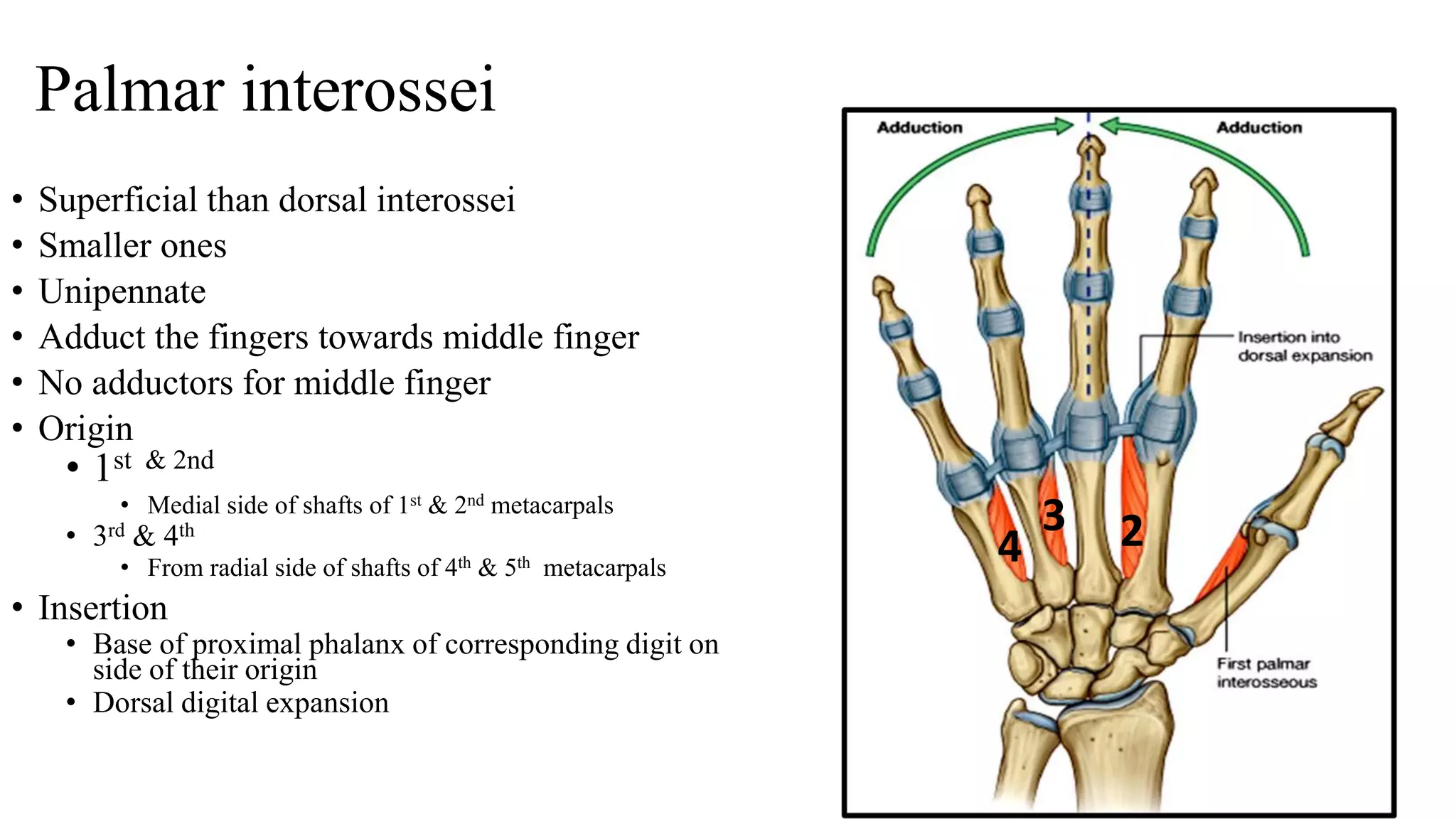
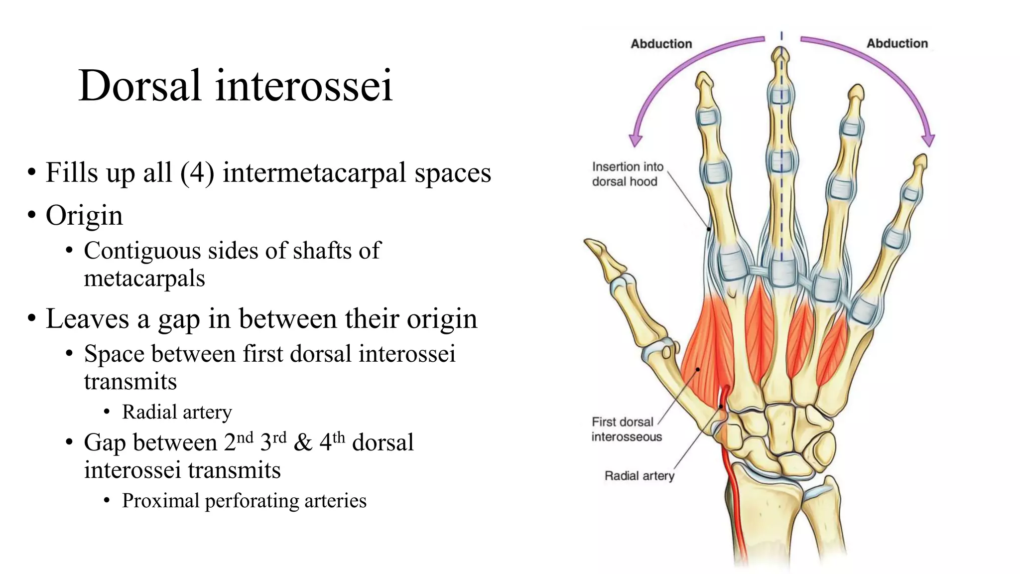
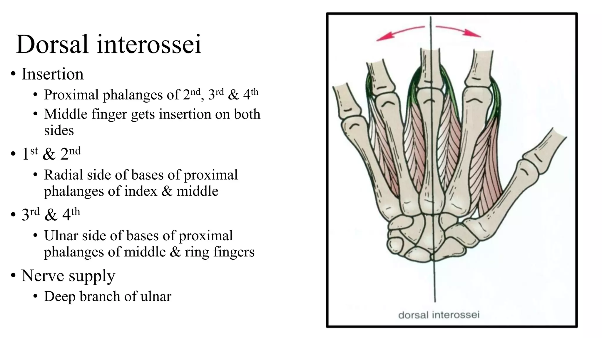
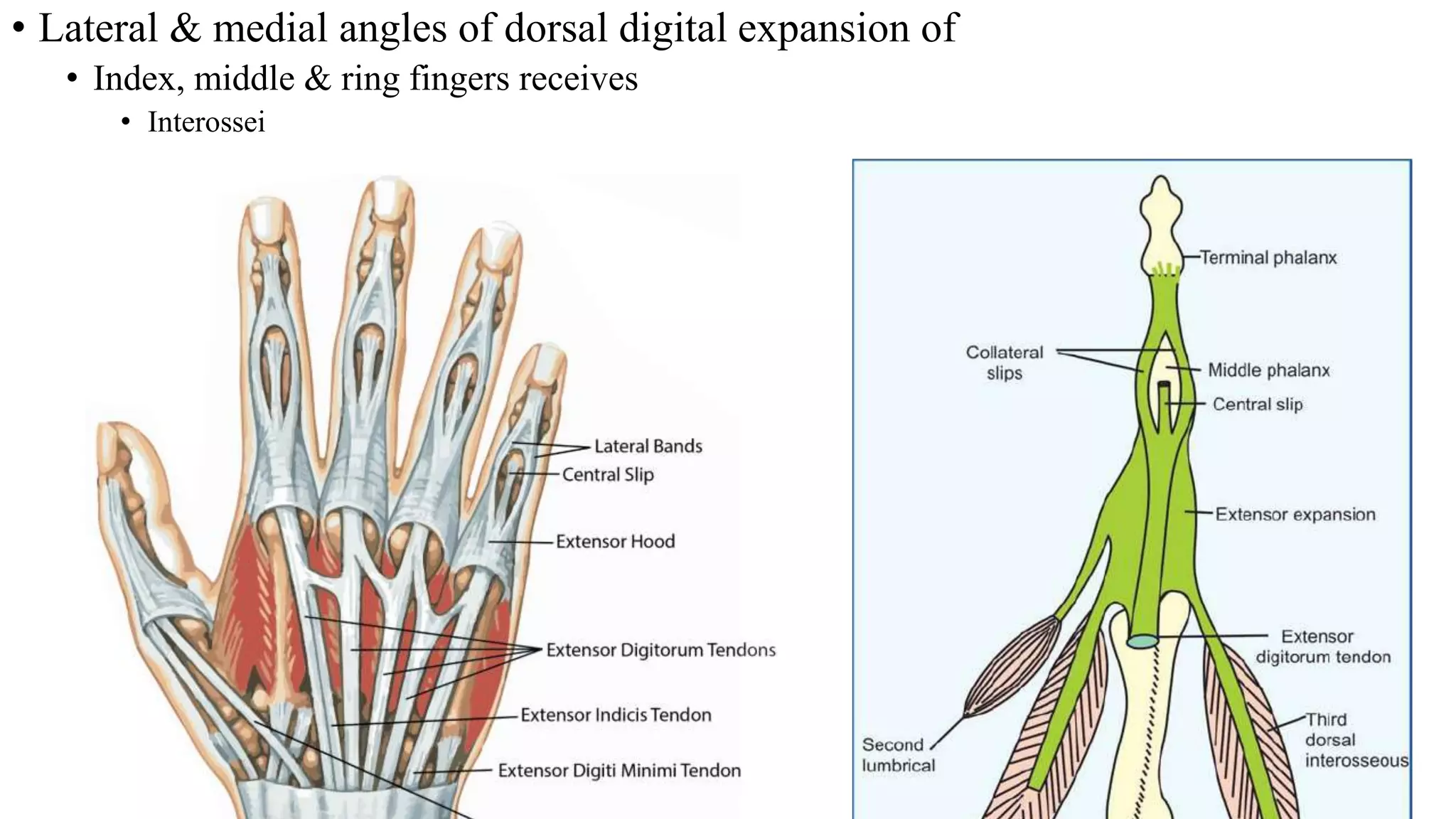
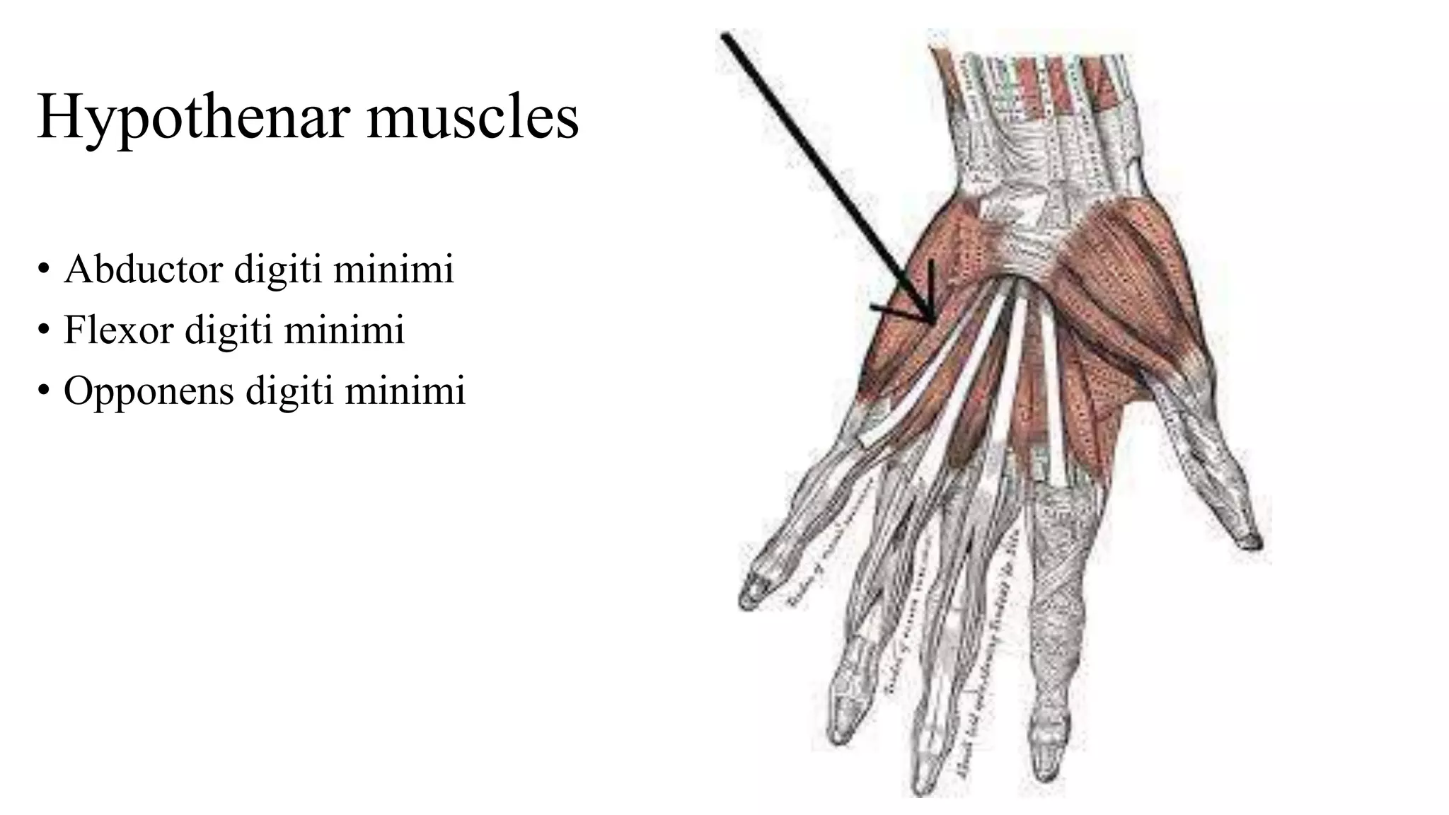
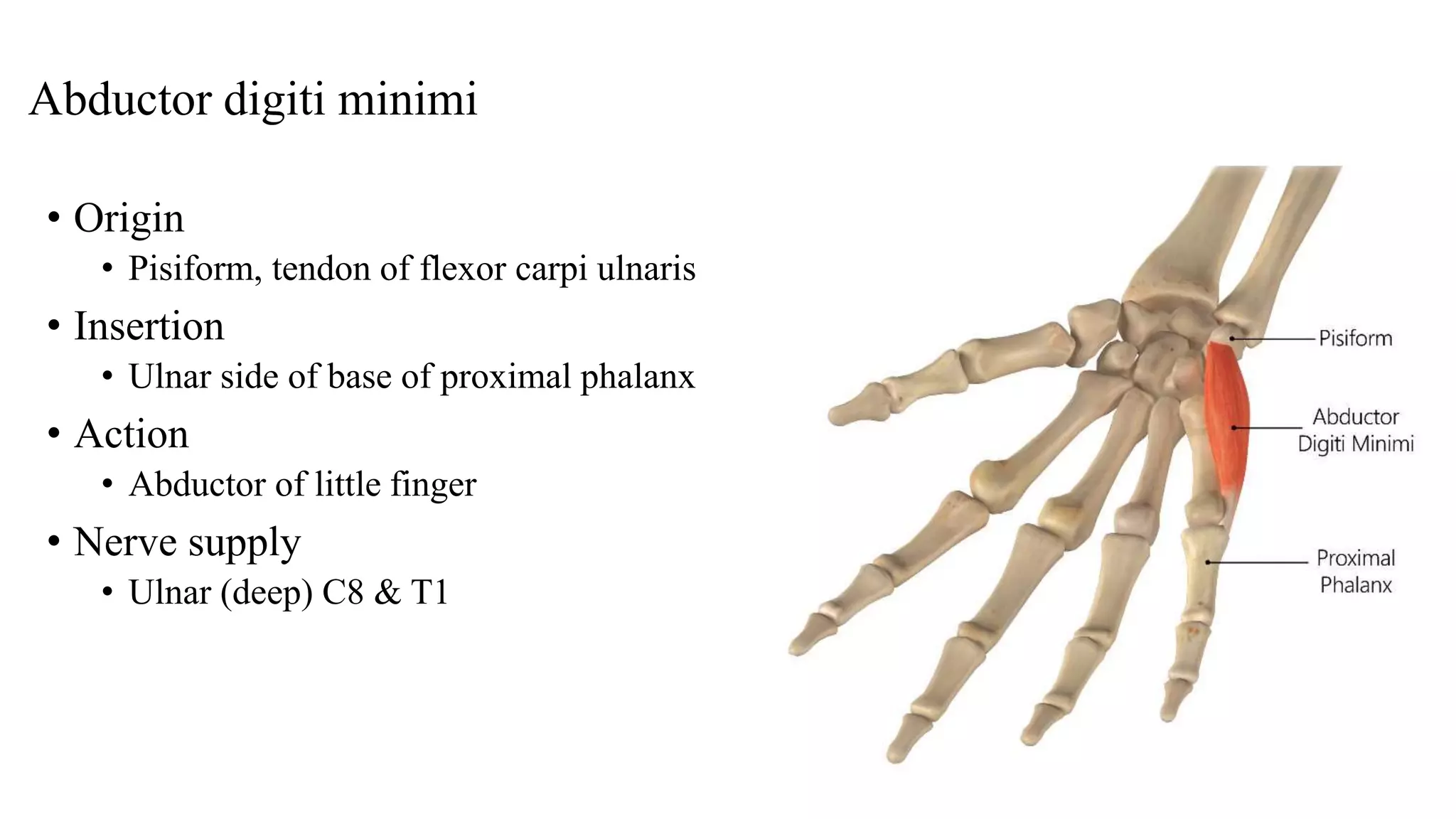
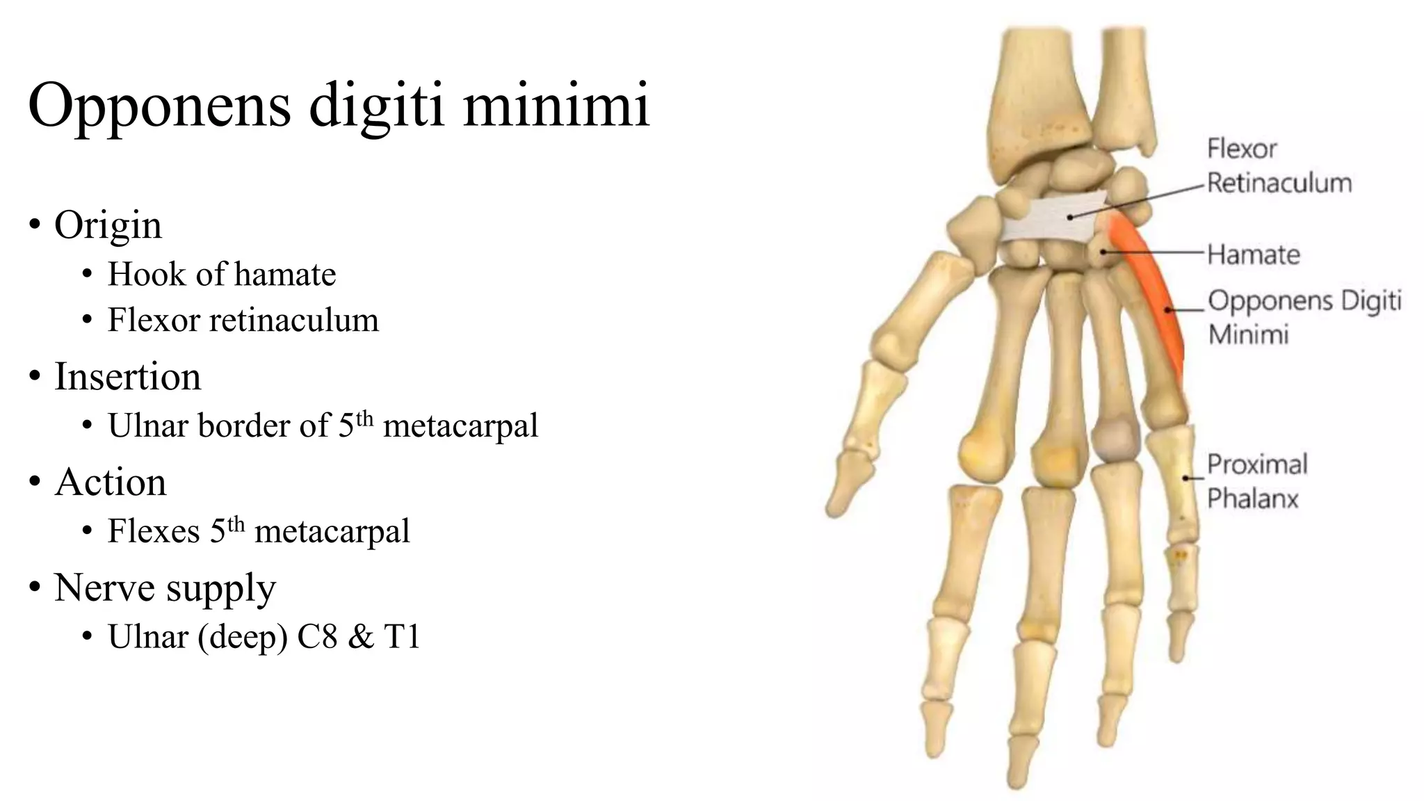
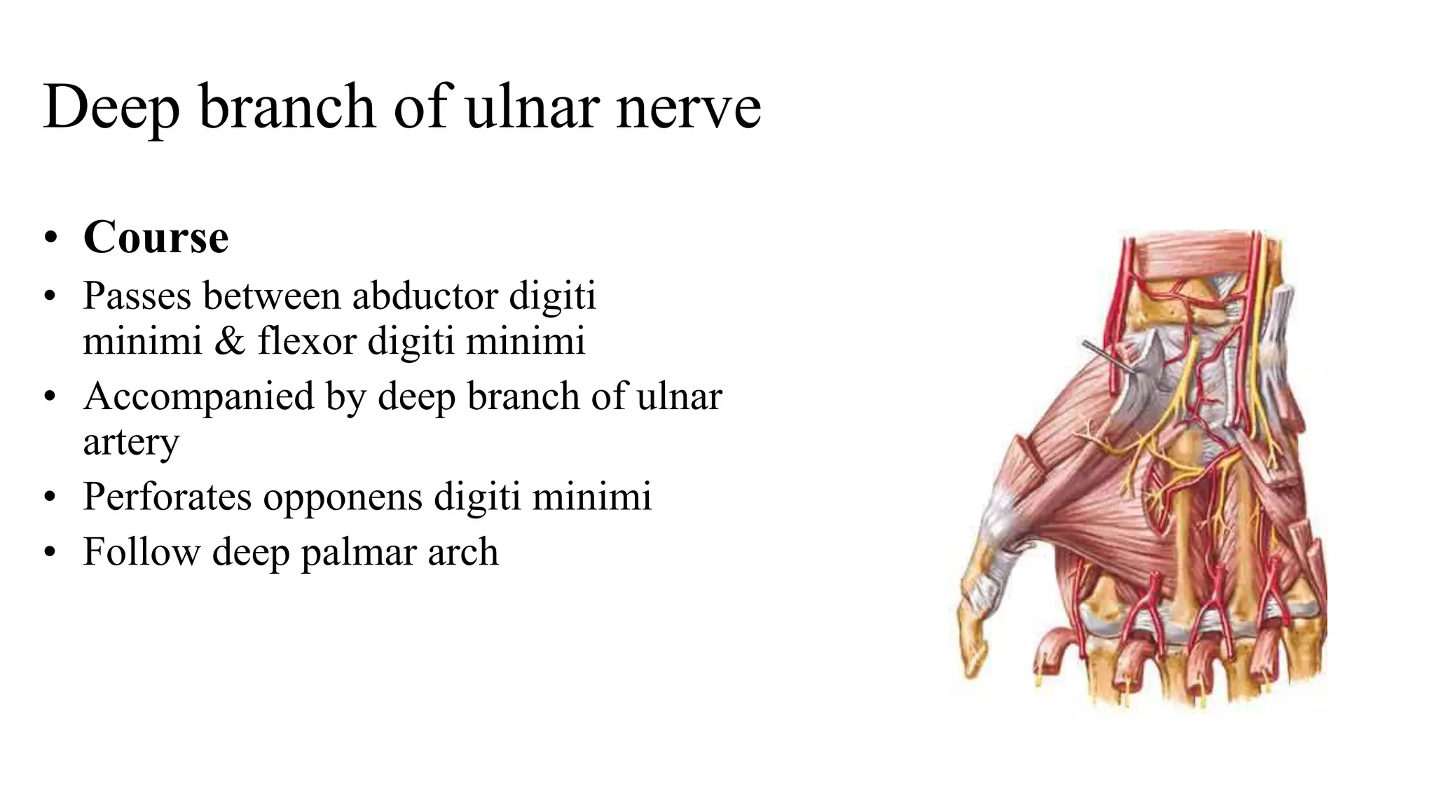
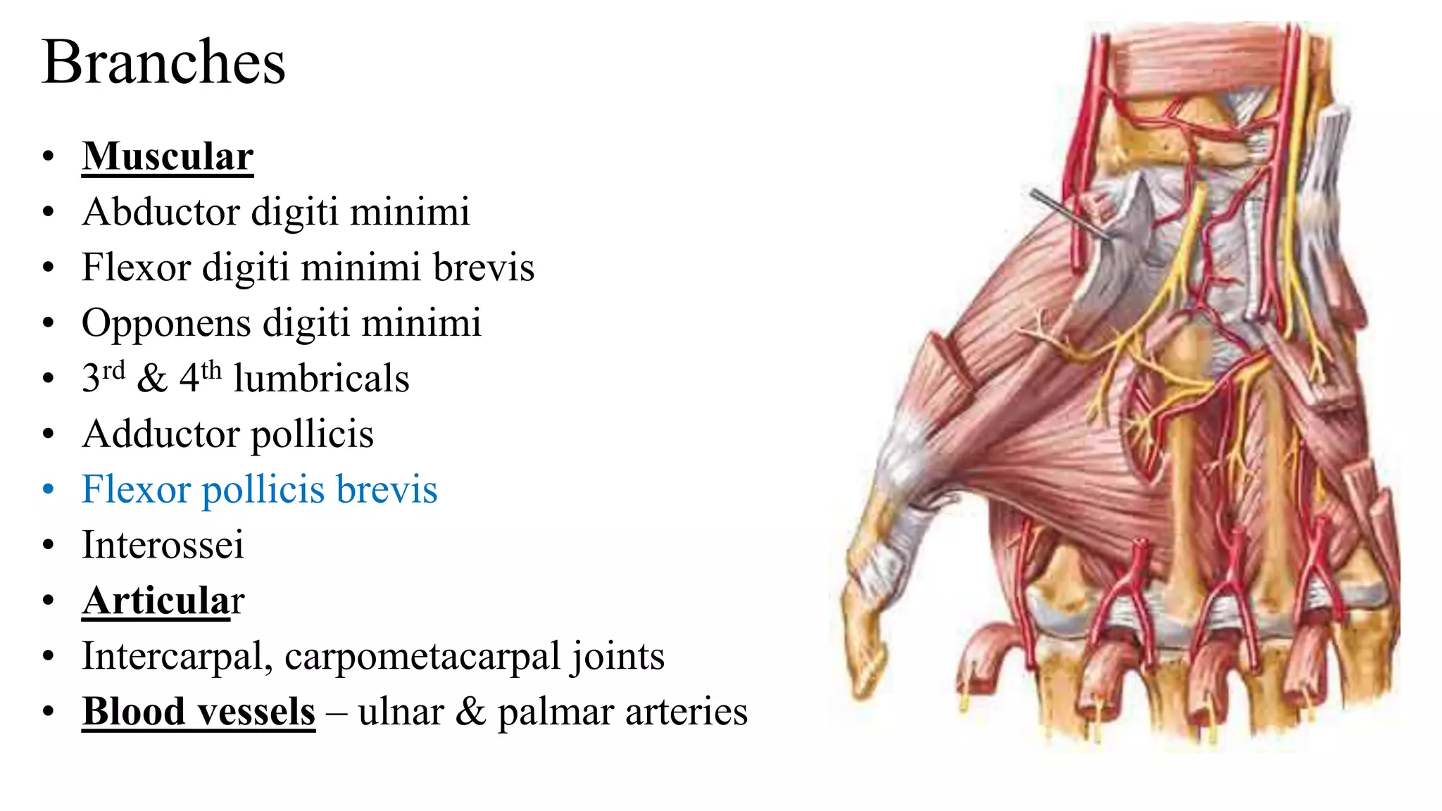
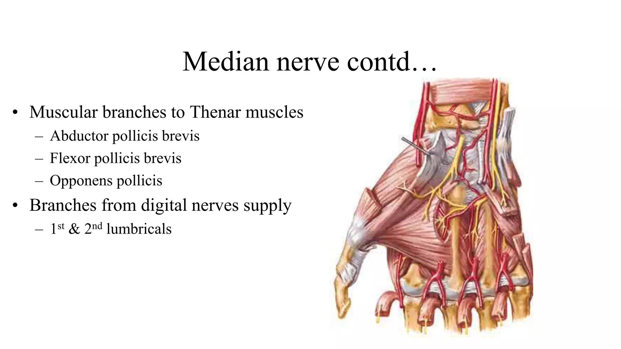
Hand
Hand
• Composed of
• Bones
• Muscles
• Ligaments
• To allow a large amount of
movements
Hand
• Has 2 surfaces
• Palmar
• Dorsal
Palm – Features
• Hollow
• Eminence
• Creases
Palm – Features
• Palmar surface
• Thick skin
• Devoid of
• Hairs & sebaceous glands
• Connected with deep fascia by
• Many fibrous septa
• Helps grip by restricting mobility of skin
• Has fat
• Act as cushion
• To withstand pressure
Palm - features
• Has
• Creases or lines
• 3 types
• Flexure lines
• Papillary ridges
• Langer lines
Creases - Flexure lines
• Visible marking
• Lies close to joints
• On medial 4 fingers
• 2 interphalangeal creases
• Proximal crease only marks joint
• Crease between palm & fingers
• Do not represent the metacarpo-
phalangeal joint
• Joint lies 2 cm proximal to crease
• Distal skin crease of wrist
• Represents proximal border of flexor
retinaculum
• At the midpoint
• Median nerve passes behind
Creases - Flexure lines
• Wrist
• Proximal
• Distal
• Palm
• Longitudinal
• Mid-palmar
• Radial
• Transverse
• Proximal
• Longitudinal
• Applied anatomy – Simian’s crease
• Phalanges
• Proximal
• Middle
• Distal
Creases – Papillary ridges
• Otherwise - Finger prints
• Peculiar over the flexor surface of
distal phalanges
• Corresponds to
• Pattern of underlying dermal papilla
• Ducts of sweat gland opens
• 3 types
• Whorl, loop & arch
• Helps in personal identity
• Dermatoglyphics
• Science of finger prints
Creases – Langer lines
• Produced by
• Bundles of collagen fibres of dermis
• On dorsum
• Transverse
• On palm
• Longitudinal
Hand - Dorsum
• Dorsal surface
• Thin skin
• Presence of
• Hair & sebaceous & sweat glands
• Beneath skin
• Dorsal digital nerves
• Dorsal venous plexus
• Drains not only
• Dorsal surface but also palm
• To avoid the pressure of grip
Superficial fascia
• Subcutaneous tissue
• Contains
• Fat
• Palmaris brevis
• Lies across the base of hypothenar
eminence
• Superficial transverse ligament of palm
• Across roots of medial 4 fingers
Palmaris brevis
• Lies across the base of hypothenar eminence
• Origin
• Flexor retinaculum & central part of palmar
aponeurosis
• Insertion
• Dermis of ulnar border of hand
• Nerve supply
• Superficial branch of ulnar nerve
• Action
• Improves the grip
• Morphology
• Remnant of Panniculus carnosus
•
Superficial transverse ligament of Palm
• Modification of superficial fascia
• Stretches across
• Free margin of the webs of medial four
fingers
• Structures passing deep to it
• Digital vessels & nerves
Cutaneous nerves
• Palmar cutaneous branch of
median– in lower part of
forearm – passes into hand
above flexor retinaculum –
supplying skin over thenar
eminence & middle of palm
• Palmar cutaneous of ulnar –
near lower forearm –supplies
skin of medial 1/3 of palm
Deep fascia
• Thickened at 3 sites
• Flexor retinaculum
• Palmar aponeurosis
• Fibrous flexor sheath
Palmar aponeurosis
• Triangular in shape
• Occupies the central area of the palm
• Has
• Apex
• Base
• Apex
• Blends with distal border of flexor
retinaculum
• Continuous with palmaris longus tendon
• Base
• Lies at bases of medial four fingers
• Divides into four digital slips
• Further each digital slip divided into
• Superficial & deep sets of fibres
Palmar aponeurosis – superficial & deep fibres
• Superficial fibres
• Joins with dermis
• Blend with superficial
transverse ligament of palm
• Deep fibres
• Divides into 2 bands
• Attaches to
• Deep transverse ligament of
palm
• Palmar ligament of
metacarpophalangeal joint
• Bases of proximal phalanges
• Fibrous flexor sheaths
Palmar aponeurosis – space between the slips
• Intervals between the four digital
slips
• Connected by transverse fibres
• Structures passing
• Digital vessels
• Digital nerves
• Lumbricals
Palmar aponeurosis – medial & lateral palmar septa
• Medial septum
• From medial margin
• To palmar surface of shaft of 5th
metacarpal
• Lateral septum
• From lateral margin
• To 1st metacarpal bone
• Septa subdivide the palm into
fascial spaces
Palmar aponeurosis – functions
• Powerful grip
• By firm attachment of overlying skin
• Prevents bow-stringing
• Of flexor tendons
• Protects
• Vessels & nerves
• Provides
• Origin to palmaris brevis
• Potential spaces
• Septa from aponeurosis forms
• Peculiarity from plantar aponeurosis
• Absence of digital slip to thumb
• Allows free movement of thumb
• Plantar aponeurosis
• Has 5 slips
• Morphology
• Degenerated part of palmaris longus tendon
Palmar aponeurosis
• Thickened deep fascia of the hand
• Triangular in shape
• Occupies the central area of the palm
• The apex is attached to the distal border
of flexor retinaculum and receives the
insertion of palmaris longus tendon.
• Base divides at the bases of the fingers
into four slips that pass into the fingers
• Functions
• Gives firm attachment to the overlying skin
and improves the grip.
• Protects the underlying tendons, vessels &
nerves
Palmar aponeurosis (Central part)
• Thickened deep fascia of the hand
• Triangular in shape
• Occupies the central area of the palm
• Apex is attached to the distal border of
flexor retinaculum and receives the
insertion of palmaris longus tendon.
• Base divides at the bases of the fingers
into four slips that pass into the fingers
• Functions:
• Gives firm attachment to the overlying skin
and improves the grip.
• Protects the underlying tendons, vessels &
nerves
Dupuytren’s contracture
• Hypertrophy of palmar
aponeurosis –progressive
fibrosis (ulnar side)
• Shortening & thickening of
fibrous bands from aponeurosis
to little and ring fingers
Intrinsic muscles of hand
Intrinsic muscles
• Thenar
• Hypothenar
• Lumbricals
• Interossei muscles
Intrinsic Muscles
• Situated totally within the hand
• Divided into 4 groups:-
• Lateral group
• Four thenar muscles
• Medial group
• Three hypothenar muscles
• Palmaris brevis
• Central group:
• Four lumbricals
• Four palmar interossei
• Four dorsal interossei
• All muscles are supplied by
• C 8 & T 1 spinal segments
• Through median & ulnar nerves
Lumbricals
• Earth worm
• 4 lumbricals
• Numbered from lateral to medial
• Arise from 4 tendons of FDP
• Inserted into extensor expansion
• Numbered from lateral to medial
• First & second are
• Unipennate
• Supplied by Median nerve
• Third & fourth are
• Bipennate
• Supplied by deep branch of ulnar
• Left Hand
Lumbricals - origin
• I lumbrical
• From radial side of tendon for index
finger
• II lumbrical
• From radial side of tendon of middle
finger
• III lumbrical
• From adjacent sides of the tendons for
middle & ring fingers
• IV lumbrical
• From adjacent sides of the tendons for
ring & little fingers
• Right Hand
Lumbricals
• Insertion
• All lumbricals pass backwards on radial side
of
• 2nd , 3rd , 4th & 5th metacarpo-phalangeal joints
respectively
• Insert on lateral angle of extensor expansion
• Action
• Flex the metacarpo-phalangeal joints
• Extend the interphalangeal joints
Thenar muscles
• Abductor pollicis brevis
• Flexor pollicis brevis
• Opponens pollicis
• Adductor pollicis
Abductor pollicis brevis
• Origin
• Tubercle of Scaphoid
• Crest of Trapezium
• Flexor retinaculum
• Insertion
• Lateral aspect of proximal phalanx of
thumb
• Near base
• Action
• Abduction & medial rotation
• At
• Metacarpophalangeal joint
• Carpometacarpal joint
• Right angle to the thumb
• Nerve supply
• Recurrent branch of Median (C 8 &
T1)
Flexor pollicis brevis
• Origin
• Superficial head
• Crest of Trapezium
• Flexor retinaculum
• Deep head
• Trapezoid
• Capitate
• Insertion
• Lateral aspect of proximal phalanx of
thumb
• Near base
• Action
• Flexion of carpometacarpal &
metacarpophalangeal joints
• Nerve supply
• Recurrent branch of Median (C 8 & T1)
Opponens pollicis
• Origin
• Tubercle of trapezium
• Flexor retinaculum
• Insertion
• I metacarpal
• Lateral margin & lateral
palmar aspect
• Action
• Opposition
• At carpo metacarpal joint
• Rotating & flexing the
metacarpal on trapezium
Thenar eminence
• Raised region between wrist & base of
thumb
• Muscles
• Abductor pollicis brevis(median)
• Opponens pollicis(median)
• Flexor pollicis brevis(median & ulnar)
• Adductor pollicis(ulnar)
Carpal tunnel syndrome
Adductor pollicis
• Origin
• Oblique head – capitate, trapezoid &
bases of 2nd & 3rd metacarpal bones
• Transverse head – distal 2/3rd of shaft
of 3rd metacarpal bone
• Insertion
• Ulnar side of base of proximal phalanx
of thumb
• Action
• Transverse head – adductor &flexor of
thumb
• Nerve supply – Ulnar nerve (deep)
C8 &T1
Interossei
• Deepest structure of hand
• Between the metacarpal bones
• 8 muscles present
• Arranged in 2 groups
• Palmar interossei (4)
• Dorsal interossei (4)
Palmar interossei
• Superficial than dorsal interossei
• Smaller ones
• Unipennate
• Adduct the fingers towards middle finger
• No adductors for middle finger
• Origin
• 1st & 2nd
• Medial side of shafts of 1st & 2nd metacarpals
• 3rd & 4th
• From radial side of shafts of 4th & 5th metacarpals
• Insertion
• Base of proximal phalanx of corresponding digit on
side of their origin
• Dorsal digital expansion
2
3
4
Dorsal interossei
• Deepest structure
• Fill up intermetacarpal spaces
• Bipennate
• Abductors
• Little & thumb have their own
abductors
• Middle finger require abduction on
both sides
Dorsal interossei
• Fills up all (4) intermetacarpal spaces
• Origin
• Contiguous sides of shafts of
metacarpals
• Leaves a gap in between their origin
• Space between first dorsal interossei
transmits
• Radial artery
• Gap between 2nd 3rd & 4th dorsal
interossei transmits
• Proximal perforating arteries
Dorsal interossei
• Insertion
• Proximal phalanges of 2nd, 3rd & 4th
• Middle finger gets insertion on both
sides
• 1st & 2nd
• Radial side of bases of proximal
phalanges of index & middle
• 3rd & 4th
• Ulnar side of bases of proximal
phalanges of middle & ring fingers
• Nerve supply
• Deep branch of ulnar
• Lateral & medial angles of dorsal digital expansion of
• Index, middle & ring fingers receives
• Interossei
Hypothenar muscles
• Abductor digiti minimi
• Flexor digiti minimi
• Opponens digiti minimi
Abductor digiti minimi
• Origin
• Pisiform, tendon of flexor carpi ulnaris
• Insertion
• Ulnar side of base of proximal phalanx
• Action
• Abductor of little finger
• Nerve supply
• Ulnar (deep) C8 & T1
Flexor digiti minimi brevis
• Origin
• Hook of hamate
• Flexor retinaculum
• Insertion
• Ulnar side of base of proximal phalanx
of little finger
• Action
• Flexor of proximal phalanx of little
finger
• Nerve supply
• Ulnar (deep) C8 & T1
Opponens digiti minimi
• Origin
• Hook of hamate
• Flexor retinaculum
• Insertion
• Ulnar border of 5th metacarpal
• Action
• Flexes 5th metacarpal
• Nerve supply
• Ulnar (deep) C8 & T1
Deep branch of ulnar nerve
• Course
• Passes between abductor digiti
minimi & flexor digiti minimi
• Accompanied by deep branch of ulnar
artery
• Perforates opponens digiti minimi
• Follow deep palmar arch
Branches
• Muscular
• Abductor digiti minimi
• Flexor digiti minimi brevis
• Opponens digiti minimi
• 3rd & 4th lumbricals
• Adductor pollicis
• Flexor pollicis brevis
• Interossei
• Articular
• Intercarpal, carpometacarpal joints
• Blood vessels – ulnar & palmar arteries
Median nerve contd…
• Muscular branches to Thenar muscles
– Abductor pollicis brevis
– Flexor pollicis brevis
– Opponens pollicis
• Branches from digital nerves supply
– 1st & 2nd lumbricals

palmar aponeurosis & muscles.pptx

  • 1.
  • 2.
    Hand • Composed of •Bones • Muscles • Ligaments • To allow a large amount of movements
  • 3.
    Hand • Has 2surfaces • Palmar • Dorsal
  • 4.
    Palm – Features •Hollow • Eminence • Creases
  • 5.
    Palm – Features •Palmar surface • Thick skin • Devoid of • Hairs & sebaceous glands • Connected with deep fascia by • Many fibrous septa • Helps grip by restricting mobility of skin • Has fat • Act as cushion • To withstand pressure
  • 6.
    Palm - features •Has • Creases or lines • 3 types • Flexure lines • Papillary ridges • Langer lines
  • 7.
    Creases - Flexurelines • Visible marking • Lies close to joints • On medial 4 fingers • 2 interphalangeal creases • Proximal crease only marks joint • Crease between palm & fingers • Do not represent the metacarpo- phalangeal joint • Joint lies 2 cm proximal to crease • Distal skin crease of wrist • Represents proximal border of flexor retinaculum • At the midpoint • Median nerve passes behind
  • 8.
    Creases - Flexurelines • Wrist • Proximal • Distal • Palm • Longitudinal • Mid-palmar • Radial • Transverse • Proximal • Longitudinal • Applied anatomy – Simian’s crease • Phalanges • Proximal • Middle • Distal
  • 9.
    Creases – Papillaryridges • Otherwise - Finger prints • Peculiar over the flexor surface of distal phalanges • Corresponds to • Pattern of underlying dermal papilla • Ducts of sweat gland opens • 3 types • Whorl, loop & arch • Helps in personal identity • Dermatoglyphics • Science of finger prints
  • 10.
    Creases – Langerlines • Produced by • Bundles of collagen fibres of dermis • On dorsum • Transverse • On palm • Longitudinal
  • 11.
    Hand - Dorsum •Dorsal surface • Thin skin • Presence of • Hair & sebaceous & sweat glands • Beneath skin • Dorsal digital nerves • Dorsal venous plexus • Drains not only • Dorsal surface but also palm • To avoid the pressure of grip
  • 12.
    Superficial fascia • Subcutaneoustissue • Contains • Fat • Palmaris brevis • Lies across the base of hypothenar eminence • Superficial transverse ligament of palm • Across roots of medial 4 fingers
  • 13.
    Palmaris brevis • Liesacross the base of hypothenar eminence • Origin • Flexor retinaculum & central part of palmar aponeurosis • Insertion • Dermis of ulnar border of hand • Nerve supply • Superficial branch of ulnar nerve • Action • Improves the grip • Morphology • Remnant of Panniculus carnosus •
  • 14.
    Superficial transverse ligamentof Palm • Modification of superficial fascia • Stretches across • Free margin of the webs of medial four fingers • Structures passing deep to it • Digital vessels & nerves
  • 15.
    Cutaneous nerves • Palmarcutaneous branch of median– in lower part of forearm – passes into hand above flexor retinaculum – supplying skin over thenar eminence & middle of palm • Palmar cutaneous of ulnar – near lower forearm –supplies skin of medial 1/3 of palm
  • 16.
    Deep fascia • Thickenedat 3 sites • Flexor retinaculum • Palmar aponeurosis • Fibrous flexor sheath
  • 17.
    Palmar aponeurosis • Triangularin shape • Occupies the central area of the palm • Has • Apex • Base • Apex • Blends with distal border of flexor retinaculum • Continuous with palmaris longus tendon • Base • Lies at bases of medial four fingers • Divides into four digital slips • Further each digital slip divided into • Superficial & deep sets of fibres
  • 18.
    Palmar aponeurosis –superficial & deep fibres • Superficial fibres • Joins with dermis • Blend with superficial transverse ligament of palm • Deep fibres • Divides into 2 bands • Attaches to • Deep transverse ligament of palm • Palmar ligament of metacarpophalangeal joint • Bases of proximal phalanges • Fibrous flexor sheaths
  • 19.
    Palmar aponeurosis –space between the slips • Intervals between the four digital slips • Connected by transverse fibres • Structures passing • Digital vessels • Digital nerves • Lumbricals
  • 20.
    Palmar aponeurosis –medial & lateral palmar septa • Medial septum • From medial margin • To palmar surface of shaft of 5th metacarpal • Lateral septum • From lateral margin • To 1st metacarpal bone • Septa subdivide the palm into fascial spaces
  • 21.
    Palmar aponeurosis –functions • Powerful grip • By firm attachment of overlying skin • Prevents bow-stringing • Of flexor tendons • Protects • Vessels & nerves • Provides • Origin to palmaris brevis • Potential spaces • Septa from aponeurosis forms • Peculiarity from plantar aponeurosis • Absence of digital slip to thumb • Allows free movement of thumb • Plantar aponeurosis • Has 5 slips • Morphology • Degenerated part of palmaris longus tendon
  • 22.
    Palmar aponeurosis • Thickeneddeep fascia of the hand • Triangular in shape • Occupies the central area of the palm • The apex is attached to the distal border of flexor retinaculum and receives the insertion of palmaris longus tendon. • Base divides at the bases of the fingers into four slips that pass into the fingers • Functions • Gives firm attachment to the overlying skin and improves the grip. • Protects the underlying tendons, vessels & nerves
  • 23.
    Palmar aponeurosis (Centralpart) • Thickened deep fascia of the hand • Triangular in shape • Occupies the central area of the palm • Apex is attached to the distal border of flexor retinaculum and receives the insertion of palmaris longus tendon. • Base divides at the bases of the fingers into four slips that pass into the fingers • Functions: • Gives firm attachment to the overlying skin and improves the grip. • Protects the underlying tendons, vessels & nerves
  • 24.
    Dupuytren’s contracture • Hypertrophyof palmar aponeurosis –progressive fibrosis (ulnar side) • Shortening & thickening of fibrous bands from aponeurosis to little and ring fingers
  • 25.
  • 26.
    Intrinsic muscles • Thenar •Hypothenar • Lumbricals • Interossei muscles
  • 27.
    Intrinsic Muscles • Situatedtotally within the hand • Divided into 4 groups:- • Lateral group • Four thenar muscles • Medial group • Three hypothenar muscles • Palmaris brevis • Central group: • Four lumbricals • Four palmar interossei • Four dorsal interossei • All muscles are supplied by • C 8 & T 1 spinal segments • Through median & ulnar nerves
  • 28.
    Lumbricals • Earth worm •4 lumbricals • Numbered from lateral to medial • Arise from 4 tendons of FDP • Inserted into extensor expansion • Numbered from lateral to medial • First & second are • Unipennate • Supplied by Median nerve • Third & fourth are • Bipennate • Supplied by deep branch of ulnar • Left Hand
  • 29.
    Lumbricals - origin •I lumbrical • From radial side of tendon for index finger • II lumbrical • From radial side of tendon of middle finger • III lumbrical • From adjacent sides of the tendons for middle & ring fingers • IV lumbrical • From adjacent sides of the tendons for ring & little fingers • Right Hand
  • 30.
    Lumbricals • Insertion • Alllumbricals pass backwards on radial side of • 2nd , 3rd , 4th & 5th metacarpo-phalangeal joints respectively • Insert on lateral angle of extensor expansion • Action • Flex the metacarpo-phalangeal joints • Extend the interphalangeal joints
  • 31.
    Thenar muscles • Abductorpollicis brevis • Flexor pollicis brevis • Opponens pollicis • Adductor pollicis
  • 32.
    Abductor pollicis brevis •Origin • Tubercle of Scaphoid • Crest of Trapezium • Flexor retinaculum • Insertion • Lateral aspect of proximal phalanx of thumb • Near base • Action • Abduction & medial rotation • At • Metacarpophalangeal joint • Carpometacarpal joint • Right angle to the thumb • Nerve supply • Recurrent branch of Median (C 8 & T1)
  • 33.
    Flexor pollicis brevis •Origin • Superficial head • Crest of Trapezium • Flexor retinaculum • Deep head • Trapezoid • Capitate • Insertion • Lateral aspect of proximal phalanx of thumb • Near base • Action • Flexion of carpometacarpal & metacarpophalangeal joints • Nerve supply • Recurrent branch of Median (C 8 & T1)
  • 34.
    Opponens pollicis • Origin •Tubercle of trapezium • Flexor retinaculum • Insertion • I metacarpal • Lateral margin & lateral palmar aspect • Action • Opposition • At carpo metacarpal joint • Rotating & flexing the metacarpal on trapezium
  • 35.
    Thenar eminence • Raisedregion between wrist & base of thumb • Muscles • Abductor pollicis brevis(median) • Opponens pollicis(median) • Flexor pollicis brevis(median & ulnar) • Adductor pollicis(ulnar) Carpal tunnel syndrome
  • 36.
    Adductor pollicis • Origin •Oblique head – capitate, trapezoid & bases of 2nd & 3rd metacarpal bones • Transverse head – distal 2/3rd of shaft of 3rd metacarpal bone • Insertion • Ulnar side of base of proximal phalanx of thumb • Action • Transverse head – adductor &flexor of thumb • Nerve supply – Ulnar nerve (deep) C8 &T1
  • 37.
    Interossei • Deepest structureof hand • Between the metacarpal bones • 8 muscles present • Arranged in 2 groups • Palmar interossei (4) • Dorsal interossei (4)
  • 38.
    Palmar interossei • Superficialthan dorsal interossei • Smaller ones • Unipennate • Adduct the fingers towards middle finger • No adductors for middle finger • Origin • 1st & 2nd • Medial side of shafts of 1st & 2nd metacarpals • 3rd & 4th • From radial side of shafts of 4th & 5th metacarpals • Insertion • Base of proximal phalanx of corresponding digit on side of their origin • Dorsal digital expansion 2 3 4
  • 39.
    Dorsal interossei • Deepeststructure • Fill up intermetacarpal spaces • Bipennate • Abductors • Little & thumb have their own abductors • Middle finger require abduction on both sides
  • 40.
    Dorsal interossei • Fillsup all (4) intermetacarpal spaces • Origin • Contiguous sides of shafts of metacarpals • Leaves a gap in between their origin • Space between first dorsal interossei transmits • Radial artery • Gap between 2nd 3rd & 4th dorsal interossei transmits • Proximal perforating arteries
  • 41.
    Dorsal interossei • Insertion •Proximal phalanges of 2nd, 3rd & 4th • Middle finger gets insertion on both sides • 1st & 2nd • Radial side of bases of proximal phalanges of index & middle • 3rd & 4th • Ulnar side of bases of proximal phalanges of middle & ring fingers • Nerve supply • Deep branch of ulnar
  • 42.
    • Lateral &medial angles of dorsal digital expansion of • Index, middle & ring fingers receives • Interossei
  • 43.
    Hypothenar muscles • Abductordigiti minimi • Flexor digiti minimi • Opponens digiti minimi
  • 44.
    Abductor digiti minimi •Origin • Pisiform, tendon of flexor carpi ulnaris • Insertion • Ulnar side of base of proximal phalanx • Action • Abductor of little finger • Nerve supply • Ulnar (deep) C8 & T1
  • 45.
    Flexor digiti minimibrevis • Origin • Hook of hamate • Flexor retinaculum • Insertion • Ulnar side of base of proximal phalanx of little finger • Action • Flexor of proximal phalanx of little finger • Nerve supply • Ulnar (deep) C8 & T1
  • 46.
    Opponens digiti minimi •Origin • Hook of hamate • Flexor retinaculum • Insertion • Ulnar border of 5th metacarpal • Action • Flexes 5th metacarpal • Nerve supply • Ulnar (deep) C8 & T1
  • 47.
    Deep branch ofulnar nerve • Course • Passes between abductor digiti minimi & flexor digiti minimi • Accompanied by deep branch of ulnar artery • Perforates opponens digiti minimi • Follow deep palmar arch
  • 48.
    Branches • Muscular • Abductordigiti minimi • Flexor digiti minimi brevis • Opponens digiti minimi • 3rd & 4th lumbricals • Adductor pollicis • Flexor pollicis brevis • Interossei • Articular • Intercarpal, carpometacarpal joints • Blood vessels – ulnar & palmar arteries
  • 49.
    Median nerve contd… •Muscular branches to Thenar muscles – Abductor pollicis brevis – Flexor pollicis brevis – Opponens pollicis • Branches from digital nerves supply – 1st & 2nd lumbricals