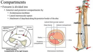
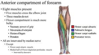
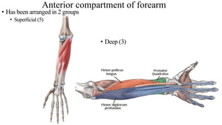
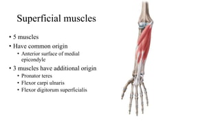
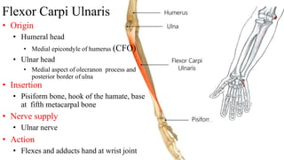
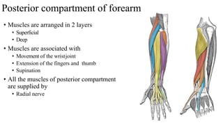
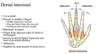
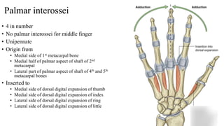
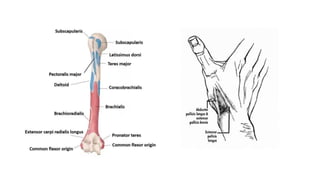
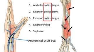
2. Compartments
⢠Forearm is divided into
⢠Anterior and posterior compartments by
⢠Aninterosseousmembrane
⢠Lateral intermuscular septum
⢠Attachment of deepfasciaalongtheposterior border of theulna
3. Anterior compartment of forearm
⢠Eight muscles present
⢠Five muscles cross the elbow joint
⢠Three musclesdonot
⢠Flexor compartment is much more
bulky
⢠Necessary power of grip
⢠Movementsofwristjoint
⢠Flexionoffingers
⢠Pronation
⢠All are innervated by median nerve
⢠Except
⢠Flexor carpi ulnaris muscle
⢠Medial half of Flexor digitorum profundus muscle
⢠Innervated byulnarnerve
4. Anterior compartment of forearm
⢠Deep (3)
⢠Has been arranged in 2 groups
⢠Superficial (5)
5. Superficial muscles
⢠5 muscles
⢠Have common origin
⢠Anterior surface of medial
epicondyle
⢠3 muscles have additional origin
⢠Pronator teres
⢠Flexor carpi ulnaris
⢠Flexor digitorum superficialis
6. Pronator Teres
⢠Origin
⢠Humeral head
⢠Medial epicondyle of humerus (CFO)
⢠Ulnar head
⢠Medial border of coronoid process of ulna
⢠Insertion
⢠Middle of lateral aspect of shaft of radius
⢠Nerve supply
⢠Median nerve
⢠Action
⢠Pronation and flexion of elbow
7. Flexor carpi radialis
⢠Origin
⢠Medial epicondyle of humerus (CFO)
⢠Insertion
⢠Bases of second and third metacarpal bones
⢠Nerve supply
⢠Median nerve
⢠Action
⢠Flexes elbow & wrist
⢠Abducts hand at wrist joint
9. Flexor Carpi Ulnaris
⢠Origin
⢠Humeral head
⢠Medial epicondyle of humerus (CFO)
⢠Ulnar head
⢠Medial aspect of olecranon process and
posterior border of ulna
⢠Insertion
⢠Pisiform bone, hook of the hamate, base
at fifth metacarpal bone
⢠Nerve supply
⢠Ulnar nerve
⢠Action
⢠Flexes and adducts hand at wrist joint
10. Flexor Digitorum Superficialis
⢠Origin
⢠Humeroulnar head
⢠Medial epicondyle of humerus (CFO);
medial border of coronoid process of ulna
⢠Radial head
⢠Oblique line on anterior surface of shaft
of radius
⢠Insertion
⢠Middle phalanx of medial four fingers
⢠Nerve supply
⢠Median nerve
⢠Action
⢠Flexes middle phalanx of fingers and
assists in flexing proximal phalanx and
hand
11. Deep group
⢠3 muscles
⢠Flexor digitorum profundus
⢠Flexor pollicis longus
⢠Pronator quadratus
12. Flexor digitorum profundus
⢠Origin
⢠Anteromedial surface of shaft of
ulna
⢠Insertion
⢠Base of distal phalanges
⢠of medial four fingers
⢠Action
⢠Flexion of middle and proximal
phalanges and wrist
⢠Nerve supply
⢠Ulnar (medial half)
⢠Anterior interosseous branch of
median nerve (lateral half)
13. Flexor pollicis longus
⢠Origin
⢠Anterior surface of shaft of
radius
⢠Insertion
⢠Distal phalanx of thumb
⢠Action
⢠Flexes distal phalanx of
thumb
⢠Nerve supply
⢠Anterior interosseous branch
of median nerve
14. Pronator quadratus
⢠Origin
⢠Lower Ÿ of anterior surface of
shaft of ulna
⢠Insertion
⢠Lower Ÿ of anterior surface of
shaft of radius
⢠Action
⢠Pronates radioulnar joint
⢠Nerve Supply
⢠Anterior interosseous
⢠Branch of median nerve
15. Space of Parona
⢠Space in front of pronator quadratus
⢠Deep to long flexor tendons of fingers and their synovial
sheaths
⢠Limited proximally by
⢠Oblique origin of flexor digitorum superficialis
⢠Space involved in proximal extensions of synovial
sheath infections
⢠Can be drained through radial and ulnar incisions to the
side of the flexor tendons
16. ⢠Flexors of Wrist
⢠Fl. Carpi Radialis
⢠Fl. Carpi Ulnaris
⢠Flexors of Middle Phalanges
⢠Fl. Digitorum Superficialis
⢠Flexors of Distal Phalanges
⢠Fl. Digitorum Profundus
⢠Fl. Pollicis Longus
⢠Pronator of the Forearm
⢠Pronator Teres
⢠Pronator Quadratus
17. Flexor Retinaculum
⢠Strong, fibrous band that covers the
carpal bones on palmar side of hand
near wrist
⢠Attachment
⢠Ulnar side
⢠Pisiform bone and hook of hamate bone
⢠Radial side
⢠Tubercle of scaphoid bone and ridge of
trapezium bone
18. Structures passing superficial to flexor retinaculum
⢠Tendon of Palmaris longus
⢠Palmer cutaneous branch of Median
nerve
⢠Palmer cutaneous branch of Ulnar
nerve
⢠Ulnar vessels
⢠Ulnar nerve
19. Structures passing deep to Flexor retinaculum
ď§ Median nerve
ď§ Tendon of flexor digitorum
superficialis
ď§ Tendon of flexor digitorum profundus
ď§ Tendon of flexor pollicis longus
ď§ Ulnar bursa
ď§ Radial bursa
20. Posterior compartment of forearm
⢠Muscles are arranged in 2 layers
⢠Superficial
⢠Deep
⢠Muscles are associated with
⢠Movement of the wristjoint
⢠Extension of the fingers and thumb
⢠Supination
⢠All the muscles of posterior compartment
are supplied by
⢠Radial nerve
21. Arrangement of posterior compartment muscles
⢠Upper part
⢠Anconeus (superficial)
⢠Supinator (deep)
⢠Lateral part of humerus
⢠Pass along the radial side
⢠Brachioradialis
⢠Extensors carpi radialis longus
⢠Extensors carpi radialis brevis
⢠Passing along posterior surface of the
forearm
⢠Extensor digitorum
⢠Extensor digiti minimi and
⢠Extensor carpi ulnaris
⢠At lower end of forearm
⢠These two groups are separated by
three muscles
⢠They emerge from deeply in between
them and go to the thumb
⢠Abductor pollicis longus
⢠Extensors pollicis longus and
⢠Extensors pollicis longus brevis
⢠One muscle for the forefinger runs
deeply to reach the back of the hand
⢠Extensor indicis
22. Superficial muscles of the back of forearm
⢠There are 7 muscles
⢠Common extensor origin
⢠Four of the superficialmuscles
⢠Arise from the tip of lateral epicondyle of the
humerus
⢠Extensor carpi radialis brevis
⢠Extensor digitorum
⢠Extensor digiti minimi, and
⢠Extensor carpi ulnaris
⢠All seven muscles cross elbow joint
23. Superficial muscles of back of forearm
⢠Categorized into two groups
⢠Lateral and posterior
⢠Each group consists of three muscles
⢠Lateral group
⢠Brachioradialis
⢠Extensor carpi radialis longus
⢠Extensor carpi radialis brevis
⢠Posterior group
⢠Extensor digitorum
⢠Extensor digiti minimi
⢠Extensor carpi ulnaris
⢠In the upper end
⢠Anconeus
24. Brachioradialis
⢠Origin
⢠Upper 2/3rd of lateral supracondylar ridge of
humerus
⢠Insertion
⢠Into base of styloid process of radius
⢠Nerve Supply
⢠Radial nerve
⢠Action
⢠Flexion of the forearm (specially in midprone
position)
25. Extensor carpi radialis longus
⢠Origin
⢠From the lower 1/3rd of the lateral
supracondylar ridge of the humerus
⢠Insertion
⢠Into posterior surface of base of 2nd
metacarpal bone
⢠Nerve Supply
⢠Radial nerve
⢠Action
⢠Extension & Abduction of wrist
joint
⢠Extension of elbow joint
26. ⢠Common extensor origin
⢠4 out of 7 superficial muscles
⢠Extensor carpi radialis brevis
⢠Extensor digitorum
⢠Extensor digiti minimi
⢠Extensor carpi ulnaris
27. Extensor carpi radialis brevis
⢠Origin
⢠Lateral epicondyle of humerus
(common extensor origin)
⢠Insertion:
⢠Posterior surface of base of 3rd
metacarpal bone
⢠Nerve Supply
⢠Posterior interosseous nerve
⢠Action
⢠Extension & abduction of the
hand at wrist joint
28. Extensor digitorum
⢠Origin
⢠Lateral epicondyle of humerus
(common extensor origin)
⢠Insertion
⢠Extensor expansion of the medial
4 fingers
⢠Nerve Supply
⢠Posterior interosseous nerve
⢠Action
⢠Extension of
metacarpophalangeal and
interphalangeal joints of the
medial 4 fingers
⢠Extension of the hand at wrist
joint
29. Dorsal digital expansion
⢠Tendons of extensor digitorum
expands
⢠Covers dorsal surface of
⢠Head of metacarpal & proximal phalanx
⢠Triangular shape
⢠Base
⢠Forms a hood over metacarpal head
⢠Apex
⢠At proximal phalanx
⢠Trifurcates into median & two lateral bands
30. Dorsal digital expansion
ď The tendon splits into three parts:
ď One Central: inserted into the base of Middle phalanx
ď Two laterals: inserted into the base of the Distal phalanx
ď Receives the insertions of:
ď Corresponding Interosseous muscle (on each side)
ď Lumbrical muscle (on the lateral side)
ď Also receives
ď Extensor indicis
ď Extensor digiti minimi
33. Extensor digiti minimi
⢠Origin
⢠Lateral epicondyle of the humerus
(common extensor origin)
⢠Insertion
⢠Extensor expansion of the little finger
⢠Nerve Supply
⢠Posterior interosseous nerve
⢠Action
⢠Extension of metacarpophalangeal
and interphalangeal joints of the
little finger
34. Extensor carpi ulnaris
⢠Origin
⢠Lateral epicondyle of the humerus
(common extensor origin)
⢠Insertion
⢠Posterior surface of the base of the 5th
metacarpal bone
⢠Nerve Supply
⢠Posterior interosseous nerve
⢠Action
⢠Extension & Adduction of the
hand at wrist joint
35. Anconeus
⢠Origin
⢠Posterior aspect of the lateral
epicondyle of the humerus
⢠Insertion
⢠Lateral surface of olecranon
process of ulna
⢠Nerve Supply
⢠Posterior interosseous nerve
⢠Action
⢠Helps the triceps in extension of
the elbow joint
36. Deep Layer
⢠There are five deep muscles
⢠Three deep extensorsof the forearm
⢠Function on thumb
⢠Abductor pollicis longus(APL)
⢠Extensor pollicis brevis (EPB)
⢠Extensor pollicis longus (EPL)
⢠To acquire insertion on the three bones of
thumb âcrop outâ
⢠From furrow in lateralelementof forearmbetweenlateral
andposterior groupsof superficialextensor
⢠One muscle
⢠Functiononindexfinger
⢠Extensor indicis
⢠In the upper part
⢠Supinator
37. Supinator
⢠Origin
⢠Lateral epicondyle of the humerus
⢠Radial collateral lig
⢠Annular ligament of the superior
radio-ulnar joint
⢠Supinator fossa and crest of the ulna
⢠Insertion
⢠Upper 1/3rd of the lateral surface of
shaft of radius
⢠Nerve Supply
⢠Posterior interosseous nerve
⢠Action
⢠Supination of the forearm
⢠At radio-ulnar joint
38. Abductor pollicis longus
⢠Origin
⢠Middle of the posterior surface of radius and ulna
⢠Interosseus membrane
⢠Insertion
⢠Posterior surface of the base of the 1st metacarpal
bone
⢠Nerve Supply
⢠Posterior interosseous nerve
⢠Action
⢠Abduction and extension of the thumb at
the carpo-metacarpal joint
39. Extensor pollicis brevis
⢠Origin
⢠Posterior surface of the shaft of radius
⢠Insertion
⢠Posterior surface of the base of the proximal
phalanx of the thumb
⢠Nerve Supply
⢠Posterior interosseous nerve
⢠Action
⢠Extension of the metacarpophalangeal joint of
the thumb
40. Extensor pollicis longus
⢠Origin
⢠Posterior surface of ulna
⢠Insertion
⢠Posterior surface of the base of the distal
phalanx of the thumb
⢠Nerve Supply
⢠Posterior interosseous nerve
⢠Action
⢠Extension of the interphalangeal joints of the
thumb
42. Extensor indicis
⢠Origin
⢠Posterior surface of ulna (below extensor
pollicis longus)
⢠Insertion
⢠Extensor expansion of the index finger.
⢠Nerve Supply
⢠Posterior interosseous nerve
⢠Action
⢠Extension of the interphalangeal joints of
the thumb
43. Wrist Drop
⢠Sign of radial nerve injury
⢠There are two common characteristic sites
of damage
⢠Axilla
⢠Injured viahumeral dislocationsor fractures of the
proximalhumerus
⢠Radial groove of the humerus
⢠Injured via a humeral shaft fracture
⢠Radial nerve innervates
⢠All muscles in extensor compartment of
forearm
⢠In radial nerve lesion
⢠These muscles are paralysed
⢠Muscles that flex the wrist
⢠Innervated by median nerve
⢠Flexor muscles produces unopposed flexion at
the wrist joint â wrist drop
44. Supination and pronation
⢠These movements occur at
⢠Radioulnar joint
⢠Head of radius & ulna rotate in their
sockets
⢠Supination
⢠Movement of forearm
⢠Radius is rotating along a longitudinal axis
⢠Pronation
⢠Movement of forearm
⢠Radius is rotating along the longitudinal axis
⢠Thumb directed medially
⢠Palm faces backwards
⢠Body of radius lies obliquely across ulna
45. Dorsal interossei
⢠4 in number
⢠Present in middle 3 fingers
⢠Middle finger has 2 muscles
⢠Ring and index finger has one each
⢠Numbered from lateral to medial
⢠Bipennate in nature
⢠Origin from adjacent sides of shaft of
metacarpals
⢠Inserts on dorsal digital expansion and
base of proximal phalanx
⢠Abductors
⢠Supplies by deep branch of ulnar nerve
46. Palmar interossei
⢠4 in number
⢠No palmar interossei for middle finger
⢠Unipennate
⢠Origin from
⢠Medial side of 1st metacarpal bone
⢠Medial half of palmar aspect of shaft of 2nd
metacarpal
⢠Lateral part of palmar aspect of shaft of 4th and 5th
metacarpal bones
⢠Inserted to
⢠Medial side of dorsal digital expansion of thumb
⢠Medial side of dorsal digital expansion of index
⢠Lateral side of dorsal digital expansion of ring
⢠Lateral side of dorsal digital expansion of little