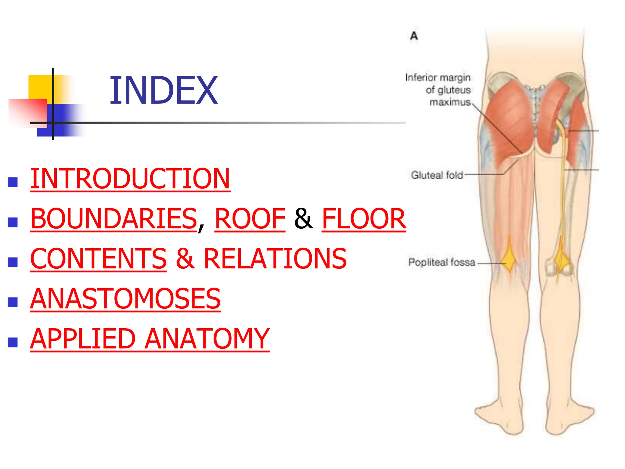
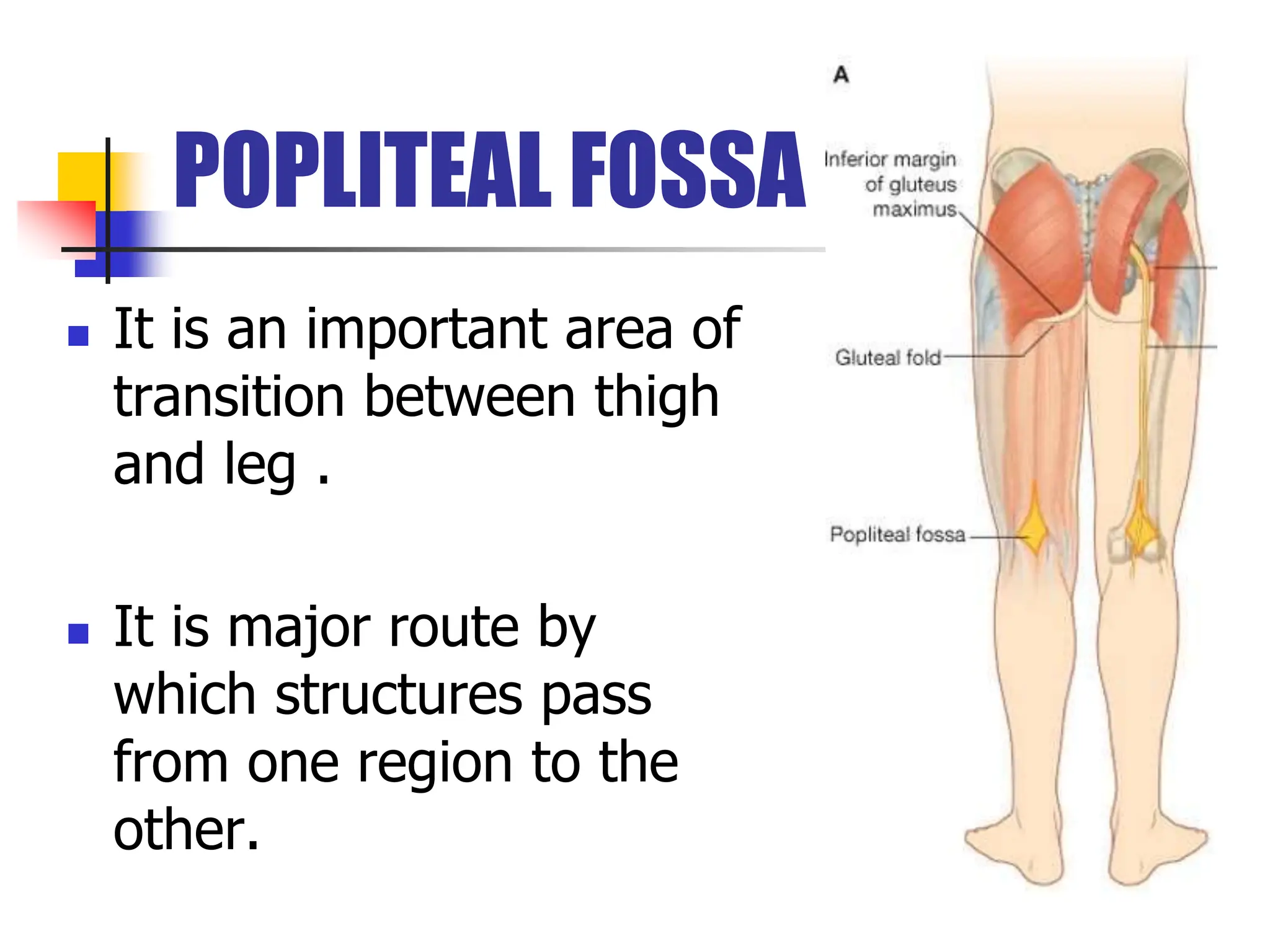
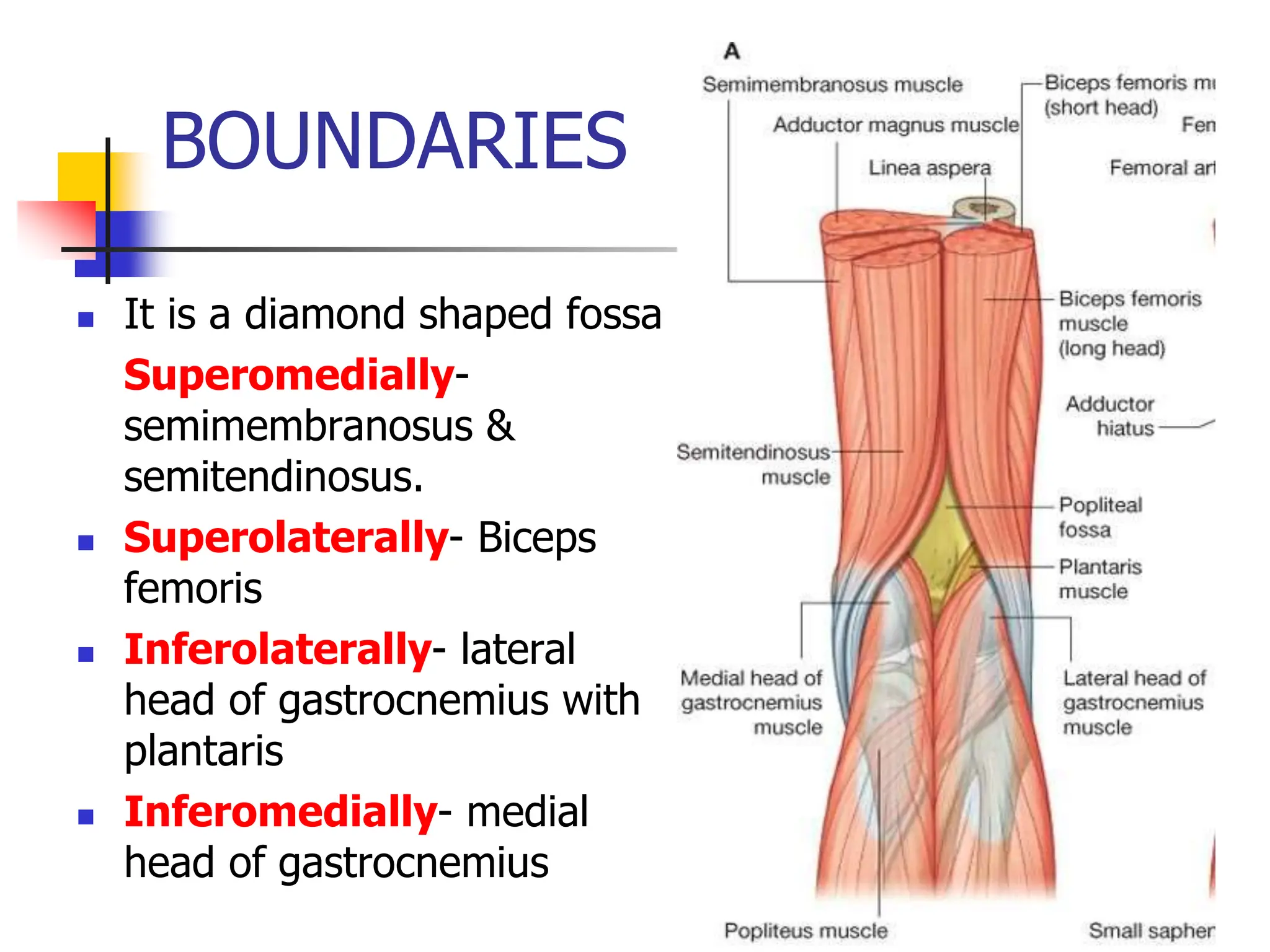
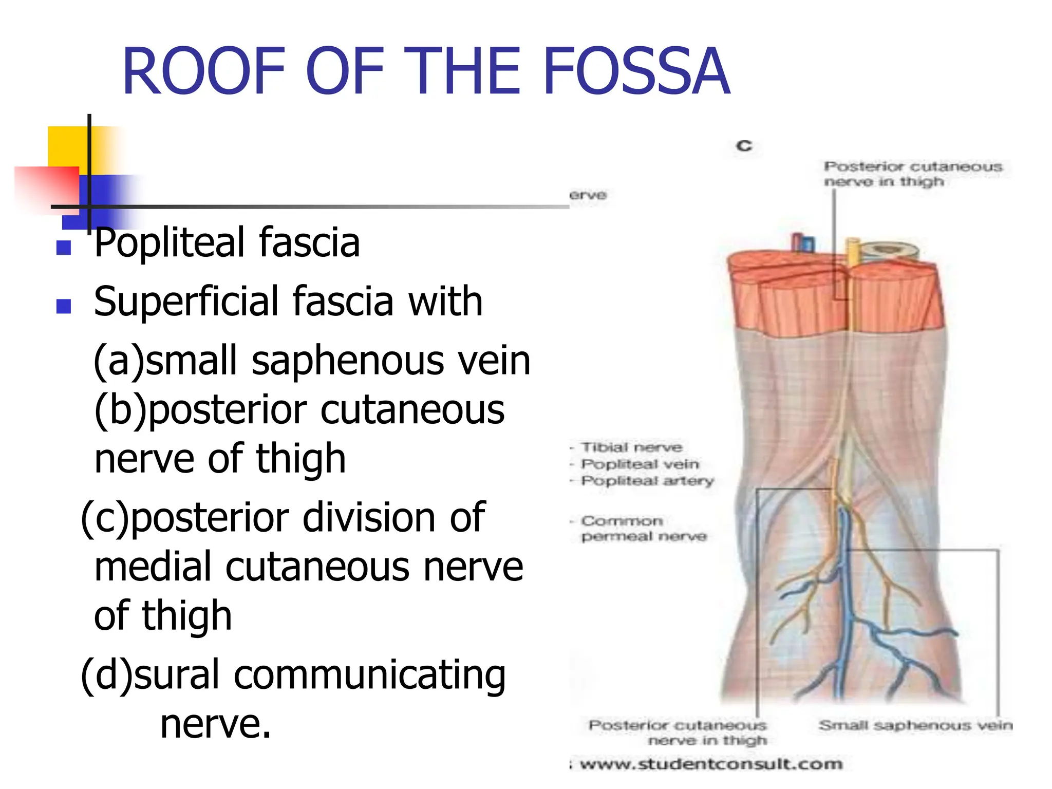
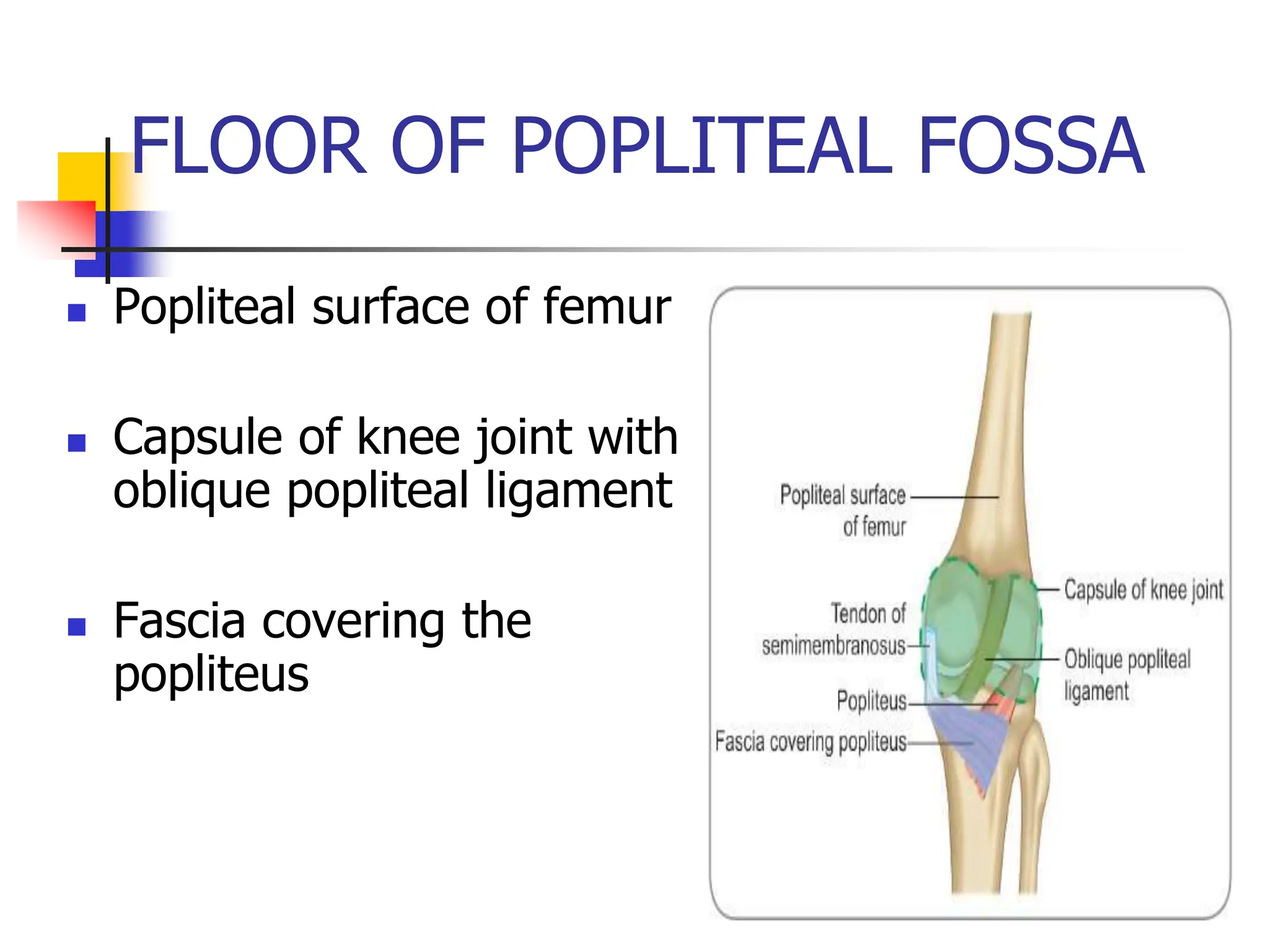
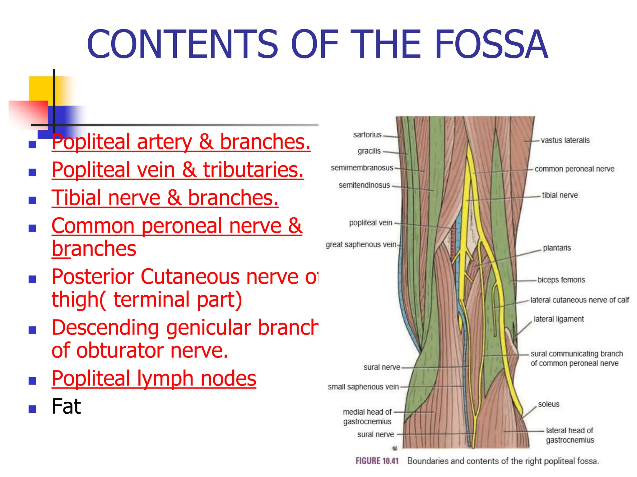
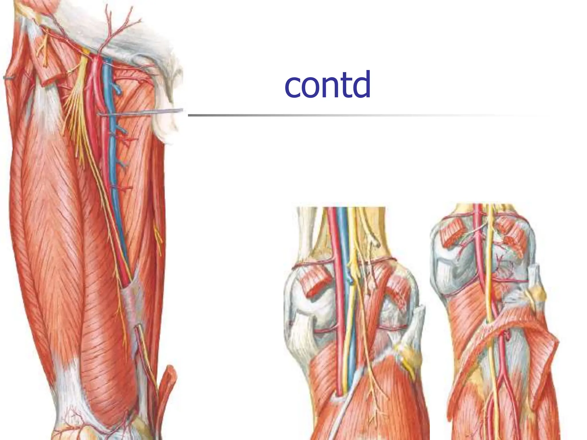
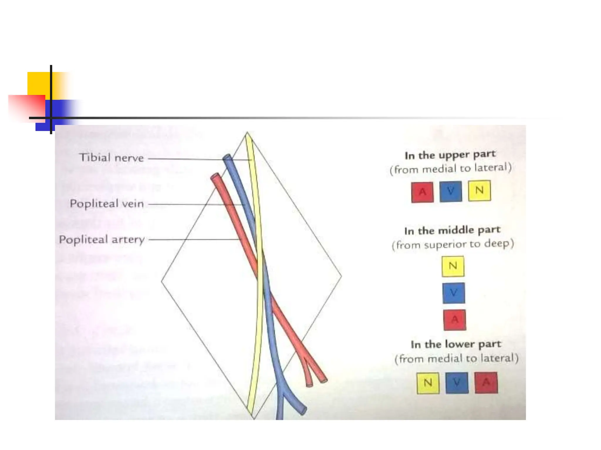
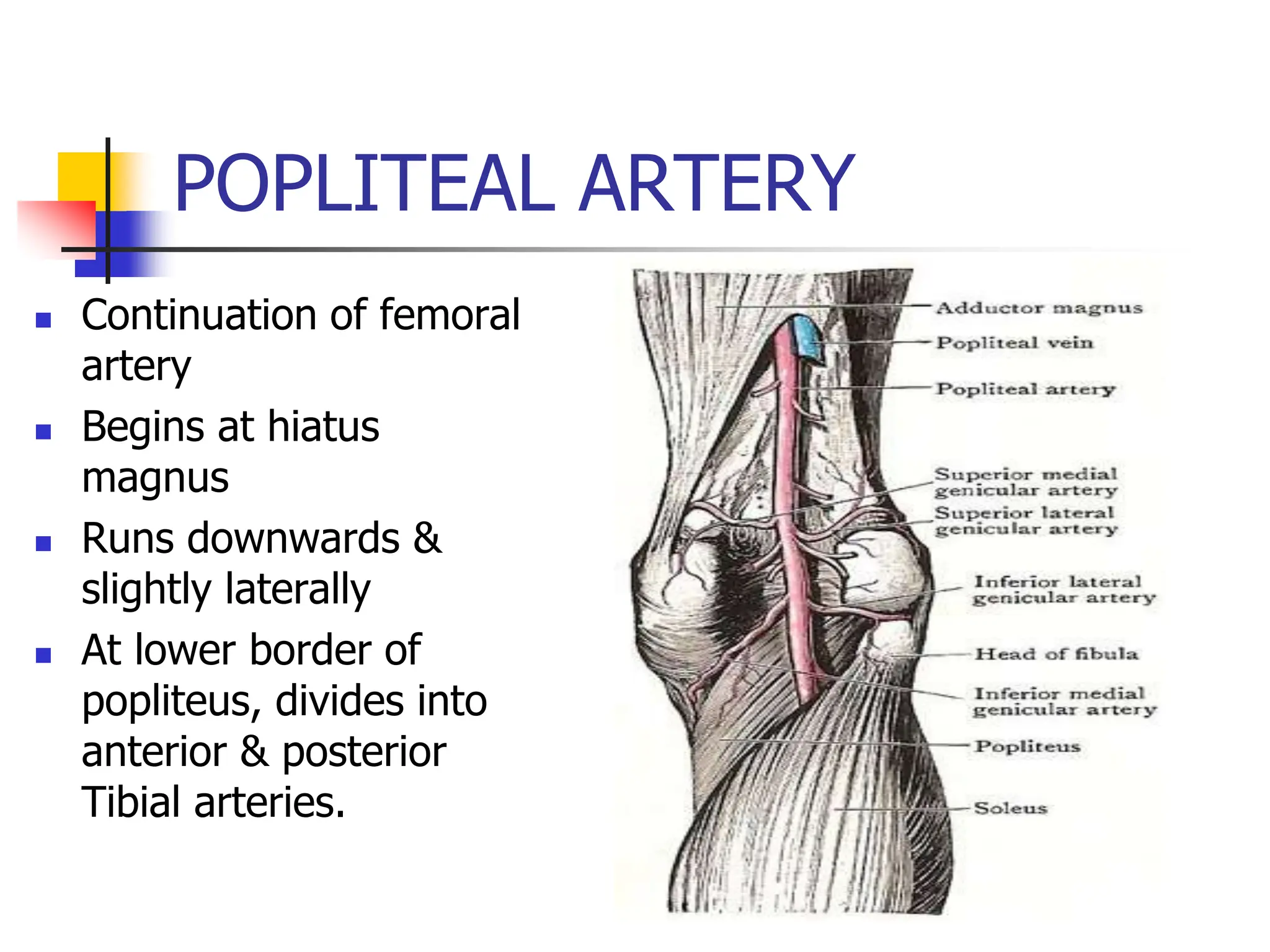
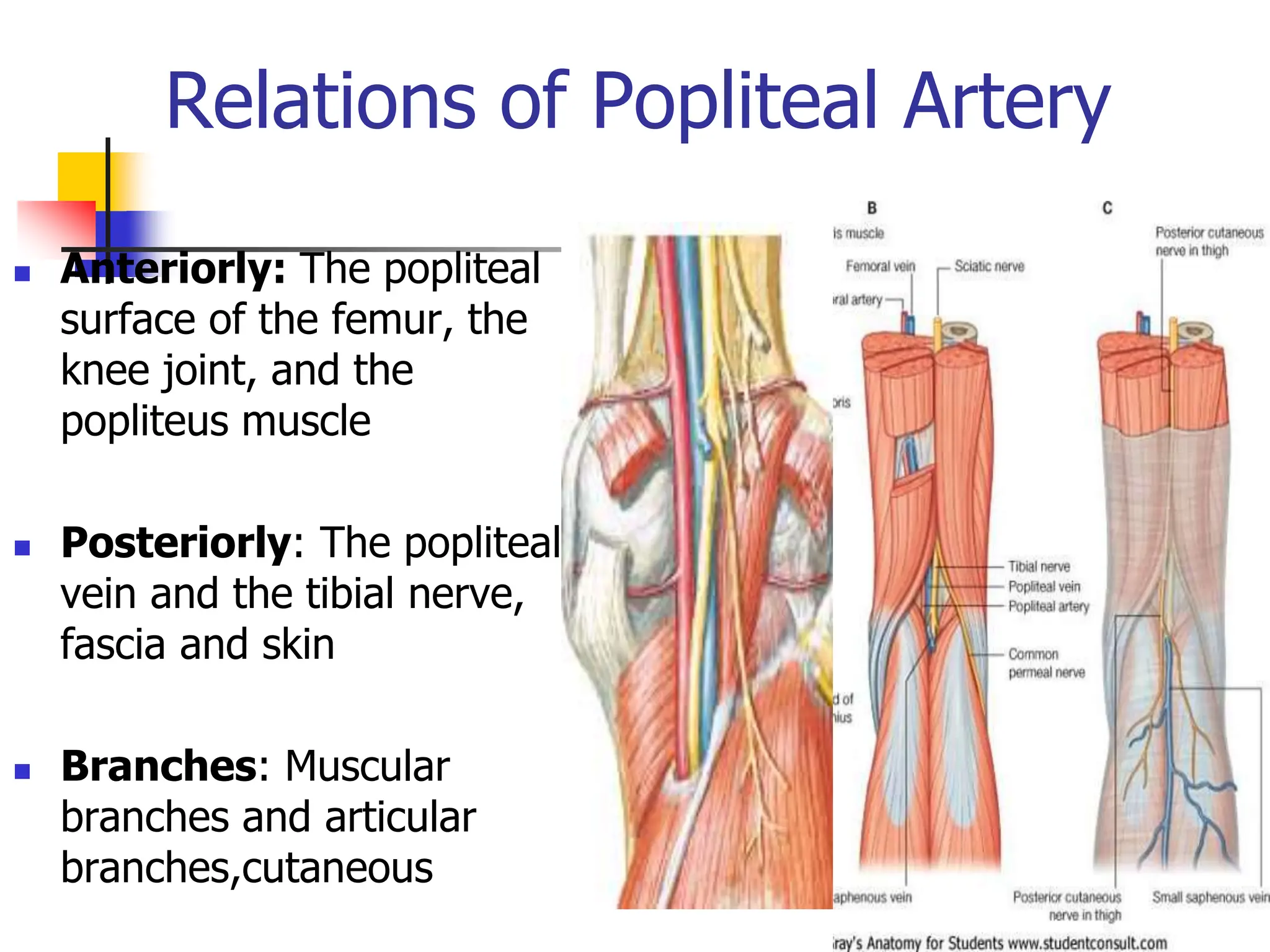
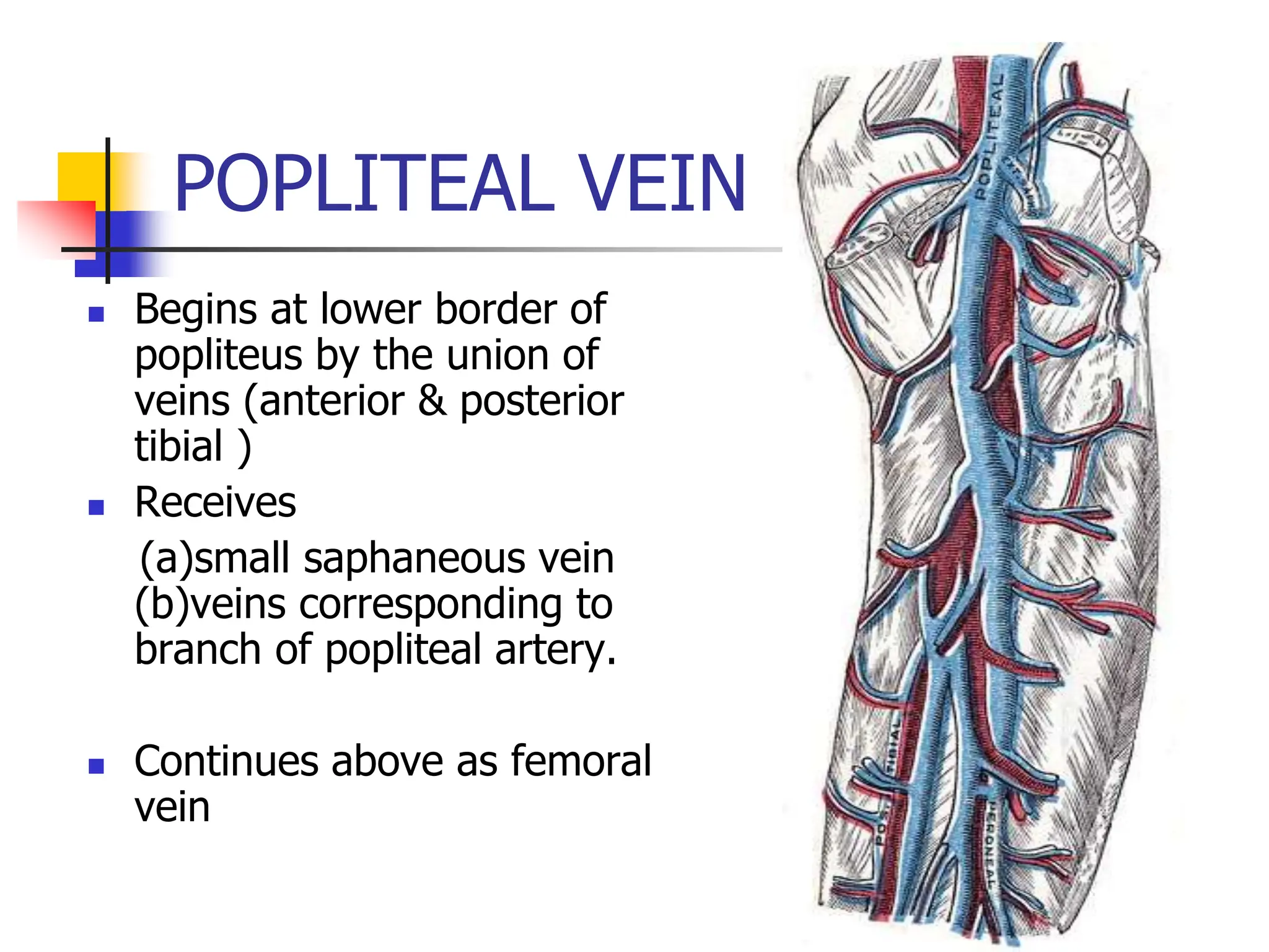
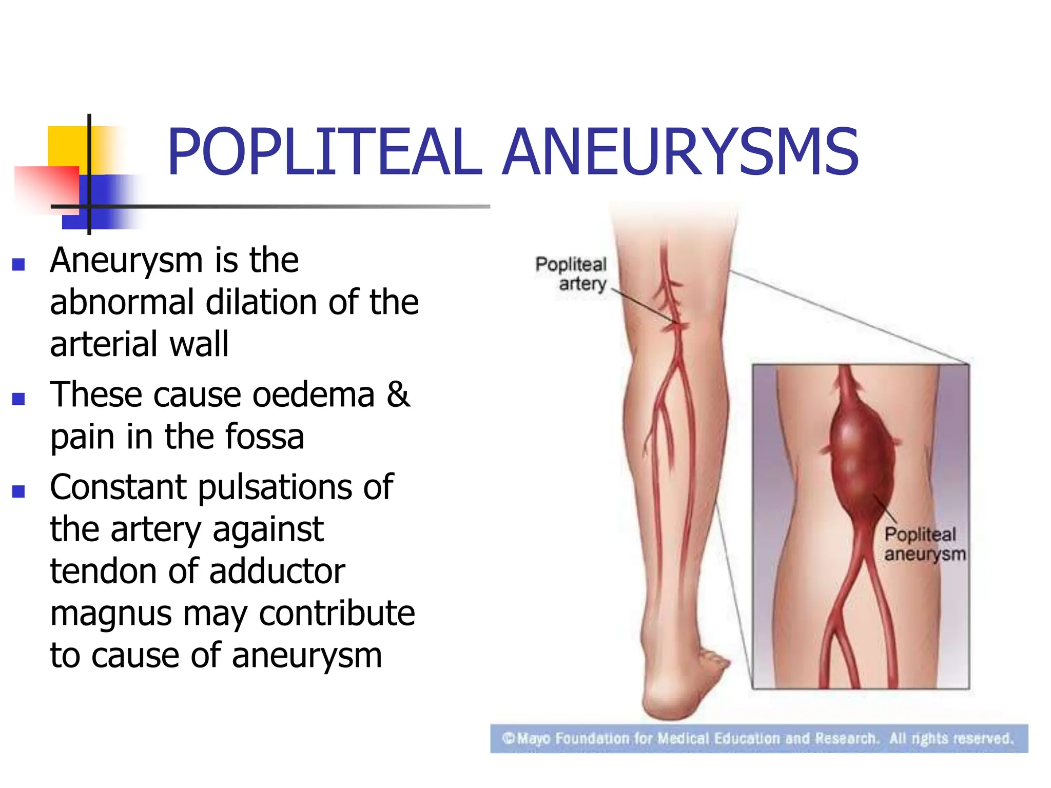
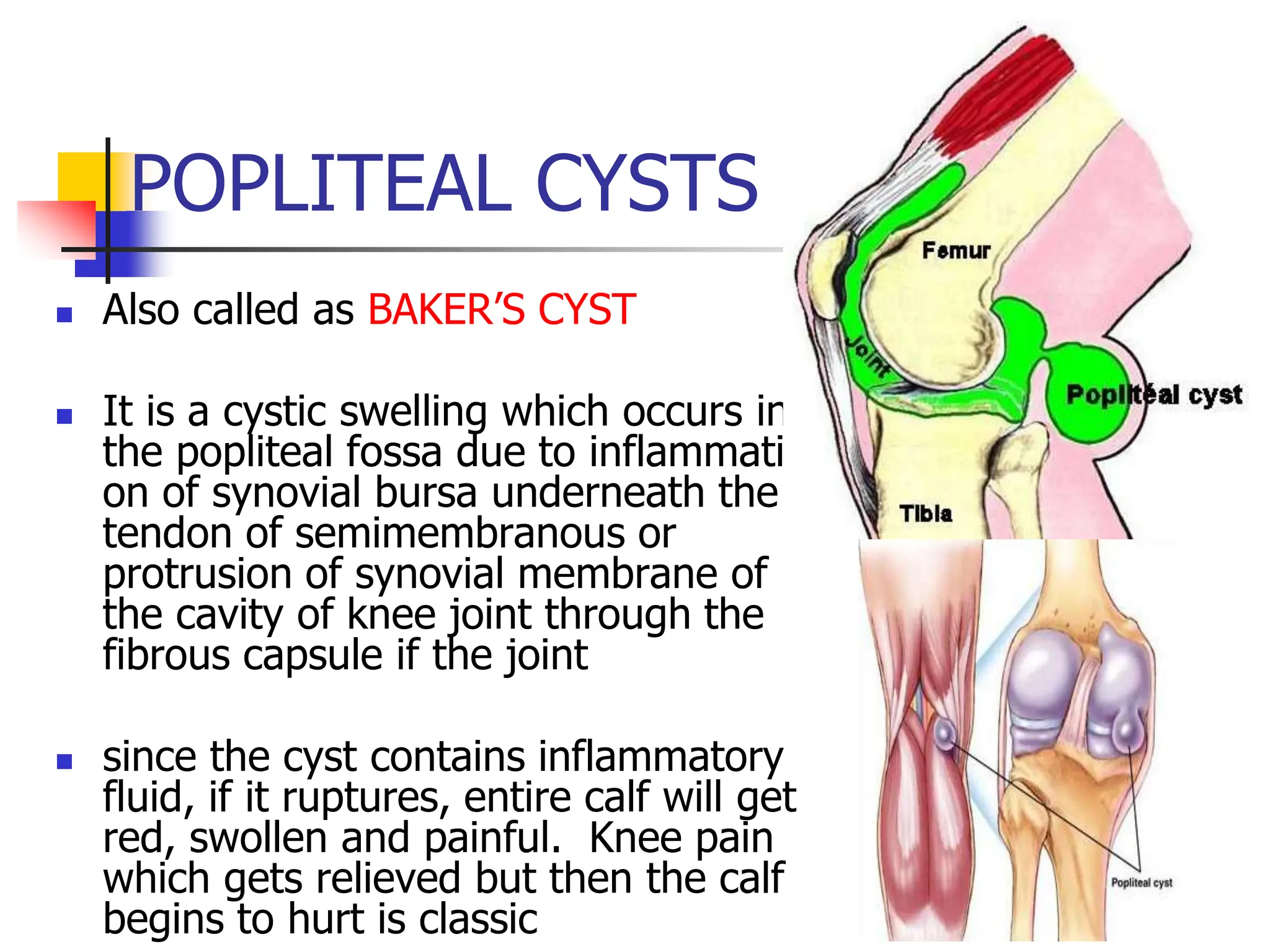
POPLITEAL FOSSA
Dr.M.VASANTHAKOHILA
POSTGRADUATE
DEPARTMENT OF ANATOMY-KMC
AN 16.6 (ECE)
DATE: 22.11.23
INDEX
 INTRODUCTION
 BOUNDARIES, ROOF & FLOOR
 CONTENTS & RELATIONS
 ANASTOMOSES
 APPLIED ANATOMY
POPLITEAL FOSSA
 It is an important area of
transition between thigh
and leg .
 It is major route by
which structures pass
from one region to the
other.
BOUNDARIES NEXT
 It is a diamond shaped fossa
Superomedially-
semimembranosus &
semitendinosus.
 Superolaterally- Biceps
femoris
 Inferolaterally- lateral
head of gastrocnemius with
plantaris
 Inferomedially- medial
head of gastrocnemius
ROOF OF THE FOSSA
 Popliteal fascia
 Superficial fascia with
(a)small saphenous vein
(b)posterior cutaneous
nerve of thigh
(c)posterior division of
medial cutaneous nerve
of thigh
(d)sural communicating
nerve.
FLOOR OF POPLITEAL FOSSA
 Popliteal surface of femur
 Capsule of knee joint with
oblique popliteal ligament
 Fascia covering the
popliteus
CONTENTS OF THE FOSSA
 Popliteal artery & branches.
 Popliteal vein & tributaries.
 Tibial nerve & branches.
 Common peroneal nerve &
branches
 Posterior Cutaneous nerve of
thigh( terminal part)
 Descending genicular branch
of obturator nerve.
 Popliteal lymph nodes
 Fat
Conti contd
POPLITEAL ARTERY
 Continuation of femoral
artery
 Begins at hiatus
magnus
 Runs downwards &
slightly laterally
 At lower border of
popliteus, divides into
anterior & posterior
Tibial arteries.
BRANCHES OF POPLITEAL ART.
 Several muscular branch
to hamstrings, adductor
magnus, gastrocnemius,
soleus & plantaris.
 Genicular- 2 superior, 2
inferior &1 middle
genicular arteries.
 Cutaneous branch arising
either directly from the
artery Or from the
muscular branches
Relations of Popliteal Artery
 Anteriorly: The popliteal
surface of the femur, the
knee joint, and the
popliteus muscle
 Posteriorly: The popliteal
vein and the tibial nerve,
fascia and skin
 Branches: Muscular
branches and articular
branches,cutaneous
POPLITEAL VEIN
 Begins at lower border of
popliteus by the union of
veins (anterior & posterior
tibial )
 Receives
(a)small saphaneous vein
(b)veins corresponding to
branch of popliteal artery.
 Continues above as femoral
vein
Tributaries of Popliteal Vein
 Veins that
correspond to
branches given off
by the popliteal
artery
 Small saphenous
vein
TIBIAL NERVE
 Larger terminal branch
of sciatic nerve.
 Superficial or posterior
to popliteal vessels
crossing them lateral to
medial side.
Branches of Tibial Nerve
 Cutaneous: Sural Nerve
 Muscular
 Gastrocnemius
 Plantaris,
 Soleus
 Nerve to Popliteus
 Genicular – 3 branches
 Vascular – T10 - L2
Common Peroneal
Nerve
 Smaller terminal branch
of sciatic nerve.
 Extends from superior
angle to lateral angle of
the fossa.
 Winds around the
posterolateral aspect of
neck of fibula.
Branches of Common
Peroneal Nerve
 Cutaneous:
1. Sural communicating
nerve
2. Lateral cutaneous
nerve/lateral sural
nerve.
 Articular branches :
1. Superior lateral
2. Inferior lateral
3. Recurrent genicular
nerves.
ANASTOMOSIS AROUND
KNEE JOINT
ANASTOMOSES
 Complicated arterial network around patella, lower end of
femur & upper end of tibia
 Formed, medially by
(a)descending genicular branch of femoral artery
(b)superior Medial genicular artery
(c)inferior Medial genicular artery
 Laterally by
(a)descending branch of lateral circumflex femoral artery
(b)superior Lateral genicular artery
(c)inferior lateral genicular artery
 These longitudinal anastomoses are interconnected to form
transverse anastomoses
ANASTOMOSES
 Inferior lateral genicular artery anastomosis with
a) Superior lateral genicular artery
b) Anterior and posterior recurrent branches of
posterior tibial artery
c) Circumflex branch of posterior tibial artery
Inferior medial genicular artery anastomosis with
a) Superior medial genicular artery
b) Saphaneous artery – a branch of descending
genicular artery (branch of femoral artery)
Arterial Anastomosis Around
Knee Joint
 To compensate for the narrowing of
the popliteal artery which occurs
during extreme flexion of the knee
POPLITEAL LYMPH NODES
 3 to 4 nodes lie at the
termination of small
saphenous vein
 Afferents
(a) territory of small
saphaneous Vein.
(b) deep parts of leg
(c) knee jt.
 Efferents run along
popliteal & femoral; vessels
to terminate into deep
inguinal lymph nodes
APPLIED ANATOMY
 Popliteal aneurysms
 Popliteal abscess & tumors
 cysts
 Foot drop
 Semimembranosus bursa swelling
 Palpation of popliteal pulse
 Damage to tibial nerve
POPLITEAL ANEURYSMS
 Aneurysm is the
abnormal dilation of the
arterial wall
 These cause oedema &
pain in the fossa
 Constant pulsations of
the artery against
tendon of adductor
magnus may contribute
to cause of aneurysm
Popliteal abscess
 Because the fascia over
the popliteus is strong &
limits expansion, pain
from an abscess or tumor
is severe
 It tends to spread
superiorly or inferiorly
because of toughness of
popliteal fascia
POPLITEAL CYSTS
 Also called as BAKER’S CYST
 It is a cystic swelling which occurs in
the popliteal fossa due to inflammation
on of synovial bursa underneath the
tendon of semimembranous or
protrusion of synovial membrane of
the cavity of knee joint through the
fibrous capsule if the joint
 since the cyst contains inflammatory
fluid, if it ruptures, entire calf will get
red, swollen and painful. Knee pain
which gets relieved but then the calf
begins to hurt is classic
BURSA SWELLING
 Semimembranosus bursa swelling is the most
common swelling in the fossa
 Made tense by extending knee joint &
become flaccid on flexing
 Should be distiguished from baker’s cysts,
which is a centrally located pathologic
diverticulum of synovial membrane through a
hole in the back of the knee joint.
PALPATION OF POPLITEAL
ARTERY
 Popliteal artery is palpated
for pulse & also used for
measuring B.P in the lower
limb.
 As it is one of the deepest
area in the fossa it is best
felt by pressing the patella
from the front &fingertips
of both the hands in to
middle of the fossa
Damage to Tibial nerve
 This is usually rare because of
its protected position in the
fossa.
 However the nerve may be
injured by deep lacerations or
wounds in the fossa. Also
posterior dislocation of knee jt
may damage tibial nerve
 Damage may cause paralysis of
flexor muscles in the leg &
intrinsic muscles of the sole.
Injury to common peroneal
nerve
 Can get easily injured at
the neck of fibula
 Paralysis of evertors of
foot & dorsiflexors
 Unapposed action of
plantar flexors leading to
foot drop
 CPN. can be palpated at
the neck of the fibula. In
leprosy it becomes
thickened, nodular &
hypersensitive.
ANATOMY AND APPLIED ASPECTS OF POPLITEAL FOSSA FINAL.ppt

ANATOMY AND APPLIED ASPECTS OF POPLITEAL FOSSA FINAL.ppt

  • 1.
  • 2.
    INDEX  INTRODUCTION  BOUNDARIES,ROOF & FLOOR  CONTENTS & RELATIONS  ANASTOMOSES  APPLIED ANATOMY
  • 3.
    POPLITEAL FOSSA  Itis an important area of transition between thigh and leg .  It is major route by which structures pass from one region to the other.
  • 4.
    BOUNDARIES NEXT  Itis a diamond shaped fossa Superomedially- semimembranosus & semitendinosus.  Superolaterally- Biceps femoris  Inferolaterally- lateral head of gastrocnemius with plantaris  Inferomedially- medial head of gastrocnemius
  • 5.
    ROOF OF THEFOSSA  Popliteal fascia  Superficial fascia with (a)small saphenous vein (b)posterior cutaneous nerve of thigh (c)posterior division of medial cutaneous nerve of thigh (d)sural communicating nerve.
  • 6.
    FLOOR OF POPLITEALFOSSA  Popliteal surface of femur  Capsule of knee joint with oblique popliteal ligament  Fascia covering the popliteus
  • 7.
    CONTENTS OF THEFOSSA  Popliteal artery & branches.  Popliteal vein & tributaries.  Tibial nerve & branches.  Common peroneal nerve & branches  Posterior Cutaneous nerve of thigh( terminal part)  Descending genicular branch of obturator nerve.  Popliteal lymph nodes  Fat
  • 8.
  • 10.
    POPLITEAL ARTERY  Continuationof femoral artery  Begins at hiatus magnus  Runs downwards & slightly laterally  At lower border of popliteus, divides into anterior & posterior Tibial arteries.
  • 11.
    BRANCHES OF POPLITEALART.  Several muscular branch to hamstrings, adductor magnus, gastrocnemius, soleus & plantaris.  Genicular- 2 superior, 2 inferior &1 middle genicular arteries.  Cutaneous branch arising either directly from the artery Or from the muscular branches
  • 12.
    Relations of PoplitealArtery  Anteriorly: The popliteal surface of the femur, the knee joint, and the popliteus muscle  Posteriorly: The popliteal vein and the tibial nerve, fascia and skin  Branches: Muscular branches and articular branches,cutaneous
  • 13.
    POPLITEAL VEIN  Beginsat lower border of popliteus by the union of veins (anterior & posterior tibial )  Receives (a)small saphaneous vein (b)veins corresponding to branch of popliteal artery.  Continues above as femoral vein
  • 14.
    Tributaries of PoplitealVein  Veins that correspond to branches given off by the popliteal artery  Small saphenous vein
  • 15.
    TIBIAL NERVE  Largerterminal branch of sciatic nerve.  Superficial or posterior to popliteal vessels crossing them lateral to medial side.
  • 16.
    Branches of TibialNerve  Cutaneous: Sural Nerve  Muscular  Gastrocnemius  Plantaris,  Soleus  Nerve to Popliteus  Genicular – 3 branches  Vascular – T10 - L2
  • 17.
    Common Peroneal Nerve  Smallerterminal branch of sciatic nerve.  Extends from superior angle to lateral angle of the fossa.  Winds around the posterolateral aspect of neck of fibula.
  • 18.
    Branches of Common PeronealNerve  Cutaneous: 1. Sural communicating nerve 2. Lateral cutaneous nerve/lateral sural nerve.  Articular branches : 1. Superior lateral 2. Inferior lateral 3. Recurrent genicular nerves.
  • 19.
  • 20.
    ANASTOMOSES  Complicated arterialnetwork around patella, lower end of femur & upper end of tibia  Formed, medially by (a)descending genicular branch of femoral artery (b)superior Medial genicular artery (c)inferior Medial genicular artery  Laterally by (a)descending branch of lateral circumflex femoral artery (b)superior Lateral genicular artery (c)inferior lateral genicular artery  These longitudinal anastomoses are interconnected to form transverse anastomoses
  • 21.
    ANASTOMOSES  Inferior lateralgenicular artery anastomosis with a) Superior lateral genicular artery b) Anterior and posterior recurrent branches of posterior tibial artery c) Circumflex branch of posterior tibial artery Inferior medial genicular artery anastomosis with a) Superior medial genicular artery b) Saphaneous artery – a branch of descending genicular artery (branch of femoral artery)
  • 22.
    Arterial Anastomosis Around KneeJoint  To compensate for the narrowing of the popliteal artery which occurs during extreme flexion of the knee
  • 23.
    POPLITEAL LYMPH NODES 3 to 4 nodes lie at the termination of small saphenous vein  Afferents (a) territory of small saphaneous Vein. (b) deep parts of leg (c) knee jt.  Efferents run along popliteal & femoral; vessels to terminate into deep inguinal lymph nodes
  • 24.
    APPLIED ANATOMY  Poplitealaneurysms  Popliteal abscess & tumors  cysts  Foot drop  Semimembranosus bursa swelling  Palpation of popliteal pulse  Damage to tibial nerve
  • 25.
    POPLITEAL ANEURYSMS  Aneurysmis the abnormal dilation of the arterial wall  These cause oedema & pain in the fossa  Constant pulsations of the artery against tendon of adductor magnus may contribute to cause of aneurysm
  • 26.
    Popliteal abscess  Becausethe fascia over the popliteus is strong & limits expansion, pain from an abscess or tumor is severe  It tends to spread superiorly or inferiorly because of toughness of popliteal fascia
  • 27.
    POPLITEAL CYSTS  Alsocalled as BAKER’S CYST  It is a cystic swelling which occurs in the popliteal fossa due to inflammation on of synovial bursa underneath the tendon of semimembranous or protrusion of synovial membrane of the cavity of knee joint through the fibrous capsule if the joint  since the cyst contains inflammatory fluid, if it ruptures, entire calf will get red, swollen and painful. Knee pain which gets relieved but then the calf begins to hurt is classic
  • 28.
    BURSA SWELLING  Semimembranosusbursa swelling is the most common swelling in the fossa  Made tense by extending knee joint & become flaccid on flexing  Should be distiguished from baker’s cysts, which is a centrally located pathologic diverticulum of synovial membrane through a hole in the back of the knee joint.
  • 29.
    PALPATION OF POPLITEAL ARTERY Popliteal artery is palpated for pulse & also used for measuring B.P in the lower limb.  As it is one of the deepest area in the fossa it is best felt by pressing the patella from the front &fingertips of both the hands in to middle of the fossa
  • 30.
    Damage to Tibialnerve  This is usually rare because of its protected position in the fossa.  However the nerve may be injured by deep lacerations or wounds in the fossa. Also posterior dislocation of knee jt may damage tibial nerve  Damage may cause paralysis of flexor muscles in the leg & intrinsic muscles of the sole.
  • 31.
    Injury to commonperoneal nerve  Can get easily injured at the neck of fibula  Paralysis of evertors of foot & dorsiflexors  Unapposed action of plantar flexors leading to foot drop  CPN. can be palpated at the neck of the fibula. In leprosy it becomes thickened, nodular & hypersensitive.