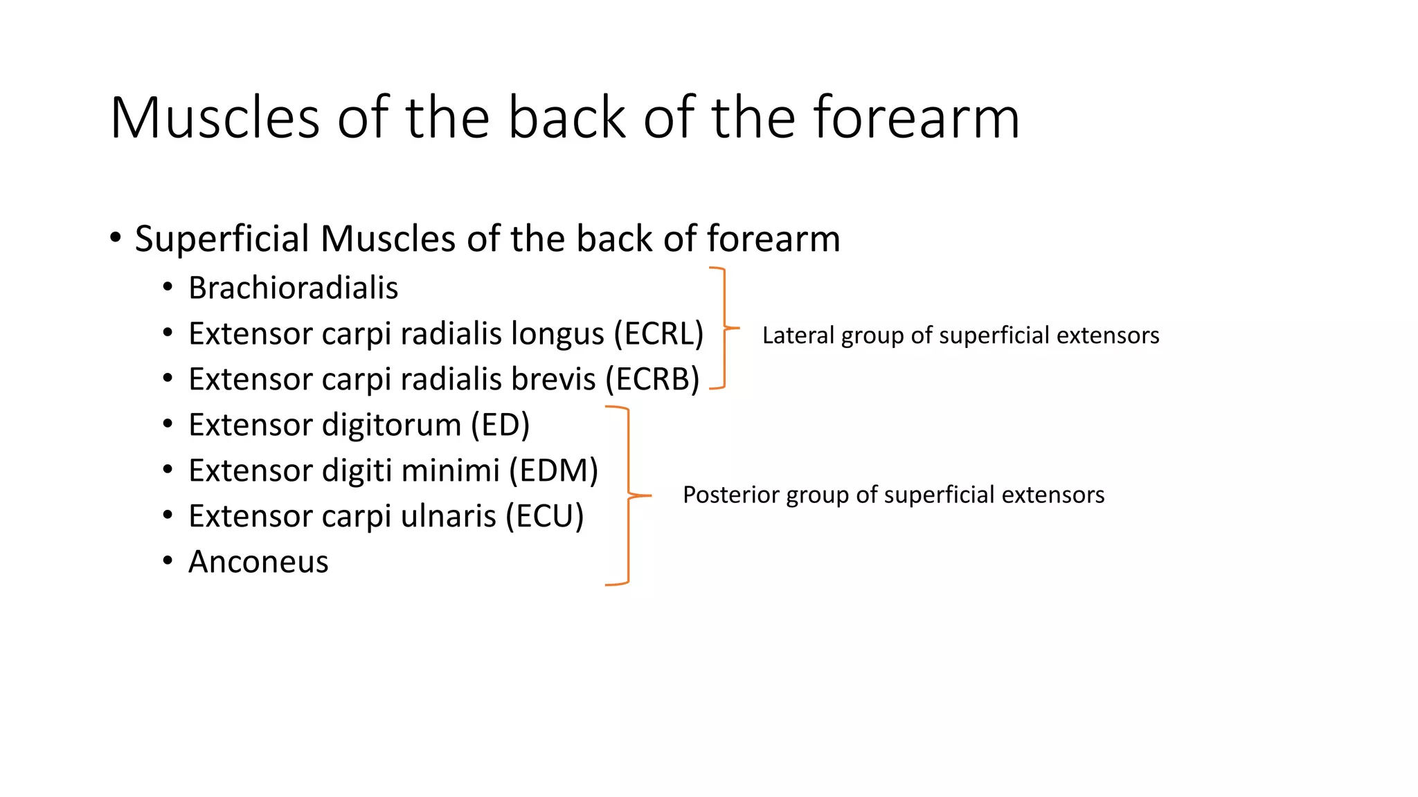
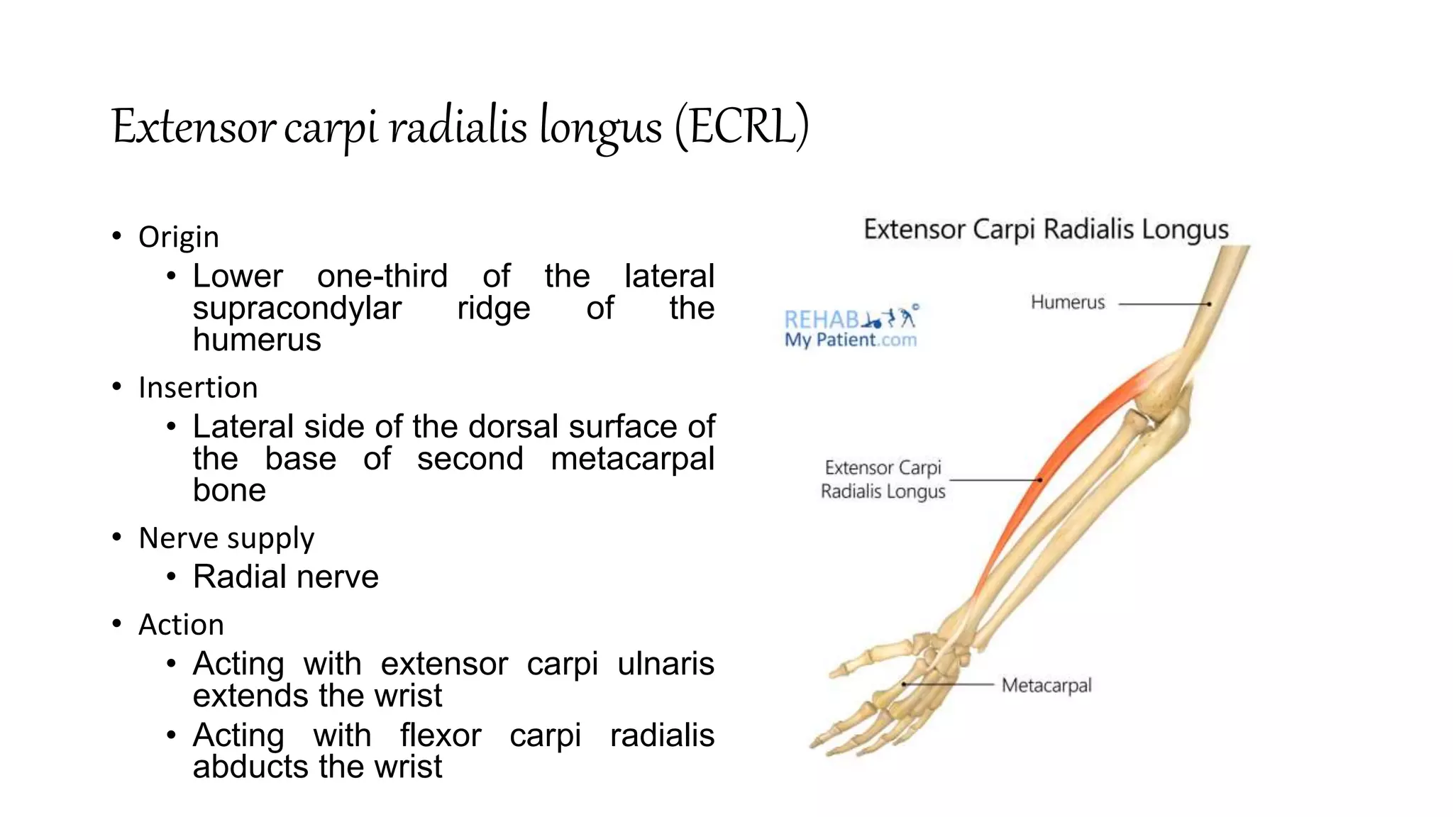
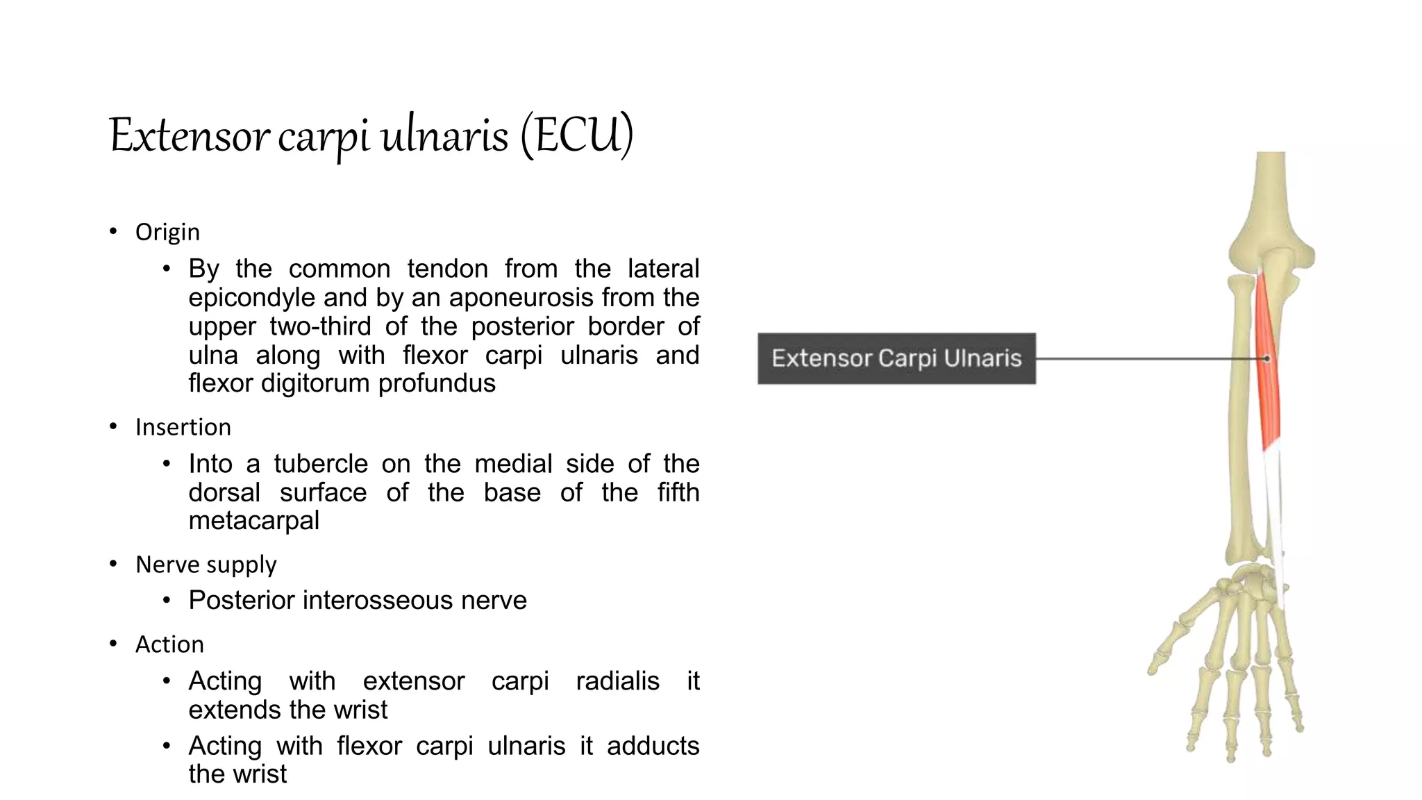
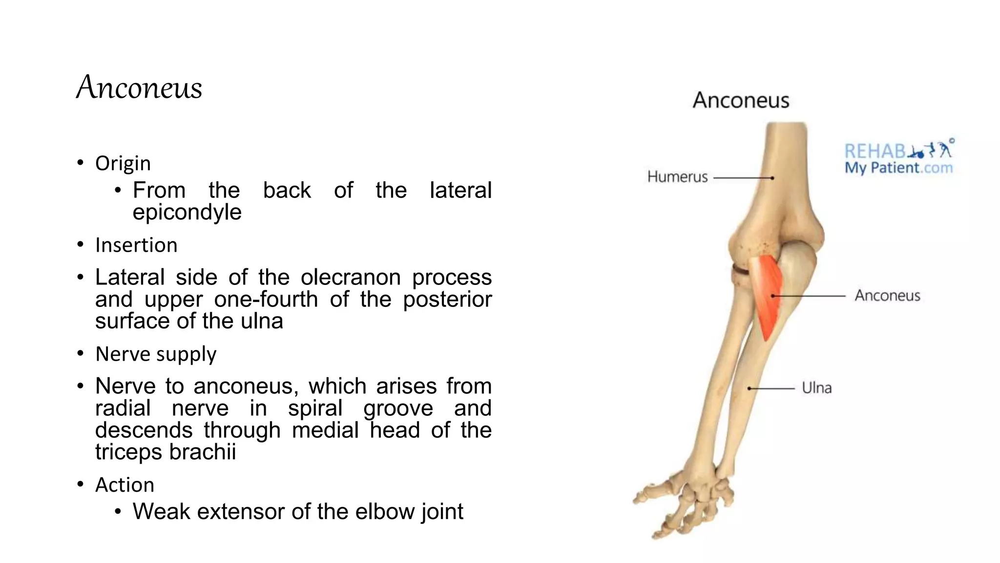
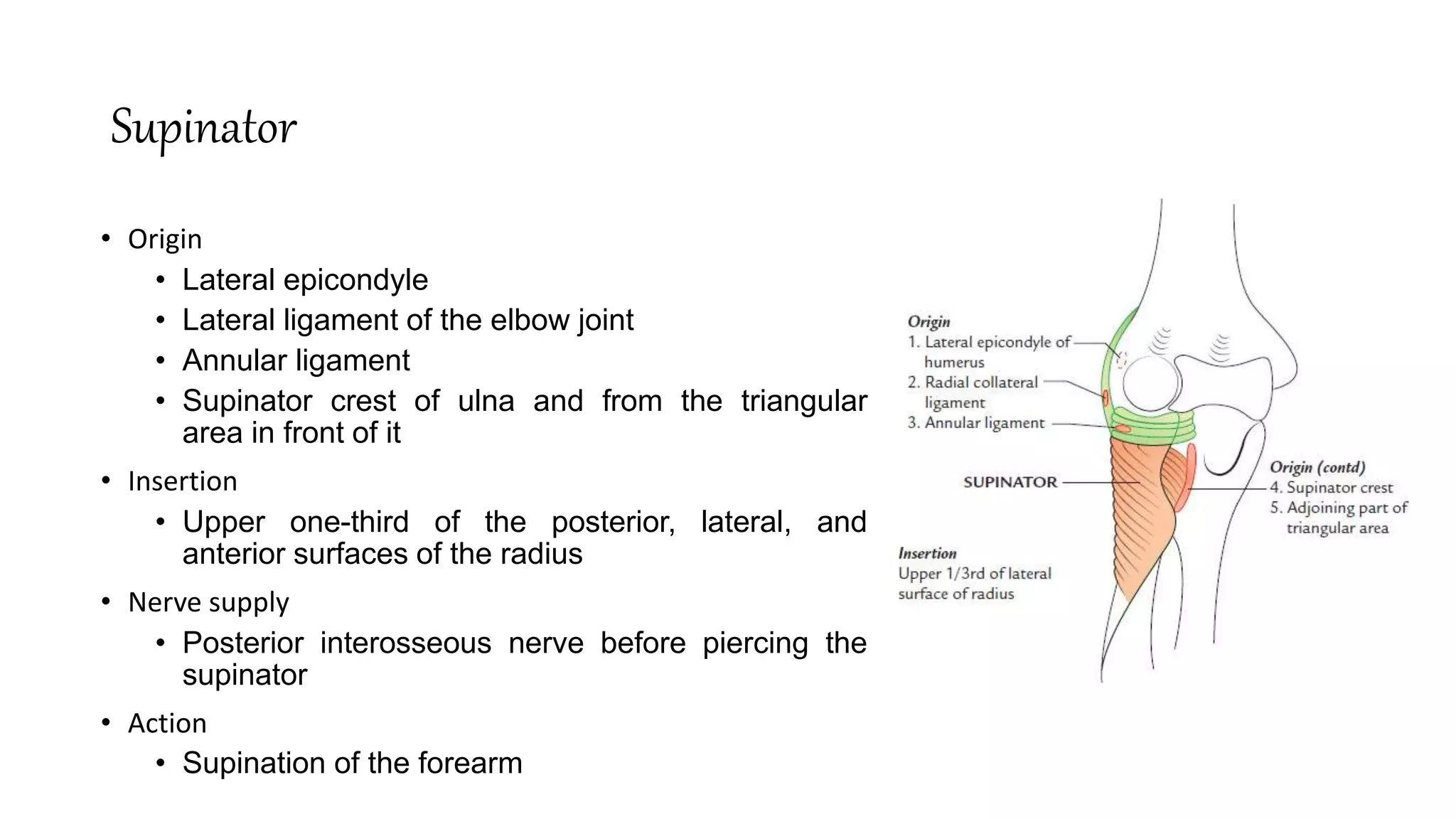
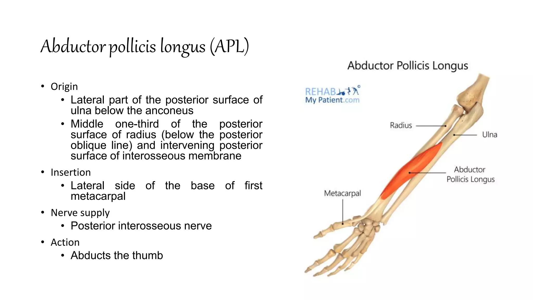
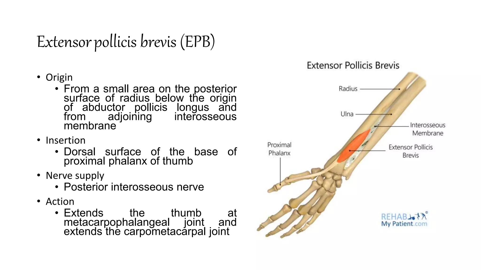
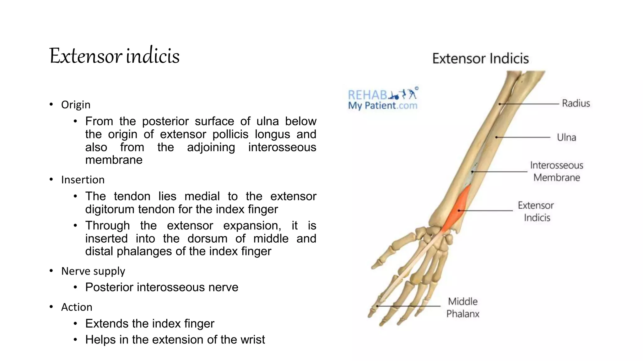
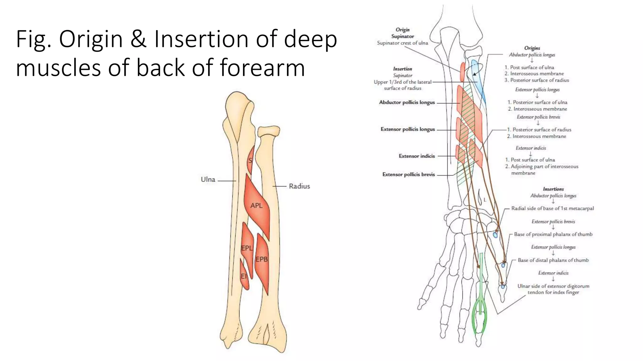
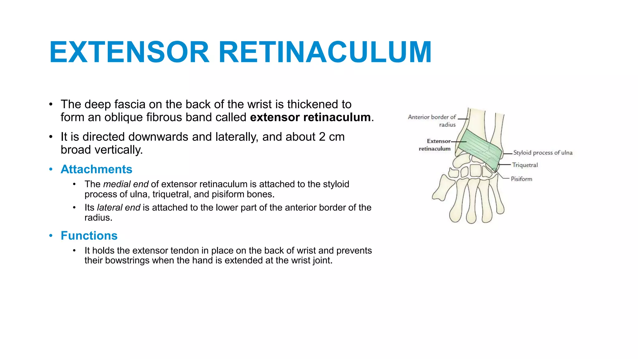
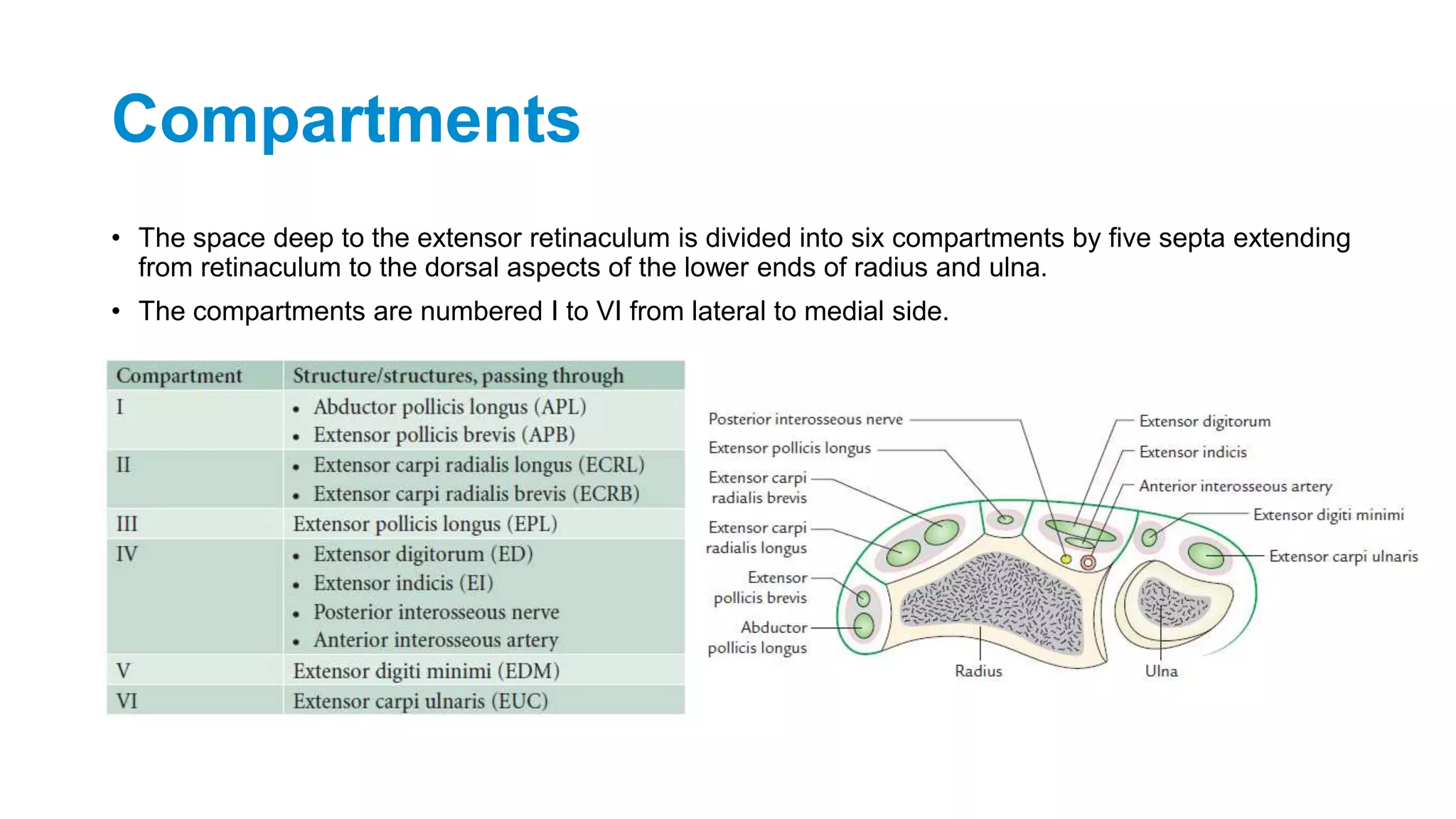
Back of Forearm
Dr. Diwakar Kumar Shah
Assistant Professor
Department of Anatomy
Nobel Medical College Teaching Hospital
Introduction to the forearm
• The forearm extends from the elbow to the wrist and contains two bones, which are tied
together by the thin strong fibrous membrane—the interosseous membrane.
• The head of radius is at the proximal end of the forearm whereas the head of ulna is at
the distal end of the forearm.
• The radius and ulna at both their ends articulate with each other to form the superior and
inferior radio-ulnar joints.
• All important movements of supination and pronation of the forearm occur at these joints.
• The upper ends of radius and ulna articulate with the lower end of humerus to form elbow
joint.
• The main purpose of the movements of the forearm at elbow and radio-ulnar joints is to
place the hand at the desired place.
• The muscles, nerves, and vessels are present both on the front and back of the forearm.
FASCIAL COMPARTMENTS OF THE FOREARM
• The forearm is enclosed in sheath of deep fascia of the forearm (antebrachial fascia).
• It is attached to the posterior subcutaneous border of the ulna.
• From the deep surface fascia, septa pass between the muscles and some of these septa
reach the bone.
• This deep fascia, together with interosseous membrane and fibrous intermuscular septa
divide the forearm into several compartments, each having its own muscles, nerves, and
blood supply.
• Classically, the forearm is divided into the two compartments:
• Anterior compartment
• Posterior compartment
• The anterior compartment contains the structures on the front of the forearm and the
posterior compartment contains the structure on the back of the forearm.
• Near the wrist, the deep fascia presents two localized thickenings, the flexor and the
extensor retinacula, which retain the digital tendons in position during hand movements.
Back of Forearm
• The following structures are to be studied on the back of the forearm:
• Muscles of the back of the forearm
• Posterior interosseous nerve
• Posterior and anterior interosseous arteries
Muscles of the back of the forearm
• Superficial Muscles of the back of forearm
• Brachioradialis
• Extensor carpi radialis longus (ECRL)
• Extensor carpi radialis brevis (ECRB)
• Extensor digitorum (ED)
• Extensor digiti minimi (EDM)
• Extensor carpi ulnaris (ECU)
• Anconeus
Lateral group of superficial extensors
Posterior group of superficial extensors
Brachioradialis
• Origin
• Upper two-third of the lateral supracondylar ridge
of the humerus
• Insertion
• Lateral surface of the distal end of radius just
above the styloid process
• Nerve supply
• Radial nerve
• Action
• Flexes the elbow joint.
• Pronates the supinated forearm to midprone
position
• Supinates the pronated forearm to midprone
position
Extensorcarpi radialis longus (ECRL)
• Origin
• Lower one-third of the lateral
supracondylar ridge of the
humerus
• Insertion
• Lateral side of the dorsal surface of
the base of second metacarpal
bone
• Nerve supply
• Radial nerve
• Action
• Acting with extensor carpi ulnaris
extends the wrist
• Acting with flexor carpi radialis
abducts the wrist
Extensorcarpi radialis brevis (ECRB)
• Origin
• By a common tendon from the
lateral epicondyle of the humerus
and lateral ligament of the elbow
joint
• Insertion
• Lateral side of the dorsal surface of
the base of third metacarpal bone
• Nerve supply
• Posterior interosseous nerve
before piercing the supinator
• Action
• Acting with extensor carpi ulnaris
extends the wrist
• Acting with flexor carpi radialis
abducts the wrist
Extensor digitorum
• Origin
• By a common tendon from the lateral
epicondyle
• Insertion
• Gives rise to four tendons for medial
four digits.
• By the extensor expansion it is
inserted into the dorsum of middle and
terminal phalanges
• Nerve supply
• Posterior interosseous nerve
• Action
• Extends the medial four digits.
• Can also extend the wrist
Extensor digiti minimi
• Origin
• By the common tendon from the lateral
epicondyle
• Insertion
• Lies medial to the extensor digitorum
tendon for the little finger.
• Through the extensor expansion, it is
inserted into the dorsum of middle and
terminal phalanges of little finger
• Nerve supply
• Posterior interosseous nerve
• Action
• Extends the little finger
• Helps in the extension of the wrist
Extensorcarpi ulnaris (ECU)
• Origin
• By the common tendon from the lateral
epicondyle and by an aponeurosis from the
upper two-third of the posterior border of
ulna along with flexor carpi ulnaris and
flexor digitorum profundus
• Insertion
• Into a tubercle on the medial side of the
dorsal surface of the base of the fifth
metacarpal
• Nerve supply
• Posterior interosseous nerve
• Action
• Acting with extensor carpi radialis it
extends the wrist
• Acting with flexor carpi ulnaris it adducts
the wrist
Anconeus
• Origin
• From the back of the lateral
epicondyle
• Insertion
• Lateral side of the olecranon process
and upper one-fourth of the posterior
surface of the ulna
• Nerve supply
• Nerve to anconeus, which arises from
radial nerve in spiral groove and
descends through medial head of the
triceps brachii
• Action
• Weak extensor of the elbow joint
Fig. showing the origin of superficial muscles of the back
of forearm
Fig. Insertion of Superficial Muscles of back of forearm
Deep Muscles of the back of forearm
• There are five deep muscles of the back of forearm, from above downwards they
are:
• Supinator
• Abductor pollicis longus (APL).
• Extensor pollicis brevis (EPB).
• Extensor pollicis longus (EPL).
• Extensor indicis.
• The three deep extensors of the forearm, which act on thumb (abductor pollicis longus,
extensor pollicis brevis, and extensor pollicis longus) lie deep to the superficial extensors
and in order to gain insertion on the three short long bones of thumb ‘crop out’ (emerge)
from the furrow in the lateral part of the forearm between lateral and posterior groups of
superficial extensor.
• These three muscles are therefore termed outcropping muscles.
Supinator
• Origin
• Lateral epicondyle
• Lateral ligament of the elbow joint
• Annular ligament
• Supinator crest of ulna and from the triangular
area in front of it
• Insertion
• Upper one-third of the posterior, lateral, and
anterior surfaces of the radius
• Nerve supply
• Posterior interosseous nerve before piercing the
supinator
• Action
• Supination of the forearm
Abductorpollicis longus (APL)
• Origin
• Lateral part of the posterior surface of
ulna below the anconeus
• Middle one-third of the posterior
surface of radius (below the posterior
oblique line) and intervening posterior
surface of interosseous membrane
• Insertion
• Lateral side of the base of first
metacarpal
• Nerve supply
• Posterior interosseous nerve
• Action
• Abducts the thumb
Extensorpollicis brevis (EPB)
• Origin
• From a small area on the posterior
surface of radius below the origin
of abductor pollicis longus and
from adjoining interosseous
membrane
• Insertion
• Dorsal surface of the base of
proximal phalanx of thumb
• Nerve supply
• Posterior interosseous nerve
• Action
• Extends the thumb at
metacarpophalangeal joint and
extends the carpometacarpal joint
Extensorpollicis longus
• Origin
• From lateral part of middle one-
third of the posterior surface of
ulna and adjoining interosseous
membrane
• Insertion
• Dorsal surface of the base of distal
phalanx of thumb
• Nerve supply
• Posterior interosseous nerve
• Action
• Extends the joints of thumb
• Helps in the extension of the wrist
Extensorindicis
• Origin
• From the posterior surface of ulna below
the origin of extensor pollicis longus and
also from the adjoining interosseous
membrane
• Insertion
• The tendon lies medial to the extensor
digitorum tendon for the index finger
• Through the extensor expansion, it is
inserted into the dorsum of middle and
distal phalanges of the index finger
• Nerve supply
• Posterior interosseous nerve
• Action
• Extends the index finger
• Helps in the extension of the wrist
Fig. Origin & Insertion of deep
muscles of back of forearm
EXTENSOR RETINACULUM
• The deep fascia on the back of the wrist is thickened to
form an oblique fibrous band called extensor retinaculum.
• It is directed downwards and laterally, and about 2 cm
broad vertically.
• Attachments
• The medial end of extensor retinaculum is attached to the styloid
process of ulna, triquetral, and pisiform bones.
• Its lateral end is attached to the lower part of the anterior border of the
radius.
• Functions
• It holds the extensor tendon in place on the back of wrist and prevents
their bowstrings when the hand is extended at the wrist joint.
Compartments
• The space deep to the extensor retinaculum is divided into six compartments by five septa extending
from retinaculum to the dorsal aspects of the lower ends of radius and ulna.
• The compartments are numbered I to VI from lateral to medial side.
POSTERIOR INTEROSSEOUS NERVE
• Origin and Course
• The posterior interosseous nerve is the deep terminal branch of the radial nerve.
• It is motor and chief nerve of the back of the forearm.
• It begins in the cubital fossa as one of the two terminal branches of radial nerve at the level of
lateral epicondyle of humerus.
• It leaves the cubital fossa by winding around the lateral side of the neck of radius in the substance
of supinator.
• After emerging from supinator, it runs in the fascial plane between superficial and deep extensor
muscles.
• At the lower border of extensor pollicis brevis, it passes deep to the extensor pollicis longus to lie on
the posterior surface of interosseous nerve, on which it runs downwards up to the wrist where it
ends into a pseudoganglion.
Branches
• Muscular branches
• Before piercing supinator, it gives branches
to the extensor carpi radialis brevis and
supinator.
• While passing through supinator, it gives
another branch to the supinator.
• After emerging from supinator, it gives
branches to three superficial extensors
(extensor digitorum, extensor digiti minimi,
and extensor carpi ulnaris) and all deep
extensors.
• Articular branches to the wrist joint,
distal radio-ulnar joint, and carpal
joints.
• Lesion of posterior interosseous
nerve:
• The posterior interosseous nerve (i.e., deep
terminal branch of the radial nerve) may be
damaged during surgical exposure of the
head of radius in fracture proximal end of
radius.
• Since the extensor carpi radialis longus is
spared wrist drop does not occur.
POSTERIOR INTEROSSEOUS ARTERY
• The posterior interosseous artery is a smaller terminal branch of the common interosseous artery
from ulnar artery.
• It begins in the cubital fossa, enters the back of the forearm by passing through the gap between
the oblique cord and upper margin of the interosseous membrane.
• From here, it passes between supinator and abductor pollicis longus to accompany the posterior
interosseous nerve.
• In the lower part of the forearm, it becomes markedly reduced and ends by anastomosing with the
anterior interosseous artery.
• In the lower part of forearm, the anterior interosseous artery enters the back of the forearm by
piercing interosseous membrane just above the pronator quadratus and supplies low one-fourth of
the back of the forearm.
• The posterior interosseous artery in the cubital fossa gives interosseous recurrent artery, which
takes part in the formation of anastomosis around the elbow joint.
Back of Forearm.pptx

Back of Forearm.pptx

  • 1.
    Back of Forearm Dr.Diwakar Kumar Shah Assistant Professor Department of Anatomy Nobel Medical College Teaching Hospital
  • 2.
    Introduction to theforearm • The forearm extends from the elbow to the wrist and contains two bones, which are tied together by the thin strong fibrous membrane—the interosseous membrane. • The head of radius is at the proximal end of the forearm whereas the head of ulna is at the distal end of the forearm. • The radius and ulna at both their ends articulate with each other to form the superior and inferior radio-ulnar joints. • All important movements of supination and pronation of the forearm occur at these joints. • The upper ends of radius and ulna articulate with the lower end of humerus to form elbow joint. • The main purpose of the movements of the forearm at elbow and radio-ulnar joints is to place the hand at the desired place. • The muscles, nerves, and vessels are present both on the front and back of the forearm.
  • 3.
    FASCIAL COMPARTMENTS OFTHE FOREARM • The forearm is enclosed in sheath of deep fascia of the forearm (antebrachial fascia). • It is attached to the posterior subcutaneous border of the ulna. • From the deep surface fascia, septa pass between the muscles and some of these septa reach the bone. • This deep fascia, together with interosseous membrane and fibrous intermuscular septa divide the forearm into several compartments, each having its own muscles, nerves, and blood supply. • Classically, the forearm is divided into the two compartments: • Anterior compartment • Posterior compartment • The anterior compartment contains the structures on the front of the forearm and the posterior compartment contains the structure on the back of the forearm. • Near the wrist, the deep fascia presents two localized thickenings, the flexor and the extensor retinacula, which retain the digital tendons in position during hand movements.
  • 4.
    Back of Forearm •The following structures are to be studied on the back of the forearm: • Muscles of the back of the forearm • Posterior interosseous nerve • Posterior and anterior interosseous arteries
  • 5.
    Muscles of theback of the forearm • Superficial Muscles of the back of forearm • Brachioradialis • Extensor carpi radialis longus (ECRL) • Extensor carpi radialis brevis (ECRB) • Extensor digitorum (ED) • Extensor digiti minimi (EDM) • Extensor carpi ulnaris (ECU) • Anconeus Lateral group of superficial extensors Posterior group of superficial extensors
  • 7.
    Brachioradialis • Origin • Uppertwo-third of the lateral supracondylar ridge of the humerus • Insertion • Lateral surface of the distal end of radius just above the styloid process • Nerve supply • Radial nerve • Action • Flexes the elbow joint. • Pronates the supinated forearm to midprone position • Supinates the pronated forearm to midprone position
  • 8.
    Extensorcarpi radialis longus(ECRL) • Origin • Lower one-third of the lateral supracondylar ridge of the humerus • Insertion • Lateral side of the dorsal surface of the base of second metacarpal bone • Nerve supply • Radial nerve • Action • Acting with extensor carpi ulnaris extends the wrist • Acting with flexor carpi radialis abducts the wrist
  • 9.
    Extensorcarpi radialis brevis(ECRB) • Origin • By a common tendon from the lateral epicondyle of the humerus and lateral ligament of the elbow joint • Insertion • Lateral side of the dorsal surface of the base of third metacarpal bone • Nerve supply • Posterior interosseous nerve before piercing the supinator • Action • Acting with extensor carpi ulnaris extends the wrist • Acting with flexor carpi radialis abducts the wrist
  • 10.
    Extensor digitorum • Origin •By a common tendon from the lateral epicondyle • Insertion • Gives rise to four tendons for medial four digits. • By the extensor expansion it is inserted into the dorsum of middle and terminal phalanges • Nerve supply • Posterior interosseous nerve • Action • Extends the medial four digits. • Can also extend the wrist
  • 11.
    Extensor digiti minimi •Origin • By the common tendon from the lateral epicondyle • Insertion • Lies medial to the extensor digitorum tendon for the little finger. • Through the extensor expansion, it is inserted into the dorsum of middle and terminal phalanges of little finger • Nerve supply • Posterior interosseous nerve • Action • Extends the little finger • Helps in the extension of the wrist
  • 12.
    Extensorcarpi ulnaris (ECU) •Origin • By the common tendon from the lateral epicondyle and by an aponeurosis from the upper two-third of the posterior border of ulna along with flexor carpi ulnaris and flexor digitorum profundus • Insertion • Into a tubercle on the medial side of the dorsal surface of the base of the fifth metacarpal • Nerve supply • Posterior interosseous nerve • Action • Acting with extensor carpi radialis it extends the wrist • Acting with flexor carpi ulnaris it adducts the wrist
  • 13.
    Anconeus • Origin • Fromthe back of the lateral epicondyle • Insertion • Lateral side of the olecranon process and upper one-fourth of the posterior surface of the ulna • Nerve supply • Nerve to anconeus, which arises from radial nerve in spiral groove and descends through medial head of the triceps brachii • Action • Weak extensor of the elbow joint
  • 14.
    Fig. showing theorigin of superficial muscles of the back of forearm
  • 15.
    Fig. Insertion ofSuperficial Muscles of back of forearm
  • 16.
    Deep Muscles ofthe back of forearm • There are five deep muscles of the back of forearm, from above downwards they are: • Supinator • Abductor pollicis longus (APL). • Extensor pollicis brevis (EPB). • Extensor pollicis longus (EPL). • Extensor indicis. • The three deep extensors of the forearm, which act on thumb (abductor pollicis longus, extensor pollicis brevis, and extensor pollicis longus) lie deep to the superficial extensors and in order to gain insertion on the three short long bones of thumb ‘crop out’ (emerge) from the furrow in the lateral part of the forearm between lateral and posterior groups of superficial extensor. • These three muscles are therefore termed outcropping muscles.
  • 17.
    Supinator • Origin • Lateralepicondyle • Lateral ligament of the elbow joint • Annular ligament • Supinator crest of ulna and from the triangular area in front of it • Insertion • Upper one-third of the posterior, lateral, and anterior surfaces of the radius • Nerve supply • Posterior interosseous nerve before piercing the supinator • Action • Supination of the forearm
  • 18.
    Abductorpollicis longus (APL) •Origin • Lateral part of the posterior surface of ulna below the anconeus • Middle one-third of the posterior surface of radius (below the posterior oblique line) and intervening posterior surface of interosseous membrane • Insertion • Lateral side of the base of first metacarpal • Nerve supply • Posterior interosseous nerve • Action • Abducts the thumb
  • 19.
    Extensorpollicis brevis (EPB) •Origin • From a small area on the posterior surface of radius below the origin of abductor pollicis longus and from adjoining interosseous membrane • Insertion • Dorsal surface of the base of proximal phalanx of thumb • Nerve supply • Posterior interosseous nerve • Action • Extends the thumb at metacarpophalangeal joint and extends the carpometacarpal joint
  • 20.
    Extensorpollicis longus • Origin •From lateral part of middle one- third of the posterior surface of ulna and adjoining interosseous membrane • Insertion • Dorsal surface of the base of distal phalanx of thumb • Nerve supply • Posterior interosseous nerve • Action • Extends the joints of thumb • Helps in the extension of the wrist
  • 21.
    Extensorindicis • Origin • Fromthe posterior surface of ulna below the origin of extensor pollicis longus and also from the adjoining interosseous membrane • Insertion • The tendon lies medial to the extensor digitorum tendon for the index finger • Through the extensor expansion, it is inserted into the dorsum of middle and distal phalanges of the index finger • Nerve supply • Posterior interosseous nerve • Action • Extends the index finger • Helps in the extension of the wrist
  • 22.
    Fig. Origin &Insertion of deep muscles of back of forearm
  • 23.
    EXTENSOR RETINACULUM • Thedeep fascia on the back of the wrist is thickened to form an oblique fibrous band called extensor retinaculum. • It is directed downwards and laterally, and about 2 cm broad vertically. • Attachments • The medial end of extensor retinaculum is attached to the styloid process of ulna, triquetral, and pisiform bones. • Its lateral end is attached to the lower part of the anterior border of the radius. • Functions • It holds the extensor tendon in place on the back of wrist and prevents their bowstrings when the hand is extended at the wrist joint.
  • 24.
    Compartments • The spacedeep to the extensor retinaculum is divided into six compartments by five septa extending from retinaculum to the dorsal aspects of the lower ends of radius and ulna. • The compartments are numbered I to VI from lateral to medial side.
  • 25.
    POSTERIOR INTEROSSEOUS NERVE •Origin and Course • The posterior interosseous nerve is the deep terminal branch of the radial nerve. • It is motor and chief nerve of the back of the forearm. • It begins in the cubital fossa as one of the two terminal branches of radial nerve at the level of lateral epicondyle of humerus. • It leaves the cubital fossa by winding around the lateral side of the neck of radius in the substance of supinator. • After emerging from supinator, it runs in the fascial plane between superficial and deep extensor muscles. • At the lower border of extensor pollicis brevis, it passes deep to the extensor pollicis longus to lie on the posterior surface of interosseous nerve, on which it runs downwards up to the wrist where it ends into a pseudoganglion.
  • 26.
    Branches • Muscular branches •Before piercing supinator, it gives branches to the extensor carpi radialis brevis and supinator. • While passing through supinator, it gives another branch to the supinator. • After emerging from supinator, it gives branches to three superficial extensors (extensor digitorum, extensor digiti minimi, and extensor carpi ulnaris) and all deep extensors. • Articular branches to the wrist joint, distal radio-ulnar joint, and carpal joints. • Lesion of posterior interosseous nerve: • The posterior interosseous nerve (i.e., deep terminal branch of the radial nerve) may be damaged during surgical exposure of the head of radius in fracture proximal end of radius. • Since the extensor carpi radialis longus is spared wrist drop does not occur.
  • 27.
    POSTERIOR INTEROSSEOUS ARTERY •The posterior interosseous artery is a smaller terminal branch of the common interosseous artery from ulnar artery. • It begins in the cubital fossa, enters the back of the forearm by passing through the gap between the oblique cord and upper margin of the interosseous membrane. • From here, it passes between supinator and abductor pollicis longus to accompany the posterior interosseous nerve. • In the lower part of the forearm, it becomes markedly reduced and ends by anastomosing with the anterior interosseous artery. • In the lower part of forearm, the anterior interosseous artery enters the back of the forearm by piercing interosseous membrane just above the pronator quadratus and supplies low one-fourth of the back of the forearm. • The posterior interosseous artery in the cubital fossa gives interosseous recurrent artery, which takes part in the formation of anastomosis around the elbow joint.