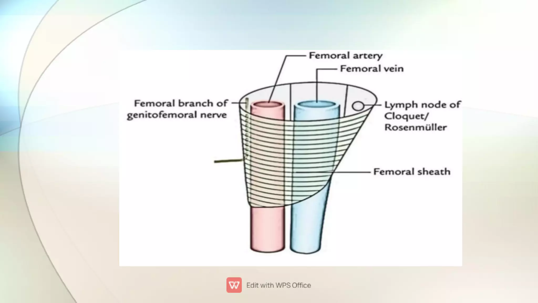
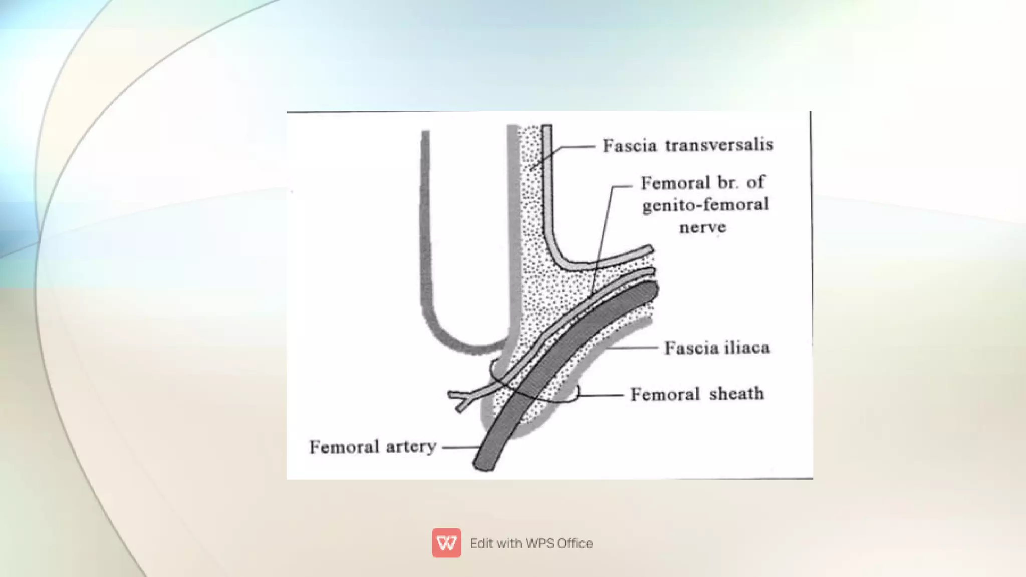
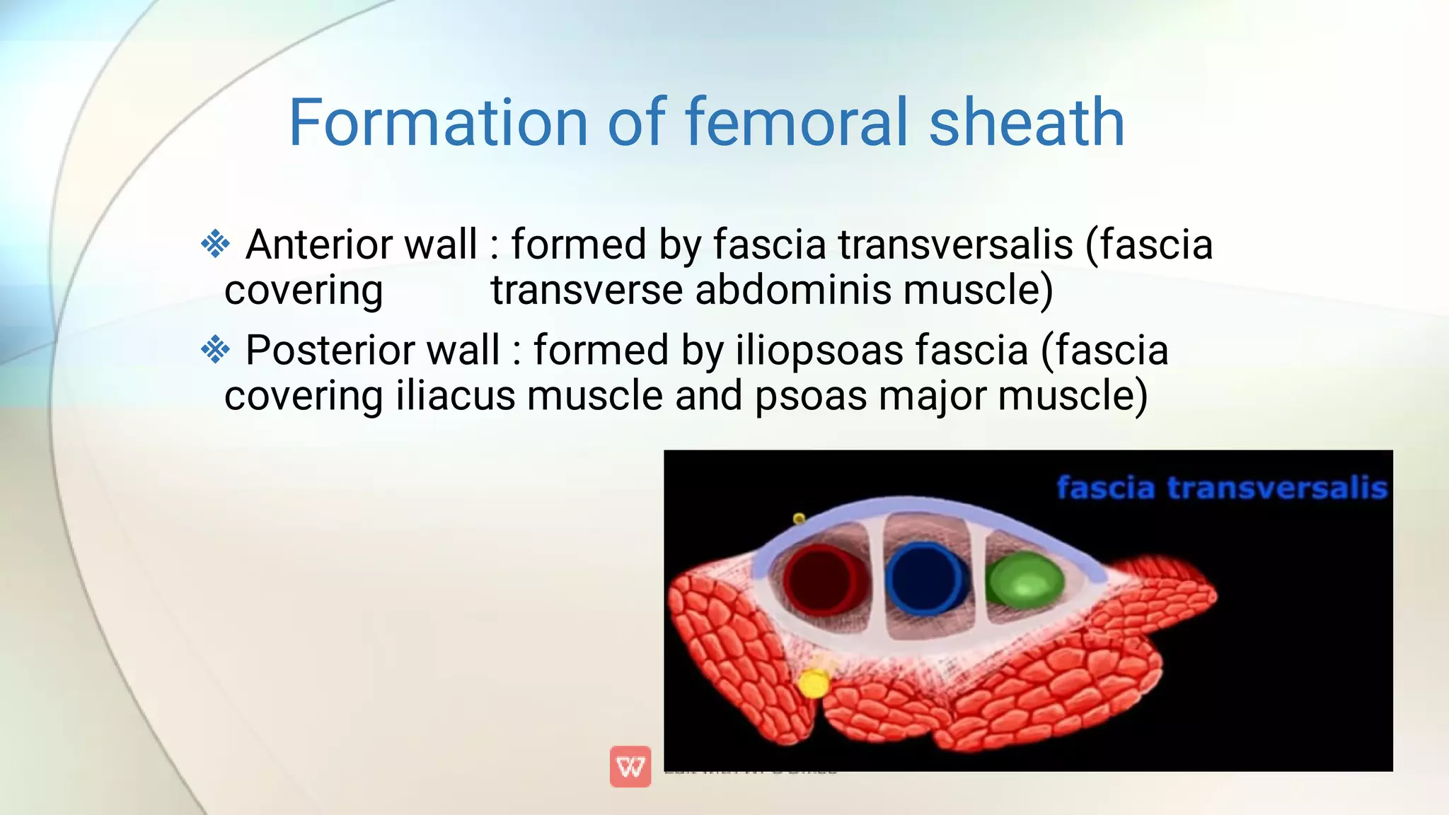
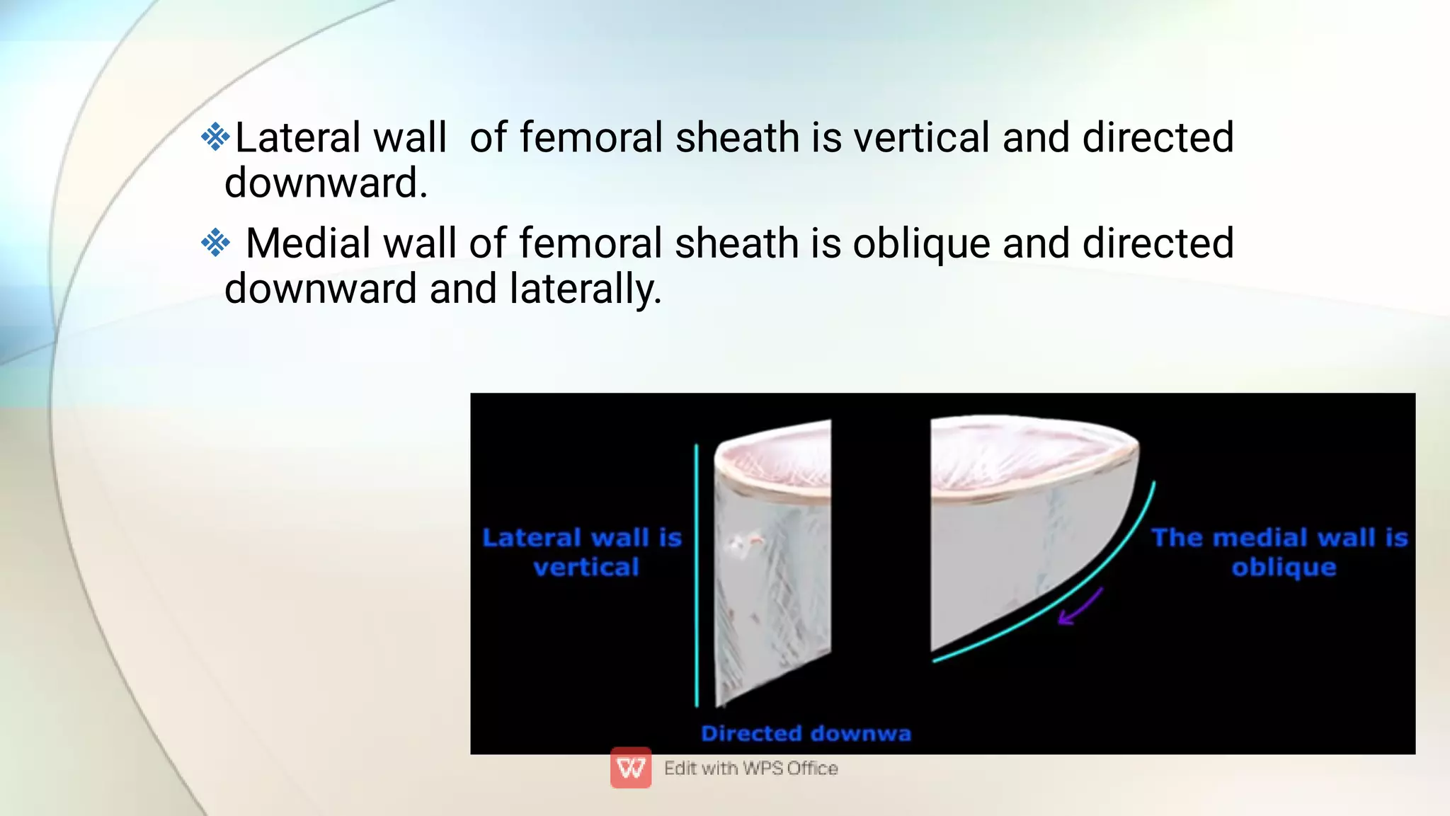
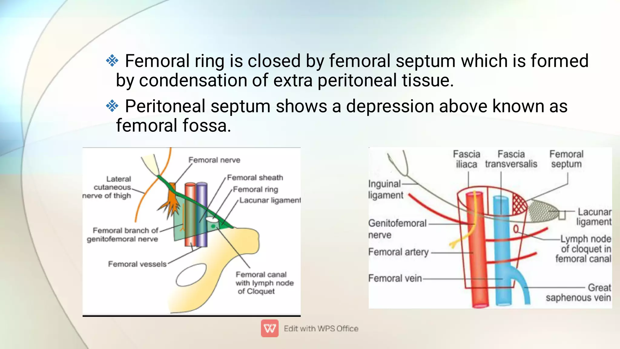
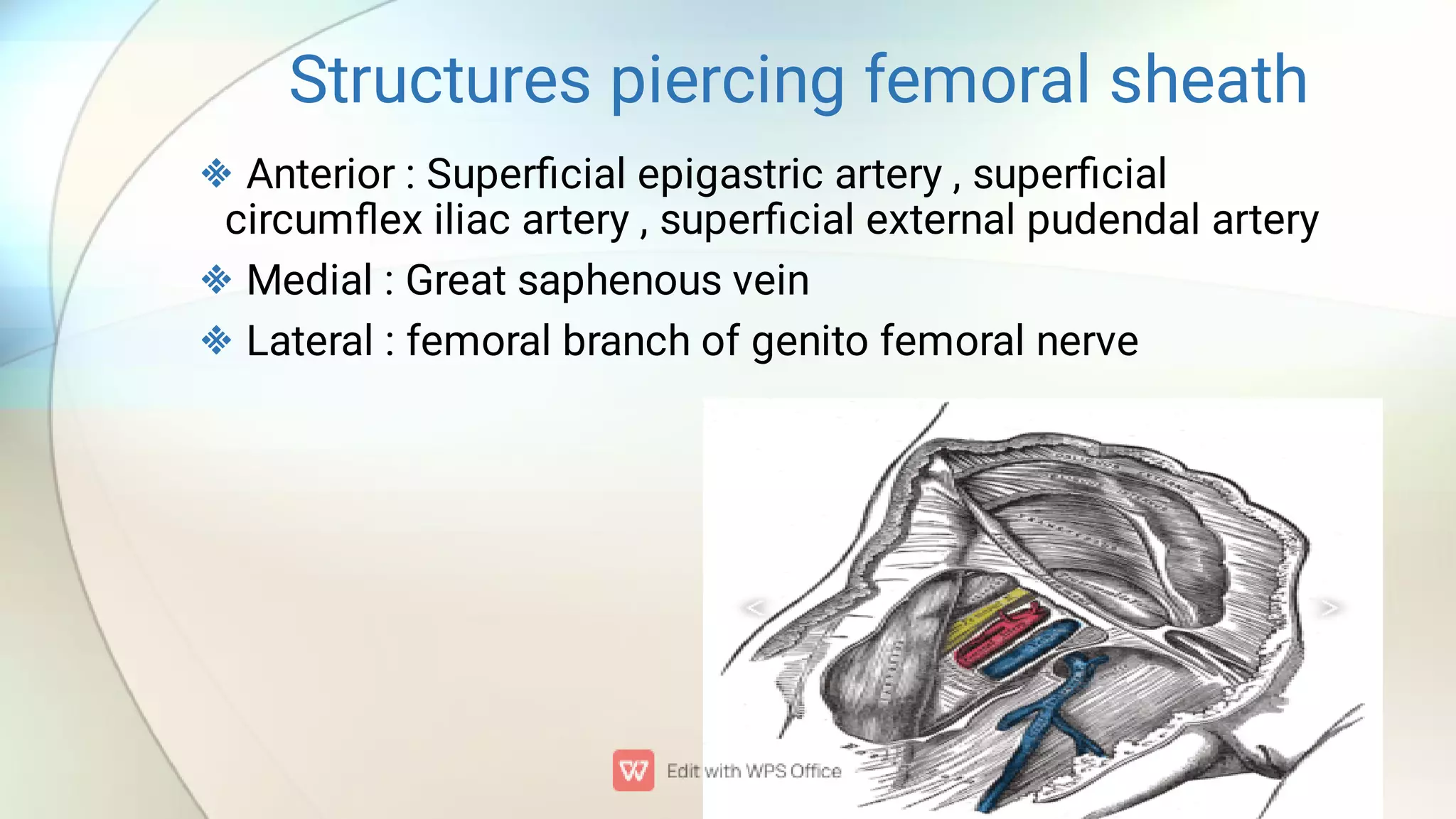
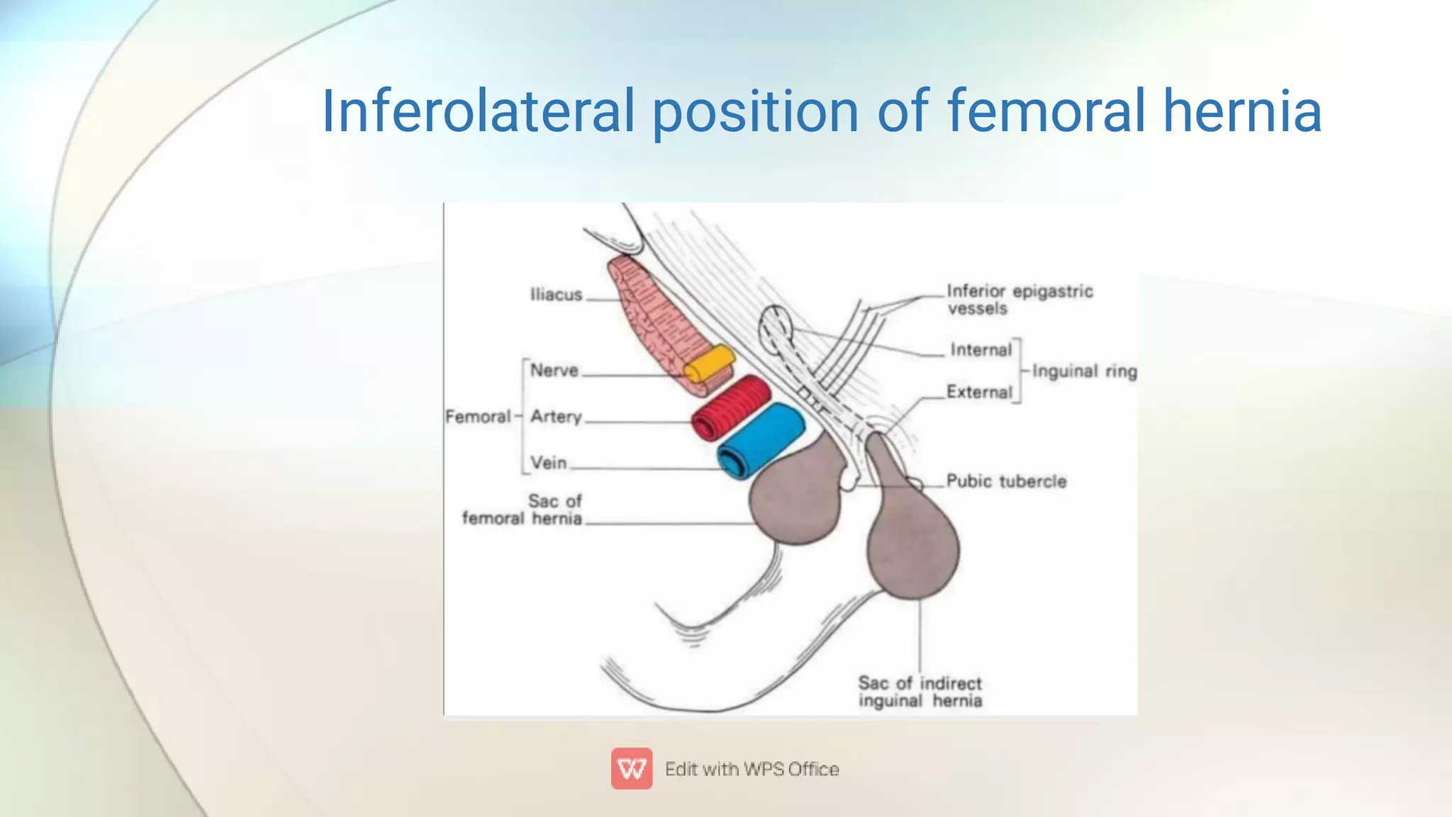
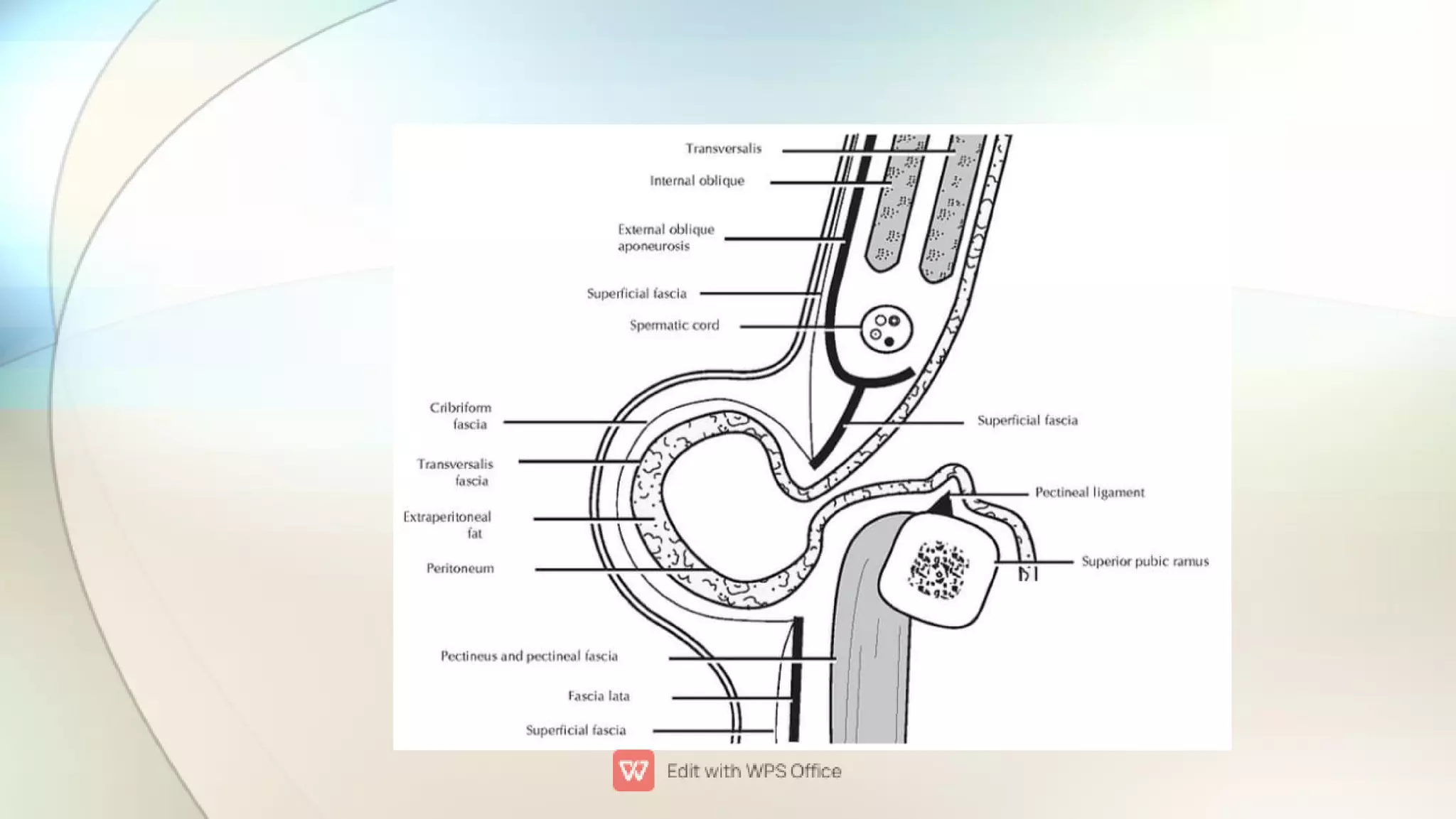
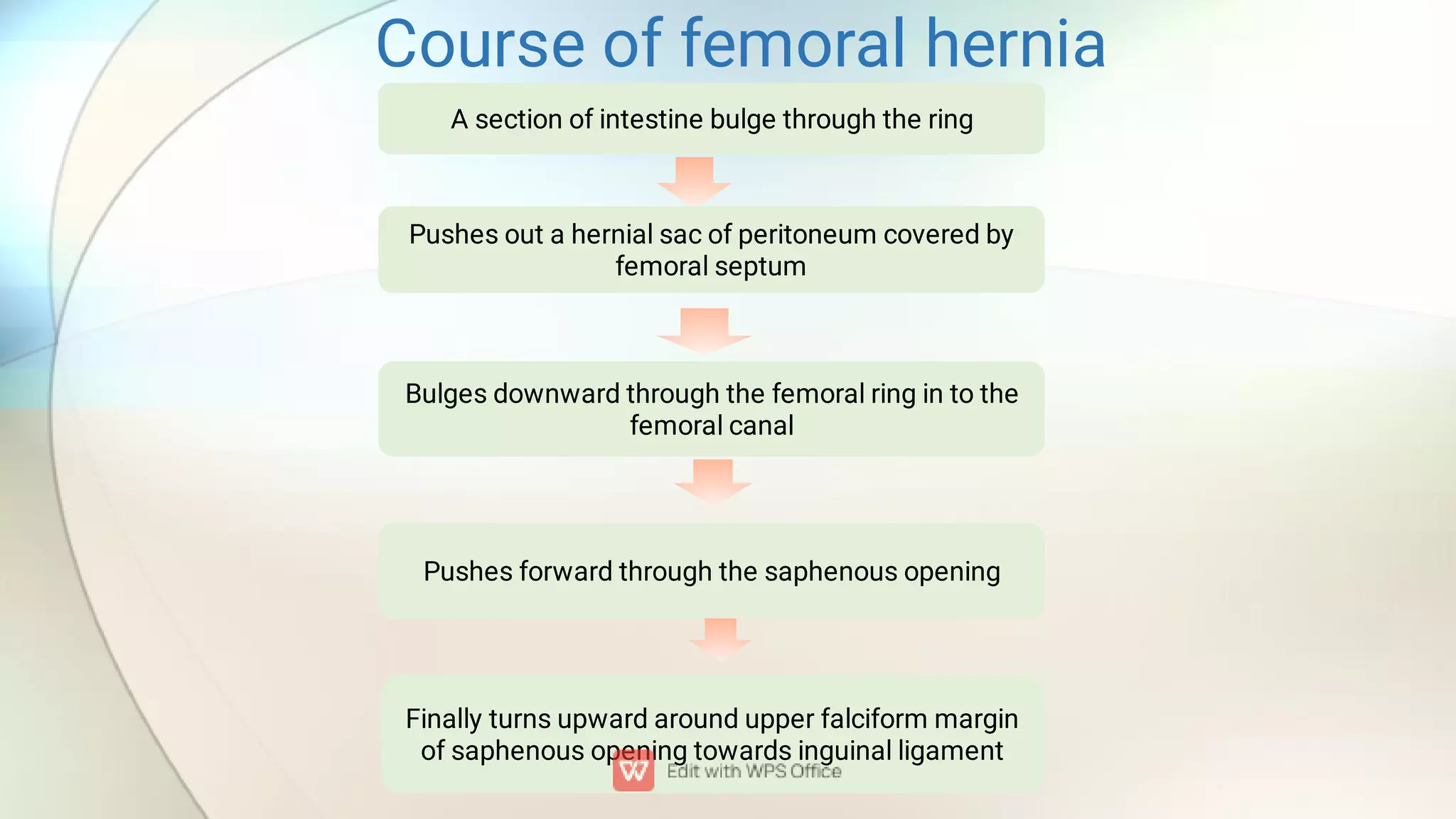
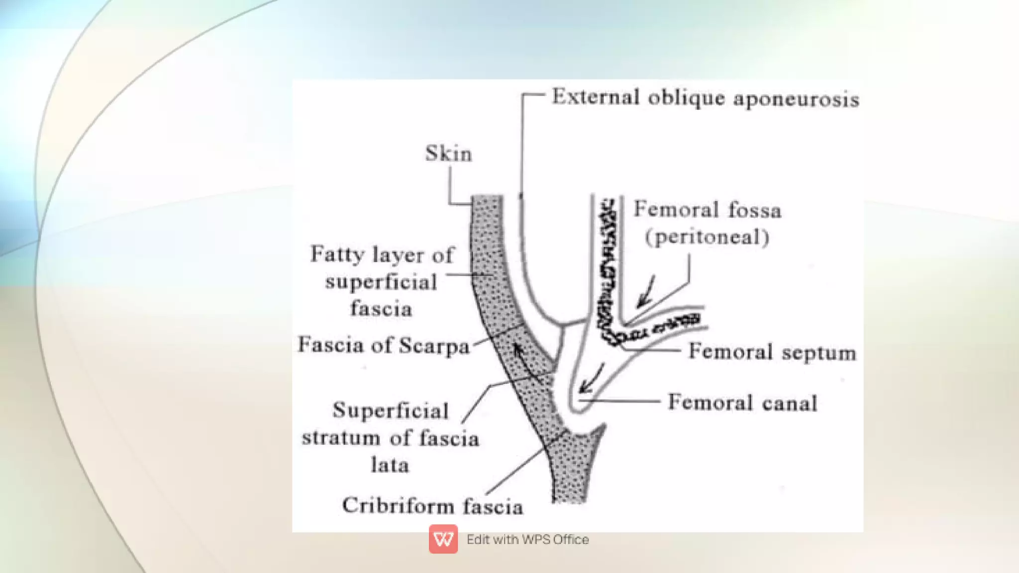
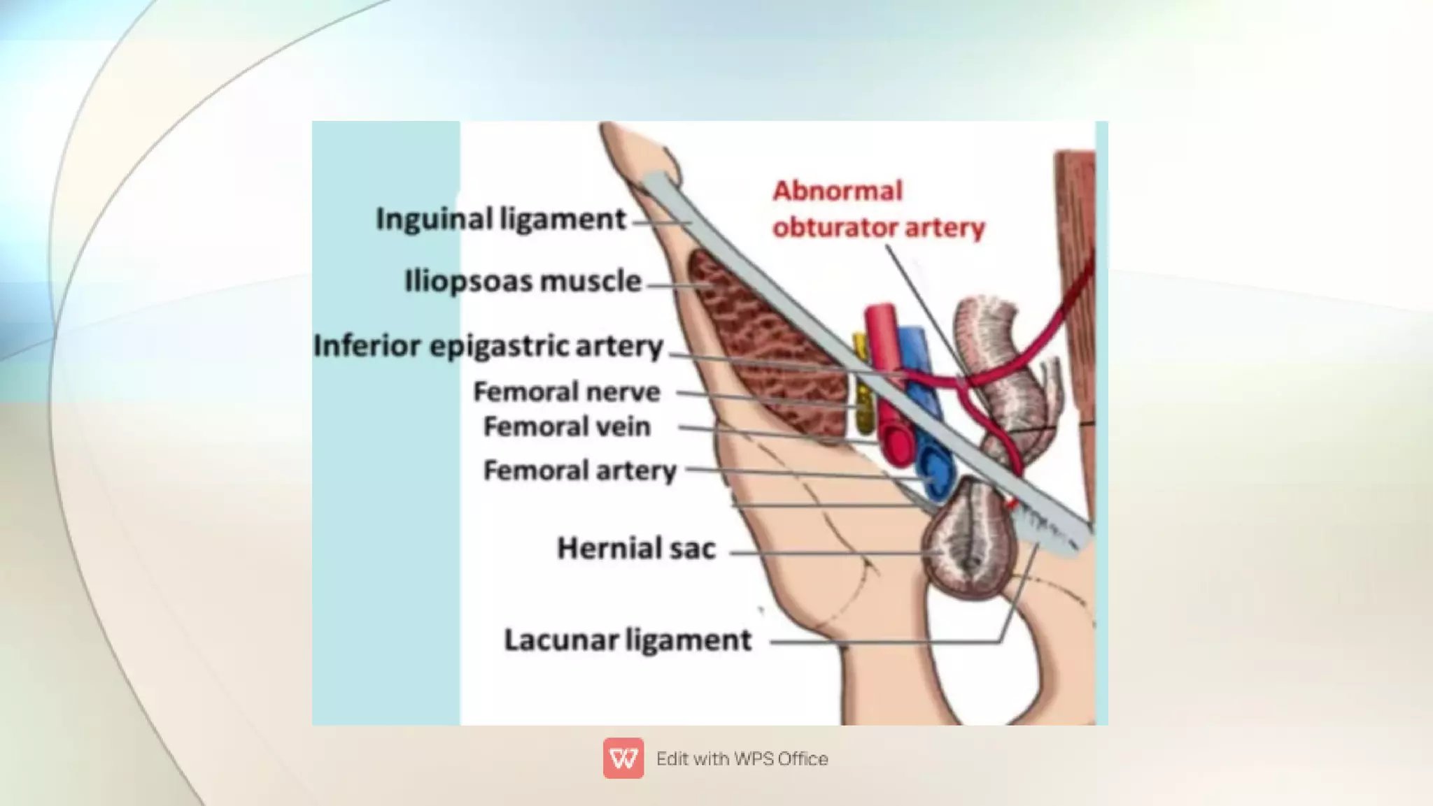
FEMORAL SHEATH AND
FEMORAL HERNIA
Presened by :
D.Bhuvanesh
Roll no : 63.
OBJECTIVES :






Introduction
Formation of femoral sheath
Gross anatomy
Structures piercing femoral sheath
Relation with saphenous opening
Femoral hernia
Introduction



Femoral sheath is a funnel shaped fibro areolar structure
that wraps around the proximal part of femoral vessels in
the upper part of thigh.
It is an funnel shaped sleeve of fascia enclosing tunica
adventitia of femoral vessels about 3 to 4 cm below the
inguinal ligament.
Femoral vessels are in the femoral sheath as they pass
through femoral triangle.
Formation of femoral sheath


Anterior wall : formed by fascia transversalis (fascia
covering transverse abdominis muscle)
Posterior wall : formed by iliopsoas fascia (fascia
covering iliacus muscle and psoas major muscle)


Lateral wall of femoral sheath is vertical and directed
downward.
Medial wall of femoral sheath is oblique and directed
downward and laterally.
Gross anatomy

1.
2.
3.
Anterior and posterior walls are interconnected by two
partitions(called as antero posterior septa) thus creating
three compartments.
Lateral compartment : femoral artery and femoral branch
of genito femoral nerve
Intermediate compartment : contain femoral vein
Medial compartment ( femoral canal )
Medial compartment or femoral canal




•
•
•
It is also called as femoral canal
It is conical shaped and about 1.25 cm long
Its base is directed above and forms an oval shaped femoral
ring
It is an potential or dead space allows expansion of femoral
vein during increased venous return
CONTENTS :
Lymph node of cloquet or Rosenmuller lymph node – drains
Glans penis in males
Clitoris in females
Lymphatic vessels
Small amount of areolar tissue
Femoral ring


•
•
•
•
Proximal end of the femoral canal is called femoral ring.
It is an area of connection between abdominal cavity and
femoral canal.
BOUNDARIES
Anterior : Inguinal ligament
Posterior : pectineus muscle and its covering fascia
Medial : Crescentic edge of lacunar ligament
Lateral : Septum separating it from femoral vein


Femoral ring is closed by femoral septum which is formed
by condensation of extra peritoneal tissue.
Peritoneal septum shows a depression above known as
femoral fossa.
Structures piercing femoral sheath



Anterior : Superficial epigastric artery , superficial
circumflex iliac artery , superficial external pudendal artery
Medial : Great saphenous vein
Lateral : femoral branch of genito femoral nerve
Relationship with saphenous opening





Medial portion of femoral sheath lies behind the saphenous
opening.
Lateral portion of femoral sheath lies behind the fascia lata.
Falciform margin of saphenous opening lies infront of
femoral sheath.
Medial ill defined margin of saphenous opening passes
behind the femoral sheath.
Cribriform fascia covers antero medial surface of femoral
sheath.
Function of femoral sheath
 Allows the femoral vessels to glide freely in and out
beneath the inguinal ligament during movements of hip
joint.
CLINICAL ANATOMY
WHAT IS HERNIA ?
ABNORMAL PROTRUSION
OF
A VISCUS OR PART OF VISCUS
THROUGH
NORMAL OR ABNORMAL OPENING
FROM
ITS WALL CONTAINING CAVITY
PARTS OF HERNIA



In a femoral hernia part of the small intestine protrudes
through the femoral ring into femoral canal.
It presents as a lump situated inferolaterally to pubic
tubercle.
Neck of sac is below and lateral the pubic tubercle.
Inferolateral position of femoral hernia

•
•
•
Femoral hernia is more common in females than men
because
Wider femoral ring
Wider pelvis
Small size of femoral vessels
Covering of femoral hernia






From inside to outside
Peritoneum of hernial sac
Femoral septum
Anterior wall of femoral sheath
Cribriform fascia
Superficial fascia and skin
Course of femoral hernia
A section of intestine bulge through the ring
Pushes out a hernial sac of peritoneum covered by
femoral septum
Bulges downward through the femoral ring in to the
femoral canal
Pushes forward through the saphenous opening
Finally turns upward around upper falciform margin
of saphenous opening towards inguinal ligament
ETIOLOGY(study of causation)






Wide femoral canal
Increased abdominal pressure
Natural weakening of tissue
More commonly seen in multiparous women
Being over weight
Over straining while coughing
Strangulation of femoral hernia




More common to obstruction and strangulated.
Boundaries of femoral ring are rigid and the medial margin
of lacunar ligament is sharp.
This sharpness can cut through the tissue or impede the
blood vessel.
Then the blood supply to the intestinal contents reduces
then results in ischemia leads to death.
Treatment




In case of strangulation surgeon have to wide femoral ring.
Widening is possible only by cutting through the sharp
concave border of lacunar ligament.
Normally obturator artery is away from lacunar ligament
so cutting lacunar ligament is not danger.
BUT IN RARE CASES aberrant or abnormal obturator artery
lie at edge of lacunar ligament and cutting it during hernial
repair cause extensive bleeding(hemorrhage).
Repair of hernia
 Normally if abdominal content not passed through neck
into body of sac it can be pushed back in opposite direction
to its course.
BIBILIOGRAPHY




Essentials of human anatomy – By Dr.A.K.Dutta.
Inderbir singh textbook of anatomy.
B.D.Chaurasia’s Human anatomy.
Netter atlas of anatomy.
SPREAD GRATITUDE NOT CORONA
THANK YOU

FEMORAL_SHEATH_AND_FEMORAL_HERNIA(2).pdf

  • 1.
    FEMORAL SHEATH AND FEMORALHERNIA Presened by : D.Bhuvanesh Roll no : 63.
  • 2.
    OBJECTIVES :       Introduction Formation offemoral sheath Gross anatomy Structures piercing femoral sheath Relation with saphenous opening Femoral hernia
  • 3.
    Introduction    Femoral sheath isa funnel shaped fibro areolar structure that wraps around the proximal part of femoral vessels in the upper part of thigh. It is an funnel shaped sleeve of fascia enclosing tunica adventitia of femoral vessels about 3 to 4 cm below the inguinal ligament. Femoral vessels are in the femoral sheath as they pass through femoral triangle.
  • 6.
    Formation of femoralsheath   Anterior wall : formed by fascia transversalis (fascia covering transverse abdominis muscle) Posterior wall : formed by iliopsoas fascia (fascia covering iliacus muscle and psoas major muscle)
  • 7.
      Lateral wall offemoral sheath is vertical and directed downward. Medial wall of femoral sheath is oblique and directed downward and laterally.
  • 8.
    Gross anatomy  1. 2. 3. Anterior andposterior walls are interconnected by two partitions(called as antero posterior septa) thus creating three compartments. Lateral compartment : femoral artery and femoral branch of genito femoral nerve Intermediate compartment : contain femoral vein Medial compartment ( femoral canal )
  • 10.
    Medial compartment orfemoral canal     • • • It is also called as femoral canal It is conical shaped and about 1.25 cm long Its base is directed above and forms an oval shaped femoral ring It is an potential or dead space allows expansion of femoral vein during increased venous return CONTENTS : Lymph node of cloquet or Rosenmuller lymph node – drains Glans penis in males Clitoris in females Lymphatic vessels Small amount of areolar tissue
  • 14.
    Femoral ring   • • • • Proximal endof the femoral canal is called femoral ring. It is an area of connection between abdominal cavity and femoral canal. BOUNDARIES Anterior : Inguinal ligament Posterior : pectineus muscle and its covering fascia Medial : Crescentic edge of lacunar ligament Lateral : Septum separating it from femoral vein
  • 15.
      Femoral ring isclosed by femoral septum which is formed by condensation of extra peritoneal tissue. Peritoneal septum shows a depression above known as femoral fossa.
  • 16.
    Structures piercing femoralsheath    Anterior : Superficial epigastric artery , superficial circumflex iliac artery , superficial external pudendal artery Medial : Great saphenous vein Lateral : femoral branch of genito femoral nerve
  • 17.
    Relationship with saphenousopening      Medial portion of femoral sheath lies behind the saphenous opening. Lateral portion of femoral sheath lies behind the fascia lata. Falciform margin of saphenous opening lies infront of femoral sheath. Medial ill defined margin of saphenous opening passes behind the femoral sheath. Cribriform fascia covers antero medial surface of femoral sheath.
  • 19.
    Function of femoralsheath  Allows the femoral vessels to glide freely in and out beneath the inguinal ligament during movements of hip joint.
  • 20.
  • 21.
    WHAT IS HERNIA? ABNORMAL PROTRUSION OF A VISCUS OR PART OF VISCUS THROUGH NORMAL OR ABNORMAL OPENING FROM ITS WALL CONTAINING CAVITY
  • 22.
  • 23.
       In a femoralhernia part of the small intestine protrudes through the femoral ring into femoral canal. It presents as a lump situated inferolaterally to pubic tubercle. Neck of sac is below and lateral the pubic tubercle.
  • 24.
  • 25.
     • • • Femoral hernia ismore common in females than men because Wider femoral ring Wider pelvis Small size of femoral vessels
  • 26.
    Covering of femoralhernia       From inside to outside Peritoneum of hernial sac Femoral septum Anterior wall of femoral sheath Cribriform fascia Superficial fascia and skin
  • 28.
    Course of femoralhernia A section of intestine bulge through the ring Pushes out a hernial sac of peritoneum covered by femoral septum Bulges downward through the femoral ring in to the femoral canal Pushes forward through the saphenous opening Finally turns upward around upper falciform margin of saphenous opening towards inguinal ligament
  • 31.
    ETIOLOGY(study of causation)       Widefemoral canal Increased abdominal pressure Natural weakening of tissue More commonly seen in multiparous women Being over weight Over straining while coughing
  • 32.
    Strangulation of femoralhernia     More common to obstruction and strangulated. Boundaries of femoral ring are rigid and the medial margin of lacunar ligament is sharp. This sharpness can cut through the tissue or impede the blood vessel. Then the blood supply to the intestinal contents reduces then results in ischemia leads to death.
  • 33.
    Treatment     In case ofstrangulation surgeon have to wide femoral ring. Widening is possible only by cutting through the sharp concave border of lacunar ligament. Normally obturator artery is away from lacunar ligament so cutting lacunar ligament is not danger. BUT IN RARE CASES aberrant or abnormal obturator artery lie at edge of lacunar ligament and cutting it during hernial repair cause extensive bleeding(hemorrhage).
  • 35.
    Repair of hernia Normally if abdominal content not passed through neck into body of sac it can be pushed back in opposite direction to its course.
  • 36.
    BIBILIOGRAPHY     Essentials of humananatomy – By Dr.A.K.Dutta. Inderbir singh textbook of anatomy. B.D.Chaurasia’s Human anatomy. Netter atlas of anatomy.
  • 37.
    SPREAD GRATITUDE NOTCORONA THANK YOU