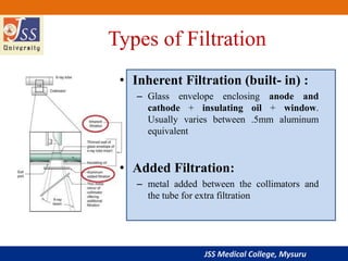
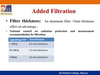
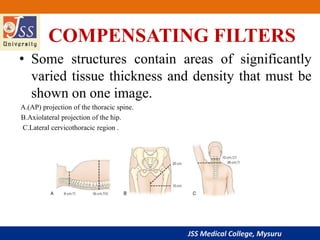
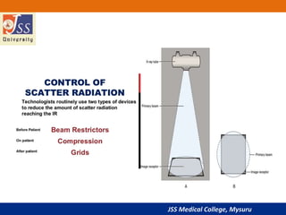
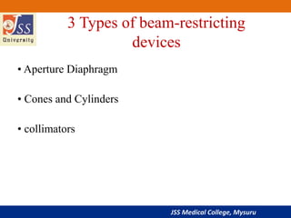
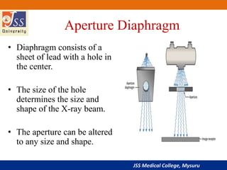
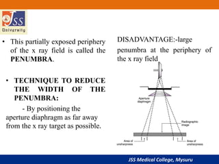
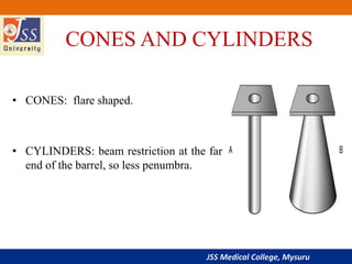
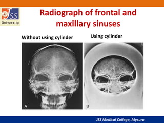
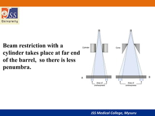
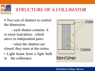
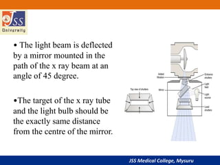
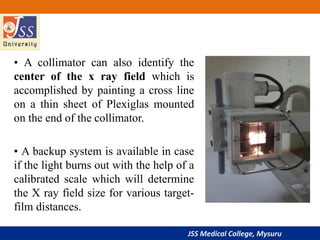
JSS Medical College, Mysuru
Filters and Beam restrictors
PRESENTER-Dr.Vishwanath patil
PG resident
MODERATOR- Dr.Vishwanath T.T
Associate professor
JSS Medical College, Mysuru
Scatter Control
JSS Medical College, Mysuru
OUTLINE
1. Filters
2. Beam restrictors
JSS Medical College, Mysuru
X-ray Filtration
• Filtration is the use of soft metal (Al ) to absorb
low energy x-rays that reach the patient and are
absorbed superficially.
• This affects both the quality and quantity of the
beam.
• Beam hardening: quality of the beam is
increased because the average energy of the
beam is increased.
• This reduces the patient exposure.
JSS Medical College, Mysuru
Types of Filtration
• Inherent Filtration (built- in) :
– Glass envelope enclosing anode and
cathode + insulating oil + window.
Usually varies between .5mm aluminum
equivalent
• Added Filtration:
– metal added between the collimators and
the tube for extra filtration
JSS Medical College, Mysuru
Added Filtration
• Filter thickness: An aluminum filter >3mm thickness
offers no advantage .
• National council on radiation protection and measurement
recommendation for filtration.
operating KVP Total filtration
< 50kVp 0.5 mm aluminum
50-70kVp 1.5 mm aluminum
>70kVp 2.5 mm aluminum
JSS Medical College, Mysuru
Added Filtration
• Effect on exposure factors: The major
disadvantage of filtration is reduction in the intensity of
X-ray beam .
• We must compensate it by increasing mAs.
• Filter absorbs many photons ,hence the total
number reaching the patient actually decreases.
JSS Medical College, Mysuru
COMPENSATING FILTERS
• Some structures contain areas of significantly
varied tissue thickness and density that must be
shown on one image.
A.(AP) projection of the thoracic spine.
B.Axiolateral projection of the hip.
C.Lateral cervicothoracic region .
JSS Medical College, Mysuru
Wedge filters
A. Thick portion of wedge partially
attenuates x-ray beam over upper thoracic
area while no filter area receives full
exposure to penetrate thick portion of spine.
An even image density results.
B, Trough filter in position for AP projection
of chest. Note how two side wedges partially
attenuate x-ray beam over lung areas while
mediastinum receives full exposure. A
better-quality image of chest and
mediastinal structures results.
JSS Medical College, Mysuru
A, Supertech wedge:AP
projection of hips, knees, and
ankles on long (51-inch) film
B,Trough,collimator-mounted
aluminum filter with double
wedge used for AP projections
of thoracic spine.
C, Boomerang contact
filter:used for AP projections of
shoulder and facial bones.
compensating filters in use today
JSS Medical College, Mysuru
compensating filters in use today
D, Ferlic collimator-mounted
filter used for AP and PA oblique
(scapular Y) projections of
shoulder.
E, Ferlic collimator-mounted
filter used for lateral projections
of cervicothoracic region
(swimmer’s technique).
F, Ferlic collimator-mounted
filter for AP axial projections of
foot.
JSS Medical College, Mysuru
• Filtration only affects patient dose and the
beam. It has no effect on the image because
those low energy photons wouldn't reach the
IR anyway.
JSS Medical College, Mysuru
BEAM RESTRICTORS
.
• X Ray beam restrictors is a device that is attached to
the X-ray tube housing, to regulating the size and
shape of an X-ray beam.
JSS Medical College, Mysuru
3 Types of beam-restricting
devices
• Aperture Diaphragm
• Cones and Cylinders
• collimators
JSS Medical College, Mysuru
Aperture Diaphragm
• Diaphragm consists of a
sheet of lead with a hole in
the center.
• The size of the hole
determines the size and
shape of the X-ray beam.
• The aperture can be altered
to any size and shape.
JSS Medical College, Mysuru
• This partially exposed periphery
of the x ray field is called the
PENUMBRA.
• TECHNIQUE TO REDUCE
THE WIDTH OF THE
PENUMBRA:
- By positioning the
aperture diaphragm as far away
from the x ray target as possible.
DISADVANTAGE:-large
penumbra at the periphery of
the x ray field
JSS Medical College, Mysuru
CONES AND CYLINDERS
• CONES: flare shaped.
• CYLINDERS: beam restriction at the far
end of the barrel, so less penumbra.
JSS Medical College, Mysuru
Radiograph of frontal and
maxillary sinuses
Without using cylinder Using cylinder
JSS Medical College, Mysuru
Beam restriction with a
cylinder takes place at far end
of the barrel, so there is less
penumbra.
JSS Medical College, Mysuru
Collimators
• Best all round x ray beam restrictor device.
TWO ADVANTAGES:
1. Provides an infinite variety of rectangular
x ray fields.
2.Light beam shows the centre and the exact
configuration of the x ray field.
JSS Medical College, Mysuru
STRUCTURE OF A COLLIMATOR
• Two sets of shutters to control
the dimension.
- each shutter contains 4
or more lead plates , which
move in independent pairs.
- when the shutters are
closed, they meet at the centre.
• Light beam from a light bulb
in the collimator.
JSS Medical College, Mysuru
• The light beam is deflected
by a mirror mounted in the
path of the x ray beam at an
angle of 45 degree.
•The target of the x ray tube
and the light bulb should be
the exactly same distance
from the centre of the mirror.
JSS Medical College, Mysuru
• A collimator can also identify the
center of the x ray field which is
accomplished by painting a cross line
on a thin sheet of Plexiglas mounted
on the end of the collimator.
• A backup system is available in case
if the light burns out with the help of a
calibrated scale which will determine
the X ray field size for various target-
film distances.
JSS Medical College, Mysuru
AUTOMATIC COLLIMATORS
• POSITIVE BEAM LIGHTING DEVICES
• Shutters are motor driven.
• When a cassette is loaded into the film holder,
sensors will identify the alignment of the cassette.
• Then the information is relayed to the collimator
motors, which will position the shutters to exactly
match the size of the film being used.
JSS Medical College, Mysuru
• These devices must be accurate to within 2%
of the source to image distance (SID).
• A perfectly aligned collimator will leave an
unexposed border on all sides of the
developed film
JSS Medical College, Mysuru
References
• Christensen's Physics of Diagnostic Radiology.
• Radiographic Imaging and Exposure4th Edition
Terri Fauber.
JSS Medical College, Mysuru
THANK YOU

Filters and beam restrictors

  • 1.
    JSS Medical College,Mysuru Filters and Beam restrictors PRESENTER-Dr.Vishwanath patil PG resident MODERATOR- Dr.Vishwanath T.T Associate professor
  • 2.
    JSS Medical College,Mysuru Scatter Control
  • 3.
    JSS Medical College,Mysuru OUTLINE 1. Filters 2. Beam restrictors
  • 4.
    JSS Medical College,Mysuru X-ray Filtration • Filtration is the use of soft metal (Al ) to absorb low energy x-rays that reach the patient and are absorbed superficially. • This affects both the quality and quantity of the beam. • Beam hardening: quality of the beam is increased because the average energy of the beam is increased. • This reduces the patient exposure.
  • 5.
    JSS Medical College,Mysuru Types of Filtration • Inherent Filtration (built- in) : – Glass envelope enclosing anode and cathode + insulating oil + window. Usually varies between .5mm aluminum equivalent • Added Filtration: – metal added between the collimators and the tube for extra filtration
  • 6.
    JSS Medical College,Mysuru Added Filtration • Filter thickness: An aluminum filter >3mm thickness offers no advantage . • National council on radiation protection and measurement recommendation for filtration. operating KVP Total filtration < 50kVp 0.5 mm aluminum 50-70kVp 1.5 mm aluminum >70kVp 2.5 mm aluminum
  • 7.
    JSS Medical College,Mysuru Added Filtration • Effect on exposure factors: The major disadvantage of filtration is reduction in the intensity of X-ray beam . • We must compensate it by increasing mAs. • Filter absorbs many photons ,hence the total number reaching the patient actually decreases.
  • 8.
    JSS Medical College,Mysuru COMPENSATING FILTERS • Some structures contain areas of significantly varied tissue thickness and density that must be shown on one image. A.(AP) projection of the thoracic spine. B.Axiolateral projection of the hip. C.Lateral cervicothoracic region .
  • 9.
    JSS Medical College,Mysuru Wedge filters A. Thick portion of wedge partially attenuates x-ray beam over upper thoracic area while no filter area receives full exposure to penetrate thick portion of spine. An even image density results. B, Trough filter in position for AP projection of chest. Note how two side wedges partially attenuate x-ray beam over lung areas while mediastinum receives full exposure. A better-quality image of chest and mediastinal structures results.
  • 10.
    JSS Medical College,Mysuru A, Supertech wedge:AP projection of hips, knees, and ankles on long (51-inch) film B,Trough,collimator-mounted aluminum filter with double wedge used for AP projections of thoracic spine. C, Boomerang contact filter:used for AP projections of shoulder and facial bones. compensating filters in use today
  • 11.
    JSS Medical College,Mysuru compensating filters in use today D, Ferlic collimator-mounted filter used for AP and PA oblique (scapular Y) projections of shoulder. E, Ferlic collimator-mounted filter used for lateral projections of cervicothoracic region (swimmer’s technique). F, Ferlic collimator-mounted filter for AP axial projections of foot.
  • 12.
    JSS Medical College,Mysuru • Filtration only affects patient dose and the beam. It has no effect on the image because those low energy photons wouldn't reach the IR anyway.
  • 13.
  • 14.
    JSS Medical College,Mysuru BEAM RESTRICTORS . • X Ray beam restrictors is a device that is attached to the X-ray tube housing, to regulating the size and shape of an X-ray beam.
  • 15.
    JSS Medical College,Mysuru 3 Types of beam-restricting devices • Aperture Diaphragm • Cones and Cylinders • collimators
  • 16.
    JSS Medical College,Mysuru Aperture Diaphragm • Diaphragm consists of a sheet of lead with a hole in the center. • The size of the hole determines the size and shape of the X-ray beam. • The aperture can be altered to any size and shape.
  • 17.
    JSS Medical College,Mysuru • This partially exposed periphery of the x ray field is called the PENUMBRA. • TECHNIQUE TO REDUCE THE WIDTH OF THE PENUMBRA: - By positioning the aperture diaphragm as far away from the x ray target as possible. DISADVANTAGE:-large penumbra at the periphery of the x ray field
  • 18.
    JSS Medical College,Mysuru CONES AND CYLINDERS • CONES: flare shaped. • CYLINDERS: beam restriction at the far end of the barrel, so less penumbra.
  • 19.
    JSS Medical College,Mysuru Radiograph of frontal and maxillary sinuses Without using cylinder Using cylinder
  • 20.
    JSS Medical College,Mysuru Beam restriction with a cylinder takes place at far end of the barrel, so there is less penumbra.
  • 21.
    JSS Medical College,Mysuru Collimators • Best all round x ray beam restrictor device. TWO ADVANTAGES: 1. Provides an infinite variety of rectangular x ray fields. 2.Light beam shows the centre and the exact configuration of the x ray field.
  • 22.
    JSS Medical College,Mysuru STRUCTURE OF A COLLIMATOR • Two sets of shutters to control the dimension. - each shutter contains 4 or more lead plates , which move in independent pairs. - when the shutters are closed, they meet at the centre. • Light beam from a light bulb in the collimator.
  • 23.
    JSS Medical College,Mysuru • The light beam is deflected by a mirror mounted in the path of the x ray beam at an angle of 45 degree. •The target of the x ray tube and the light bulb should be the exactly same distance from the centre of the mirror.
  • 24.
    JSS Medical College,Mysuru • A collimator can also identify the center of the x ray field which is accomplished by painting a cross line on a thin sheet of Plexiglas mounted on the end of the collimator. • A backup system is available in case if the light burns out with the help of a calibrated scale which will determine the X ray field size for various target- film distances.
  • 25.
    JSS Medical College,Mysuru AUTOMATIC COLLIMATORS • POSITIVE BEAM LIGHTING DEVICES • Shutters are motor driven. • When a cassette is loaded into the film holder, sensors will identify the alignment of the cassette. • Then the information is relayed to the collimator motors, which will position the shutters to exactly match the size of the film being used.
  • 26.
    JSS Medical College,Mysuru • These devices must be accurate to within 2% of the source to image distance (SID). • A perfectly aligned collimator will leave an unexposed border on all sides of the developed film
  • 27.
    JSS Medical College,Mysuru References • Christensen's Physics of Diagnostic Radiology. • Radiographic Imaging and Exposure4th Edition Terri Fauber.
  • 28.
    JSS Medical College,Mysuru THANK YOU