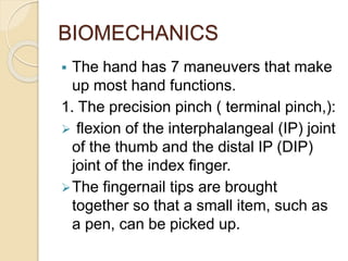
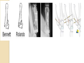
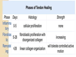
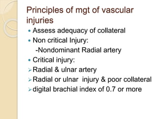
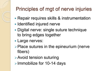
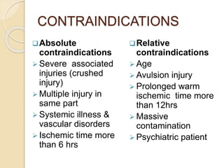
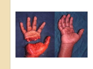
The document discusses hand injuries and their management. It covers relevant hand anatomy, types of common hand injuries including tendon injuries, fractures, and dislocations. The key principles of management are early debridement and stabilization of injuries, restoration of anatomy, and rapid mobilization. Specific techniques are described for repairing tendons, fixing fractures like Bennett's fracture, and achieving proper skeletal stabilization while preserving hand function.
Title: Sense of Smell
Presenter: Dr. Faiza, Assistant Professor of Physiology
Qualifications:
MBBS (Best Graduate, AIMC Lahore)
FCPS Physiology
ICMT, CHPE, DHPE (STMU)
MPH (GC University, Faisalabad)
MBA (Virtual University of Pakistan)
Learning Objectives:
Describe the primary categories of smells and the concept of odor blindness.
Explain the structure and location of the olfactory membrane and mucosa, including the types and roles of cells involved in olfaction.
Describe the pathway and mechanisms of olfactory signal transmission from the olfactory receptors to the brain.
Illustrate the biochemical cascade triggered by odorant binding to olfactory receptors, including the role of G-proteins and second messengers in generating an action potential.
Identify different types of olfactory disorders such as anosmia, hyposmia, hyperosmia, and dysosmia, including their potential causes.
Key Topics:
Olfactory Genes:
3% of the human genome accounts for olfactory genes.
400 genes for odorant receptors.
Olfactory Membrane:
Located in the superior part of the nasal cavity.
Medially: Folds downward along the superior septum.
Laterally: Folds over the superior turbinate and upper surface of the middle turbinate.
Total surface area: 5-10 square centimeters.
Olfactory Mucosa:
Olfactory Cells: Bipolar nerve cells derived from the CNS (100 million), with 4-25 olfactory cilia per cell.
Sustentacular Cells: Produce mucus and maintain ionic and molecular environment.
Basal Cells: Replace worn-out olfactory cells with an average lifespan of 1-2 months.
Bowman’s Gland: Secretes mucus.
Stimulation of Olfactory Cells:
Odorant dissolves in mucus and attaches to receptors on olfactory cilia.
Involves a cascade effect through G-proteins and second messengers, leading to depolarization and action potential generation in the olfactory nerve.
Quality of a Good Odorant:
Small (3-20 Carbon atoms), volatile, water-soluble, and lipid-soluble.
Facilitated by odorant-binding proteins in mucus.
Membrane Potential and Action Potential:
Resting membrane potential: -55mV.
Action potential frequency in the olfactory nerve increases with odorant strength.
Adaptation Towards the Sense of Smell:
Rapid adaptation within the first second, with further slow adaptation.
Psychological adaptation greater than receptor adaptation, involving feedback inhibition from the central nervous system.
Primary Sensations of Smell:
Camphoraceous, Musky, Floral, Pepperminty, Ethereal, Pungent, Putrid.
Odor Detection Threshold:
Examples: Hydrogen sulfide (0.0005 ppm), Methyl-mercaptan (0.002 ppm).
Some toxic substances are odorless at lethal concentrations.
Characteristics of Smell:
Odor blindness for single substances due to lack of appropriate receptor protein.
Behavioral and emotional influences of smell.
Transmission of Olfactory Signals:
From olfactory cells to glomeruli in the olfactory bulb, involving lateral inhibition.
Primitive, less old, and new olfactory systems with different path
ABDOMINAL TRAUMA in pediatrics part one.drhasanrajab
Abdominal trauma in pediatrics refers to injuries or damage to the abdominal organs in children. It can occur due to various causes such as falls, motor vehicle accidents, sports-related injuries, and physical abuse. Children are more vulnerable to abdominal trauma due to their unique anatomical and physiological characteristics. Signs and symptoms include abdominal pain, tenderness, distension, vomiting, and signs of shock. Diagnosis involves physical examination, imaging studies, and laboratory tests. Management depends on the severity and may involve conservative treatment or surgical intervention. Prevention is crucial in reducing the incidence of abdominal trauma in children.
Here is the updated list of Top Best Ayurvedic medicine for Gas and Indigestion and those are Gas-O-Go Syp for Dyspepsia | Lavizyme Syrup for Acidity | Yumzyme Hepatoprotective Capsules etc
Rasamanikya is a excellent preparation in the field of Rasashastra, it is used in various Kushtha Roga, Shwasa, Vicharchika, Bhagandara, Vatarakta, and Phiranga Roga. In this article Preparation& Comparative analytical profile for both Formulationon i.e Rasamanikya prepared by Kushmanda swarasa & Churnodhaka Shodita Haratala. The study aims to provide insights into the comparative efficacy and analytical aspects of these formulations for enhanced therapeutic outcomes.
micro teaching on communication m.sc nursing.pdfAnurag Sharma
Microteaching is a unique model of practice teaching. It is a viable instrument for the. desired change in the teaching behavior or the behavior potential which, in specified types of real. classroom situations, tends to facilitate the achievement of specified types of objectives.
Basavarajeeyam is an important text for ayurvedic physician belonging to andhra pradehs. It is a popular compendium in various parts of our country as well as in andhra pradesh. The content of the text was presented in sanskrit and telugu language (Bilingual). One of the most famous book in ayurvedic pharmaceutics and therapeutics. This book contains 25 chapters called as prakaranas. Many rasaoushadis were explained, pioneer of dhatu druti, nadi pareeksha, mutra pareeksha etc. Belongs to the period of 15-16 century. New diseases like upadamsha, phiranga rogas are explained.
Local Advanced Lung Cancer: Artificial Intelligence, Synergetics, Complex Sys...Oleg Kshivets
Overall life span (LS) was 1671.7±1721.6 days and cumulative 5YS reached 62.4%, 10 years – 50.4%, 20 years – 44.6%. 94 LCP lived more than 5 years without cancer (LS=2958.6±1723.6 days), 22 – more than 10 years (LS=5571±1841.8 days). 67 LCP died because of LC (LS=471.9±344 days). AT significantly improved 5YS (68% vs. 53.7%) (P=0.028 by log-rank test). Cox modeling displayed that 5YS of LCP significantly depended on: N0-N12, T3-4, blood cell circuit, cell ratio factors (ratio between cancer cells-CC and blood cells subpopulations), LC cell dynamics, recalcification time, heparin tolerance, prothrombin index, protein, AT, procedure type (P=0.000-0.031). Neural networks, genetic algorithm selection and bootstrap simulation revealed relationships between 5YS and N0-12 (rank=1), thrombocytes/CC (rank=2), segmented neutrophils/CC (3), eosinophils/CC (4), erythrocytes/CC (5), healthy cells/CC (6), lymphocytes/CC (7), stick neutrophils/CC (8), leucocytes/CC (9), monocytes/CC (10). Correct prediction of 5YS was 100% by neural networks computing (error=0.000; area under ROC curve=1.0).
Integrating Ayurveda into Parkinson’s Management: A Holistic ApproachAyurveda ForAll
Explore the benefits of combining Ayurveda with conventional Parkinson's treatments. Learn how a holistic approach can manage symptoms, enhance well-being, and balance body energies. Discover the steps to safely integrate Ayurvedic practices into your Parkinson’s care plan, including expert guidance on diet, herbal remedies, and lifestyle modifications.
Title: Sense of Taste
Presenter: Dr. Faiza, Assistant Professor of Physiology
Qualifications:
MBBS (Best Graduate, AIMC Lahore)
FCPS Physiology
ICMT, CHPE, DHPE (STMU)
MPH (GC University, Faisalabad)
MBA (Virtual University of Pakistan)
Learning Objectives:
Describe the structure and function of taste buds.
Describe the relationship between the taste threshold and taste index of common substances.
Explain the chemical basis and signal transduction of taste perception for each type of primary taste sensation.
Recognize different abnormalities of taste perception and their causes.
Key Topics:
Significance of Taste Sensation:
Differentiation between pleasant and harmful food
Influence on behavior
Selection of food based on metabolic needs
Receptors of Taste:
Taste buds on the tongue
Influence of sense of smell, texture of food, and pain stimulation (e.g., by pepper)
Primary and Secondary Taste Sensations:
Primary taste sensations: Sweet, Sour, Salty, Bitter, Umami
Chemical basis and signal transduction mechanisms for each taste
Taste Threshold and Index:
Taste threshold values for Sweet (sucrose), Salty (NaCl), Sour (HCl), and Bitter (Quinine)
Taste index relationship: Inversely proportional to taste threshold
Taste Blindness:
Inability to taste certain substances, particularly thiourea compounds
Example: Phenylthiocarbamide
Structure and Function of Taste Buds:
Composition: Epithelial cells, Sustentacular/Supporting cells, Taste cells, Basal cells
Features: Taste pores, Taste hairs/microvilli, and Taste nerve fibers
Location of Taste Buds:
Found in papillae of the tongue (Fungiform, Circumvallate, Foliate)
Also present on the palate, tonsillar pillars, epiglottis, and proximal esophagus
Mechanism of Taste Stimulation:
Interaction of taste substances with receptors on microvilli
Signal transduction pathways for Umami, Sweet, Bitter, Sour, and Salty tastes
Taste Sensitivity and Adaptation:
Decrease in sensitivity with age
Rapid adaptation of taste sensation
Role of Saliva in Taste:
Dissolution of tastants to reach receptors
Washing away the stimulus
Taste Preferences and Aversions:
Mechanisms behind taste preference and aversion
Influence of receptors and neural pathways
Impact of Sensory Nerve Damage:
Degeneration of taste buds if the sensory nerve fiber is cut
Abnormalities of Taste Detection:
Conditions: Ageusia, Hypogeusia, Dysgeusia (parageusia)
Causes: Nerve damage, neurological disorders, infections, poor oral hygiene, adverse drug effects, deficiencies, aging, tobacco use, altered neurotransmitter levels
Neurotransmitters and Taste Threshold:
Effects of serotonin (5-HT) and norepinephrine (NE) on taste sensitivity
Supertasters:
25% of the population with heightened sensitivity to taste, especially bitterness
Increased number of fungiform papillae
3. Learning Objectives…
To understand important anatomical
features of the hand
To appreciate the spectrum of injuries to
the hand
To understand principles of management
of hand injuries
4. INTRODUCTION
A prehensile organ with structural
adaptations to functions
Injuries to the hand are not uncommon
present as isolated injury or component of
multiple injuries.
Injury can be soft tissue or bony/skeletal
These injuries can be
definitively/temporarily managed in the
first clinical setting.
9. BIOMECHANICS
The hand has 7 maneuvers that make
up most hand functions.
1. The precision pinch ( terminal pinch,):
flexion of the interphalangeal (IP) joint
of the thumb and the distal IP (DIP)
joint of the index finger.
The fingernail tips are brought
together so that a small item, such as
a pen, can be picked up.
11. …
2. The oppositional pinch ( subterminal
pinch)
pulp of the thumb and index finger are
brought together with the IP and DIP
joints in extension
Allows for increased forces to be
generated through thumb opposition.
First dorsal interosseous contracts with
simultaneous flexion of Index finger
profundus
13. …
3. Key pinch
The thumb is adducted to the radial
aspect of the index finger’s middle
phalanx.
It also requires adequate length of the
digit and a metacarpal phalangeal joint
(MCP), which is capable of resisting
thumb adduction
15. ---
4. The chuck grip (directional grip)
allows the index finger, long finger,
and thumb to come together to
envelop a cylindrical object.
A rotational and axial force is usually
applied to the object when using this
type of grip
17. …
5. The hook grip
requires finger flexion at the IP joints
and extension at the MCP joints.
This grip is used, for example, when
one picks up a suitcase or a briefcase.
It does not require thumb function
21. ---
7. The span grasp
The DIP joints and the proximal IP (PIP)
joints flex to approximately 30 degrees
and the thumb is palmarly abducted
Forces are generated between the
thumb and fingers.
Stability is needed at the thumb, MCP,
and IP joints.
This type of grip is used, for example, to
grab a ball
23. HAND INJURIES
Hand injury: any pathological change
occurring in the part of the upper limb
distal to the wrist as a result of
exchange of energy between an
individual and his environment
(Bashiru et al )
SKELETAL
INJURIES
SOFT TISSUE
INJURIES
24. EPIDEMIOLOGY
Paucity of data locally: 2.7% of RTA
(Hassan 2000)
USA :5-10% of emergency department
(ED)
Males › Females 26.9 years
average
3.7:1 ratio at KNH (Kaisha 2006)
right hand distal phalanges
›50% right hand dominance
Commonest etiology: work related
27. TENDON INJURIES
17% reported among Hand injuries @
KNH (Kaisha 2006)
Flexor digitorum superficialis and
profundus + extensor digitorum
communis commonly injured
28. Flexor Tendon Injuries
volar lacerations plus/minus
concomitant neurovascular injury
Classified by the zone of injury
Courte of orthobullet
29. Flexor pulley system
Digits 1-4 contain
5 annular pulleys (A1 to A5)
3 cruciate pulleys (C1 to C3)
A2 and A4 are the most important
pulleys to prevent flexor tendon
bowstringing
thumb contains
2 annular pulley
interposed oblique pulley (most
important)
31. ---
Flexor digitorum profundus (FDP)
Flexor digitorum superficialis (FDS)
FDS to 5th finger (absent in 25% of
people
Flexor pollicis longus (FPL)
Flexor carpi radialis (FCR)
Flexor carpi ulnaris (FCU)
32. Extensor Tendon Injuries
caused by laceration, trauma, or
overuse
long finger (most injured) & zone VI (
most frequently injured zone)
Mechanism
Zone I : forced flexion of extended DIP
joint
Zone II :dorsal laceration or crush injury
Zone V : commonly from "fight bite"
sagittal band rupture ("flea flicker
injury")
34. Special tendon related injuries
Jersey finger:
Avulsion injury of FDP from insertion at
the base of distal phalanx
A zone I flexor tendon injury with 75%
at the tip of the ring finger
Boutonniere deformity:
Zone III extensor tendon injury resulting
to PIP flexion and DIP extension
35. VASCULAR INJURIES
Are not uncommon
Can involve Radial or ulnar arteries
and digital branches + associated
veins
37. SKELETAL INJURIES
27 bones & complex articulations
amenable to fractures and dislocations
Fractures & dislocations
Carpal: Scaphoid, perilunate
dislocation
Metacarpal FX, MCP dislocation
Phalanx FX
38. Special fractures/dislocation
Intraarticular fractures of the Thumb
Bennett FX :
Intra-articular fracture/dislocation of base of
1st metacarpal characterized by volar lip of
metacarpal based attached to volar oblique
ligament
small fragment of 1st metacarpal continues
to articulate with trapezium
Rolando FX:
Intra-articular fracture of base of 1st
metacarpal characterized by intra-articular
39. Extraarticular fx
Pseudo Bennett's fracture (also
called Epibasal fractures of the thumb)
are two piece fractures of the proximal
first metacarpal bone which is usually
stable
40. …
Fracture stability depends upon its
specific pattern, location, relationship to
tendon and ligament insertions, and any
associated Injuries
Common fracture patterns in the hand
metacarpals.
I. Spiral fracture
II. Oblique fracture
III. Multifragmentary fracture
IV. Partial articular fracture
V. Complete articular fracture
41. MANAGEMENT
HX + PE + Investigations
Basic concepts in repair are similar for
different zones
Location of laceration directly affects
healing potential
Principle of management can be tailored
to severity of injury and structure injured
(tendon, vascular, nerve, skeletal etc)
44. Basic principles
Multidisciplinary
Treatment is directed at structures
injured
In emergency situations, the goals of
treatment are
Distal circulation
Bone frame work
Skin cover
46. …
Debridement & fracture stabilization
for open injuries
Primary tendon repair reserved for
cleanly cut tendons.
Delayed primary repair is indicated for
potentially contaminated wounds
Bayonet incision
47. Principles of hand fracture
management
AO Guiding principles of hand fracture
(FX) management
I. restoration of articular anatomy;
II. correction of angular or rotational
deformity;
III. stabilization of fractures;
IV. surgical approach not compromising
hand function;
V. rapid mobilization
Open Hand fractures…management
principle remains the same
48. …
Many fXs of the hand can effectively be
treated using nonoperatively.
Stable skeletal fixation maybe required
in the following groups of fractures:
multifragmentary
severely displaced
multiple metacarpal
short oblique or spiral metacarpal with
associated soft-tissue injury.
49. …
FXs at particular anatomical locations:
subcondylar, proximal phalanx; palmar
base middle phalanx.
Displaced articular and periarticular
fractures:
Bennett’s FX
Rolando’s FX
Pseudo Bennette Fx
unicondylar and bicondylar FX
51. Management of special fx
Scaphoid fracture
Modalities:
Non-operative
Stable & nondisplaced fx:
Thumb spica cast immobilization for 12-
21 days and reevaluate
Nonunion increase with delayed
immobilization of > 4 weeks after injury
Risk of nonunion
52. …
Operative: Screw fixation
(percutaneous/ORIF)
Indications:
Unstable fractures
I. Proximal pole fractures
II. Displacement > 1 mm
III. 15° scaphoid humpback deformity
IV. Radiolunate angle > 15°
V. Intrascaphoid angle of > 35°
VI. scaphoid fractures associated with
perilunate dislocation
VII. comminuted fractures
VIII.unstable vertical or oblique fracture
53. Thumb Intraarticular fractures
I. BENNETTE
Nonoperative : closed reduction & cast
immobilization
Indications: nondisplaced fractures
Technique: reduction maneuver with
traction, extension, pronation, and
abduction
54. ..
Operative :
A. CRPP
Indications
Small volar fragment to hold a screw
anatomic reduction unstable
Technique
can attempt reduction of shaft to trapezium
to hold reduction
B. ORIF
Indications
large fragment
2mm+ joint displacement
55. ---
2. ROLANDO FX
Nonoperative : Cast immobilization
Indications : severe comminution, stable
Early ROM
Operative : external fixation, CRPP
Indications
severe comminution, unstable fx
Technique: Approximate large fragments
with k-wires
56. ---
ORIF : T-plate/k-wire
Indications
most common fixation method
Technique
use t-plate or blade plate
use k-wires for small fragments
Complications : post-traumatic
osteoarthritis
57.
58. TENDON REPAIRS
Principle
Primary repair of tendon for cleanly cut
tendons
Delayed primary repair for contaminated
wounds
Retraction after 4 weeks results in joint
flexion.
Tendon graft repair may be required
Methods:
Kessler, Bunnell, Modified Kessler,
Pulvertaft etc
Variations in methods globally with no
59. Units Core Sutures Peripheral
Sutures
Country
Mayo Clinic Modified
Pennington,
double Tsuge,
double Kessler
Simple running or
simple locking
USA
Stanford
University,
Palo
Alto (CA
Modified Kessler
plus 2-strand
horizontal
mattress suture
Simple running USA
Saint John
Regional
Hospital
Double Kessler Simple running Canada
Verona
University
4-strand Kessler Simple running Italy
Broomfield
Hospital,
Chelmsford
4-strand
Evans/Smith
6-strand Tang
Simple running
None
United Kingdom
The methods used currently in some hand surgery units in direct end-
to-end flexor tendon repairs
61. Contraindications for primary
repair
Injuries more than 12 hours old
Crush wounds with poor skin
coverage
Contaminated wounds, especially
human bites
Tendon loss more than 1 cm
Injury at multiple sites along the
tendon
Destruction of the pulley system
62.
63. Principles of mgt of vascular
injuries
Assess adequacy of collateral
Non critical Injury:
-Nondominant Radial artery
Critical injury:
Radial & ulnar artery
Radial or ulnar injury & poor collateral
digital brachial index of 0.7 or more
64. method
Control of bleeding: compression, use of
BP cuff, or tourniquet at 200-250 mmHg
≤15-20 min.
Digital arteries: repair skin lacerations
and apply pressure dressing
Large vessels: End to end anastomosis
of healthy vessels
Healthy intima must be visualized
Appropriate length of vein grafts are
used from the saphenous vein
65. Principles of mgt of nerve injuries
Repair requires skills & instrumentation
Identified injured nerve
Digital nerve: single suture technique
to bring edges together
Large nerves:
Place sutures in the epineurium (nerve
fibers)
Avoid tension suturing
Immobilize for 10-14 days
66. …
Primary nerve repair in 72 hours of
injury
Delayed primary repair 72 hours to 14
days
Secondary nerve repairs 14 days or
longer after injury.
Adequate debridement of edges
May use nylon 8-0
collagen tubes for gaps or nerve grafts
for large gaps
67. Amputation
In general if any four of the following
basic six are irreparably damaged
Skin
Extensor and
Flexor tendons
Vessels
nerves
Skeleton
Prosthesis-total, partial, digital or active
or passive
69. CONTRAINDICATIONS
Absolute
contraindications
Severe associated
injuries (crushed
injury)
Multiple injury in
same part
Systemic illness &
vascular disorders
Ischemic time more
than 6 hrs
Relative
contraindications
Age
Avulsion injury
Prolonged warm
ischemic time more
than 12hrs
Massive
contamination
Psychiatric patient
70. Transport of amputated tissue
Modality
keep amputated tissue wrapped in
moist gauze in lactate ringers solution
place in sealed plastic bag and place in
ice water (avoid direct ice or dry ice)
wrap, cover and compress stump with
moistened gauze
71. Operative sequence of
replantation
Bone fixation +/- shortening
Extensor tendon repair
Artery repair(repair second after bone if
ischemic time is >3-4 hours)
Venous anastomoses
Flexor tendon repair, Nerve repair
Skin closure +/- fasciotomy
finger order : thumb, long, ring, small,
index
72.
73. General Postoperative care
Immobilization, limb Elevation &
analgesia ± antibiotics
Early physiotherapy when edema
subsides
Rehabilitation should start early
Clinical follow-up
Revision when necessary
Prognosis is multifactorial
Watch for postop compartment
syndrome, infections and other postop
75. GENERAL COMPLICATIONS
Immediate and Early
1. Compartment syndrome
2. Infection-fasciitis, abscess, wound
dehiscence
late
1. Hand stiffness
2. Loss of function
3. Implantation cysts
4. Volkmann contractures
5. contractures
76. PROGNOSTIC FACTORS
Severity of injury
Time of presentation
Other associated injuries
Skill of the surgeon
Availability of skilled hand surgeon
Adequate physiotherapy
77. CONCLUSION
“Nowhere in the body does function follow
form as closely as in the hand. The stability
of its small articulations, the balance between
its extrinsic and intrinsic muscles, and the
complexity of the tendon systems require a
stable and well aligned supporting skeleton.
The outcome of skeletal injuries in the hand
may be judged more on the return of function
of the soft-tissue structures rather than on
skeletal union.” AO Principle of fracture
management vol 2
Therefore surgeons have to be meticulous in
managing hand injuries
78. REFERENCES
Brinker, M.R: Review of Orthopaedic Trauma 2nd
ed. Lippincott william & Wilkins 2013;p.938-1027
Duncan, S.F. M et al. Biomechanics of the Hand.
Hand Clin 29: 483-492 2013
Dy CJ, Hernandez-Soria A, Ma Y, et al.
Complications after flexor tendon repair: a
systematic review and meta-analysis. J Hand
Surg (Am) 37:543-551,2012
Karunadasa K. Management of the injured
hand-basic principles of care. Sri Lanka Journal
of Surg 2015;33(1): 9-13
Ruedi T. P et al: A O Principles of Fracture
Management 2nd Ed. Vol 2 ; 2007
www.orthobullet.com