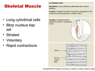
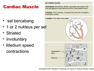
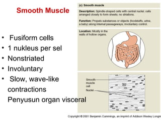
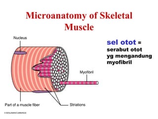
Muscle
Physiology
Muscle Tissue
• Skeletal Muscle
• Cardiac Muscle
• Smooth Muscle
Skeletal Muscle


• Long cylindrical cells
• Bbrp nucleus tiap
  sel
• Striated
• Voluntary
• Rapid contractions
Cardiac Muscle


•    sel bercabang
•   1 or 2 nukleus per sel
•   Striated
•   Involuntary
•   Medium speed
    contractions
Smooth Muscle


•    Fusiform cells
•   1 nukleus per sel
•   Nonstriated
•   Involuntary
•    Slow, wave-like
    contractions
    Penyusun organ visceral
Microanatomy of Skeletal
        Muscle
                  sel otot =
                  serabut otot
                  yg mengandung
                  myofibril
• Tiap serabut otot terdiri dari ribuan
  myofibril
• Tiap myofibril terdapat 1500 filamen
  meosin dan 3000 filamen aktin
• Tiap filamen meosin terdiri dari molekul
  meosin BM 33.200 dan molekul aktin
  BM 70.000
• Dengan mikroskup cahaya : nampak
  gambaran gelap terang kepadatan
  relatif dan ada bagian yang tumpah
  tindih dr aktin dan miosin
Pita cahaya I
• Terdiri filamen aktin saja (nampak terang)
• Sifat terhadap cahaya yg terpolarisasi
  (berkas cahaya yg datang dipancarkan ke
  semua jurusan krn kepadatan sama)


                                I band
Pita A
• Terletak dimana aktin dan meosin
  tumpang tindih (shg nampak gelap)
• Sifat anisotropik (cahaya dipancarkan
  tdk merata)


                 A band
Sarkomer
• Terbentuk oleh 2 garis Z (protein
  nonkontraktil) atau sebuah pita A dan 2
  belahan pita I

Sarkoplasma
• Sitoplasma sel2 otot
• Terdapat retikulum sarkopasma agranuler
Z line            Z line


         H band
Filamen meosin
Tediri 2 utas peptid yg satu sama lainnya saling melilit
dalam suatu heliks yg ujungnya membntuk kepala.
Filamen aktin
Terdiri 3 unsur : troponin, tropomeosin, aktin
• Active site : suatu molekul ADP dimana
 filamen meosin saling mengadakan interaksi
 dgn filamen aktin shg terjadi kontraksi otot
• Tropomeosin : merupakan protein yg akan
 “menutupi” active site shg tdk terjadi kontraksi
• Troponin : akan berikatan dgn Ca utk
 permulaan kontraksi
Ilustrasi sarkomer
                 H Band
Sarcomere Relaxed


Pita I nampak lebih lebar
Sarcomere Partially
         Contracted


Pita I lebih pendek
Sarcomere Completely
           Contracted


Kontraksi yg sempurna menyebabkan pita H tidak nampak,
pita I pendek sekali
Ilustrasi relaksasi dan kontraksi otot


Terlihat bahwa filamen meosin tidak berubah,
yg bergerak adalah aktin
Teori kontraksi
 otot:
• Ion Ca akan aktifkan
  filamen aktin
• Kepala meosin akan
  terikat pd active site
  filamen aktin, akan
  dihasilkan energi
• Kepala jadi miring ke
  pertengahan meosin
  (disebut power
  stroke)
• Kmdn secara otomatis
  kepala dilepaskan dr
  active site kembali
  tegak lurus
• Kmdn berikatan dgn
  active site berikutnya,
  kmdn miring (power
  stroke) sehingga
  filamen aktin bergerak
• Begitu seterusnya
  langkah demi langkah
  tarik filamen aktin ke
  pertengahan meosin
Binding Site        Tropomyosin


           Ion Ca
Troponin
Myosin
Neuromuscular Junction
Mikroanatomi otot skelet


Retikulum sarkoplama simpan Ca, mitokondria hasilkan ATP
utk kontraksi
Asetilkolin dilepaskan dari ujung saraf motorik
Mekanisme umum
kontraksi otot
• Potensial aksi berjalan di
  sepanjang saraf motorik
  sampai ujungnya pada
  saraf otot
• Pada setiap ujung saraf
  mensekresi neurotransmitter
  (y.i asetilkolin) dlm jumlah
  sedikit.
• Asetilkolin bekerja pd
  membran serabt otot dan
  akan membuka saluran
  bergerbang asetilkolin
• Terbukanya       saluran      asetilkolin
  memungkinkan ion Na mengalir ke bag.
  dalam membran serabut otot pada titik
  terminal saraf sehingga timbul potensial
  aksi dalm serabut otot.
• Potensial aksi berjalan di sepanjang
  membran serabut otot shg sbabkan
  retikulum sarkoplasma lepaskan ion Ca.
• ion Ca akan berikatan dgn troponin utk
  memulai terjadi kontraksi

• Kepala meosin berikatan dgn active site dan
  terjadi power stroke (lihat teori kontraksi !!)

• Setelah kurang dr 1 detik ion Ca dipompa ke
  dalam retikulum endoplasma (disimpan
  sampai potensial aksi yang baru datang lagi)
  sehingga kontraksi berhenti
Motor Unit
All the muscle cells controlled by one
              nerve cell
Muscle Fatique/kelelahan otot
• Tidak adanya oksigen sehingga ATP yg
  dihasilkan sedikit
• Respirasi anaerob menyebabkan terbentuk
  asam laktat (terasa pegal)
Muscle Atrophy
•    Mengecilnya ukuran otot
•    Dapat disebabkan karena :
3.   Immobilization
4.   Loss of neural stimulation
Muscle Hypertrophy
•   Peningkatan ukuran sel otot
•   Banyak kapiler
•   Banyak mitochondria
•   Dapat disebabkan karena :
    – Sering latihan
    – Steroid hormones
Steroid Hormones
• Stimulate muscle growth and hypertrophy
Muscle Tonus
• Tightness of a muscle
• Some fibers always contracted
Tetany
• Sustained contraction of a muscle
• Result of a rapid succession of nerve
  impulses
THE END

FISIOLOGI JARINGAN OTOT

  • 1.
  • 2.
    Muscle Tissue • SkeletalMuscle • Cardiac Muscle • Smooth Muscle
  • 3.
    Skeletal Muscle • Longcylindrical cells • Bbrp nucleus tiap sel • Striated • Voluntary • Rapid contractions
  • 4.
    Cardiac Muscle • sel bercabang • 1 or 2 nukleus per sel • Striated • Involuntary • Medium speed contractions
  • 5.
    Smooth Muscle • Fusiform cells • 1 nukleus per sel • Nonstriated • Involuntary • Slow, wave-like contractions Penyusun organ visceral
  • 6.
    Microanatomy of Skeletal Muscle sel otot = serabut otot yg mengandung myofibril
  • 7.
    • Tiap serabutotot terdiri dari ribuan myofibril • Tiap myofibril terdapat 1500 filamen meosin dan 3000 filamen aktin • Tiap filamen meosin terdiri dari molekul meosin BM 33.200 dan molekul aktin BM 70.000
  • 8.
    • Dengan mikroskupcahaya : nampak gambaran gelap terang kepadatan relatif dan ada bagian yang tumpah tindih dr aktin dan miosin
  • 9.
    Pita cahaya I •Terdiri filamen aktin saja (nampak terang) • Sifat terhadap cahaya yg terpolarisasi (berkas cahaya yg datang dipancarkan ke semua jurusan krn kepadatan sama) I band
  • 10.
    Pita A • Terletakdimana aktin dan meosin tumpang tindih (shg nampak gelap) • Sifat anisotropik (cahaya dipancarkan tdk merata) A band
  • 11.
    Sarkomer • Terbentuk oleh2 garis Z (protein nonkontraktil) atau sebuah pita A dan 2 belahan pita I Sarkoplasma • Sitoplasma sel2 otot • Terdapat retikulum sarkopasma agranuler
  • 12.
    Z line Z line H band
  • 14.
    Filamen meosin Tediri 2utas peptid yg satu sama lainnya saling melilit dalam suatu heliks yg ujungnya membntuk kepala.
  • 16.
    Filamen aktin Terdiri 3unsur : troponin, tropomeosin, aktin
  • 17.
    • Active site: suatu molekul ADP dimana filamen meosin saling mengadakan interaksi dgn filamen aktin shg terjadi kontraksi otot • Tropomeosin : merupakan protein yg akan “menutupi” active site shg tdk terjadi kontraksi • Troponin : akan berikatan dgn Ca utk permulaan kontraksi
  • 18.
  • 19.
    Sarcomere Relaxed Pita Inampak lebih lebar
  • 20.
    Sarcomere Partially Contracted Pita I lebih pendek
  • 21.
    Sarcomere Completely Contracted Kontraksi yg sempurna menyebabkan pita H tidak nampak, pita I pendek sekali
  • 22.
    Ilustrasi relaksasi dankontraksi otot Terlihat bahwa filamen meosin tidak berubah, yg bergerak adalah aktin
  • 23.
    Teori kontraksi otot: •Ion Ca akan aktifkan filamen aktin • Kepala meosin akan terikat pd active site filamen aktin, akan dihasilkan energi • Kepala jadi miring ke pertengahan meosin (disebut power stroke)
  • 24.
    • Kmdn secaraotomatis kepala dilepaskan dr active site kembali tegak lurus • Kmdn berikatan dgn active site berikutnya, kmdn miring (power stroke) sehingga filamen aktin bergerak • Begitu seterusnya langkah demi langkah tarik filamen aktin ke pertengahan meosin
  • 25.
    Binding Site Tropomyosin Ion Ca Troponin
  • 26.
  • 27.
  • 28.
    Mikroanatomi otot skelet Retikulumsarkoplama simpan Ca, mitokondria hasilkan ATP utk kontraksi
  • 29.
    Asetilkolin dilepaskan dariujung saraf motorik
  • 30.
    Mekanisme umum kontraksi otot •Potensial aksi berjalan di sepanjang saraf motorik sampai ujungnya pada saraf otot • Pada setiap ujung saraf mensekresi neurotransmitter (y.i asetilkolin) dlm jumlah sedikit. • Asetilkolin bekerja pd membran serabt otot dan akan membuka saluran bergerbang asetilkolin
  • 31.
    • Terbukanya saluran asetilkolin memungkinkan ion Na mengalir ke bag. dalam membran serabut otot pada titik terminal saraf sehingga timbul potensial aksi dalm serabut otot. • Potensial aksi berjalan di sepanjang membran serabut otot shg sbabkan retikulum sarkoplasma lepaskan ion Ca.
  • 32.
    • ion Caakan berikatan dgn troponin utk memulai terjadi kontraksi • Kepala meosin berikatan dgn active site dan terjadi power stroke (lihat teori kontraksi !!) • Setelah kurang dr 1 detik ion Ca dipompa ke dalam retikulum endoplasma (disimpan sampai potensial aksi yang baru datang lagi) sehingga kontraksi berhenti
  • 34.
    Motor Unit All themuscle cells controlled by one nerve cell
  • 35.
    Muscle Fatique/kelelahan otot •Tidak adanya oksigen sehingga ATP yg dihasilkan sedikit • Respirasi anaerob menyebabkan terbentuk asam laktat (terasa pegal)
  • 36.
    Muscle Atrophy • Mengecilnya ukuran otot • Dapat disebabkan karena : 3. Immobilization 4. Loss of neural stimulation
  • 37.
    Muscle Hypertrophy • Peningkatan ukuran sel otot • Banyak kapiler • Banyak mitochondria • Dapat disebabkan karena : – Sering latihan – Steroid hormones
  • 38.
    Steroid Hormones • Stimulatemuscle growth and hypertrophy
  • 39.
    Muscle Tonus • Tightnessof a muscle • Some fibers always contracted
  • 40.
    Tetany • Sustained contractionof a muscle • Result of a rapid succession of nerve impulses
  • 41.

Editor's Notes

  • #2   This is the presentation for Muscle Physiology for Human Anatomy and Physiology II at Oklahoma City Community College.
  • #3 There are three types of muscle tissue in the body. Skeletal muscle is the type that attaches to our bones and is used for movement and maintaining posture. Cardiac muscle is only found in the heart. It pumps blood. Smooth muscle is found in organs of the body such as the GI tract. Smooth muscle in the GI tract moves food and its digested products.
  • #4 Skeletal muscle attaches to our skeleton. *The muscle cells a long and cylindrical. *Each muscle cell has many nuclei. *Skeletal muscle tissue is striated. It has tiny bands that run across the muscle cells. *Skeletal muscle is voluntary. We can move them when we want to. *Skeletal muscle is capable of rapid contractions. It is the most rapid of the muscle types.
  • #5 Cardiac muscle tissue is only found in the heart. *Cardiac cells are arranged in a branching pattern. * Only one or two nuclei are present each cardiac cell. *Like skeletal muscle, cardiac muscle is striated. *Cardiac muscle is involuntary. *Its speed of contraction is not as fast as skeletal, but faster than that of smooth muscle.
  • #6 Smooth muscle is found in the walls of hollow organs. *Their muscle cells are fusiform in shape. *Smooth muscle cells have just on nucleus per cell. *Smooth muscle is nonstriated. *Smooth muscle is involuntary. *The contractions of smooth muscle are slow and wave-like.
  • #7 In this unit we will primarily study skeletal muscle. Each muscle cell is called a muscle fiber. Within each muscle fiber are many myofibrils.
  • #13 A small section of a myofibril is illustrated here. Note the thick myosin filaments are arranged between overlapping actin filaments. *The two Z lines mark the boundary of a sarcomere. The sarcomere is the functional unit of a muscle cell .We will examine how sarcomeres function to help us better understand how muscles work.
  • #14 Dark and light bands can be seen in the muscle fiber and also in the smaller myofibrils. An enlargement of the myofibril reveals that they are made of smaller filaments or myofilaments. *There is a thick filament called myosin and *a thin filament called actin. Note the I band, A band H zone or band and Z disc or line. These will be discussed shortly.  
  • #15 A myosin molecule is elongated with an enlarged head at the end.
  • #16 Many myosin molecules form the thick myosin filament. It has many heads projecting away from the main molecule.
  • #17 The thinner actin filament is composed of three parts: actin, tropomyosin and troponin.
  • #19 Here is a sarcomere illustrating the thin actin and thick myosin filaments. The area of the sarcomere has only myosin is called the H band.
  • #20 Here is another diagram of a sarcomere. Note the A band. It is formed by both myosin and actin filaments. The part of the sarcomere with only actin filaments is called the I band. This is a sarcomere that is relaxed.
  • #21 This sarcomere is partially contracted. Notice than the I bands are getting shorter.
  • #22 The sarcomere is completely contracted in this slide. The I and H bands have almost disappeared.
  • #23 Which filament has moved as the sarcomere contracted? Note the thick myosin filaments have not changed, but the thin actin filaments have moved closer together.
  • #26 The string of green circles represents an actin filament. There are binding sites in the filament for the attachment of myosin heads. *In a relaxed muscle the binding sites are covered by tropomyosin. The tropomyosin has molecules of troponin attached to it. *Calcium, shown in yellow, will attach to troponin. *Calcium will change the position of the troponin, tropomyosin complex. *The troponin, tropomyosin complex has now moved so that the binding sites are longer covered by the troponin, tropomyosin complex.
  • #27 The binding sites are now exposed and myosin heads are able to attach to form cross bridges.*  
  • #28 The next few slides will summarize the events of a muscle contraction. The nerve impulse reaches the neuromuscular junction (myoneural junction).
  • #29 This diagram shows the microanatomy of skeletal muscle tissue again. *The blue sarcoplasmic reticulum is actually the endoplasmic reticulum. It stores calcium. *The mitochondria are illustrated in orange. They generate ATP, which provides the energy for muscle contractions.
  • #30 Acetylcholine is released from the motor neuron.
  • #34 The influx of sodium will create an action potential in the sarcolemma. Note: This is the same mechanism for generating action potentials for the nerve impulse. The action potential travels down a T tubule. As the action potential passes through the sarcoplamic reticulum it stimulates the release of calcium ions. Calcium binds with troponin to move tropomyosin and expose the binding sites. Myosin heads attach to the binding sites of the actin filament and create a power stroke. ATP detaches the myosin heads and energizes them for another contraction. The process will continue until the action potentials cease. Without action potentials the calcium ions will return to the sarcoplasmic reticulum.  
  • #35 A motor unit is all the muscle cells controlled by one nerve cell. This diagram represents two motor units. Motor unit one illustrates two muscle cells controlled by one nerve cell. When the nerve sends a message it will cause both muscle cells to contract. Motor unit two has three muscle cells innervated by one nerve cell.
  • #36 Muscle fatigue is often due to a lack of oxygen that causes ATP deficit. Lactic acid builds up from anaerobic respiration in the absence of oxygen. Lactic acid fatigues the muscle.
  • #37 Muscle atrophy is a weakening and shrinking of a muscle. It can be caused by immobilization or loss of neural stimulation.
  • #38 Hypertrophy is the enlargement of a muscle. Hypertrophied muscles have more capillaries and more mitochondria to help them generate more energy. Strenuous exercise and steroid hormones can induce muscle hypertrophy. Since men produce more steroid hormones than women, they usually have more hypertrophied muscles.
  • #39 Steroid hormones such as testosterone stimulate muscle growth and hypertrophy.
  • #40 Muscle tonus or muscle tone refers to the tightness of a muscle. In a muscle some fibers are always contracted to add tension or tone to the muscle.
  • #41 Tetany is a sustained contraction of a muscle. It results from a rapid succession of nerve impulses delivered to the muscle.
  • #42 This concludes the presentation on Muscle Physiology