The document summarizes key aspects of secondary protein structure, including the two main types - alpha helix and beta pleated sheet. It describes the alpha helix as a right-handed spiral stabilized by hydrogen bonds between amino acids four positions apart in the sequence. The beta pleated sheet involves hydrogen bonding between adjacent protein molecules in a zigzag pattern. Both secondary structures are important for determining the 3D shape of globular proteins. The Ramachandran plot is also introduced as a way to visualize allowed backbone dihedral angles in protein structures.
INTRODUCTION
Proteins are animportant class of biological macromolecules which are the polymers
of amino acids. • Biochemists have distinguished several levels of structural
organization of proteins. They are: – Primary structure – Secondary structure –
Tertiary structure – Quaternary structure
Secondary configuration: Protein molecules of sec. structure are spirally coiled. In
addition to peptide bond, amino acids are linked by hydrogen bonds between oxygen of
one amide group and hydrogen of another amide group. This structure is of two types -
(i) Alpha–Helix
(ii) Beta- Helix or pleated sheath structure
4.
SECONDARY STRUCTURE OFPROTEIN
Localized arrangement of adjacent amino acids formed as the polypeptide chain
folds.
• It consists of
• Linus Pauling proposed some essential features of peptide units and polypeptide
backbone. They are: –
The amide group is rigid and planar as a result of resonance. So rotation about C-
N bond is not feasible.
– Rotation can take place only about N- Cα and Cα – C bonds.
– Trans configuration is more stable than cis for R grps at Cα
• From these conclusions Pauling postulated 2 ordered structures α helix and β
sheet α-helix β-pleated sheet β-bends Non repetitive structures Super secondary
structures
α-Helix
● Right handedrotation of spirally coiled chain with approximately 3.5 amino
acids in each turn. This structure has intramolecular hydrogen bonding i. e.
between two amino acids of same chain. Side chain extend outwards.
● Stabilized by H bonding that are arranged such that the peptide Carbonyl
oxygen (nth residue) and amide hydrogen(n+4 th residue).
● Amino acids per turn – 3.6 , Pitch is 5.4 A°
● Alpha helical segments are found in many globular proteins like
myoglobin,troponin C.
● Length ~12 residues and ~3 helical turns.
● phi = -60 degrees, psi = -45 degrees , falls within the fully allowed regions of the
Ramachandran diagram.
● E.g. Keratin ,Myosin, Tropomyosin.
7.
● Certain aminoacids (particularly PROLINE) disrupts the α helix. The larger number of
acidic (Asp, Glu) or Basic (Lys, Arg and His) amino acids also interfere with α helix
structure.
● In general, an α helix consists of 5 to more than 40 amino acidsAmino acids promote the
formation of α helix are Ala, Glu, Leu, Met.Amino acids are bad trainers Pro, Gly, Tyr,
Ser.
● Α helices can be hydrophilic, amphipathic or hydrophobic.It depends on the amino acid
composition of the propeller.
● Indeed, the amino acid radicals are turning out the axis of the helix, standard terms
their response to their environment. Thus, if the α helix contains only hydrophobic amino
acids, so it is put in contact with hydrophobic surfaces, such as the lipid bilayer.
● If the hydrophobic residues are positioned on one side and hydrophilic residues on the
other side, the α helix is amphipathic (or amphiphilic ).
● That is to say, we will find the interface of the hydrophilic and hydrophobic regions.
● This is the alpha-helix structure of the protein.
β -Pleated Sheet
●Protein molecule has zig - zag structure. Two or more protein molecules are held together
by intermolecular hydrogen bonding. e.g. Fibroin (silk).
● Proteins of sec. structure are insoluble in water and fibrous in appearance.
● Keratin is a fibrous , tough, resistant to digestion, scleroprotein.Hardness of keratin is
due to abundance of cysteine amino acid in its structure.
● The connection between two antiparallel strands may be just a small loop but the link
between tandem parallel strands must be a crossover connection that is out of the plane of
the β sheet.The β-pleated sheet structure (beta-sheet structure) proposed by Pauling and
Corey.
● The β-pleated sheet structure has two Polypeptide chains.
● It consists of the juxtaposition of β strands, chain conformation very stretched.
● Chains are presented in “Pleated sheet “(to take the first topographical sense- a succession of
“roofs”).
10.
B PLEATED SHEET
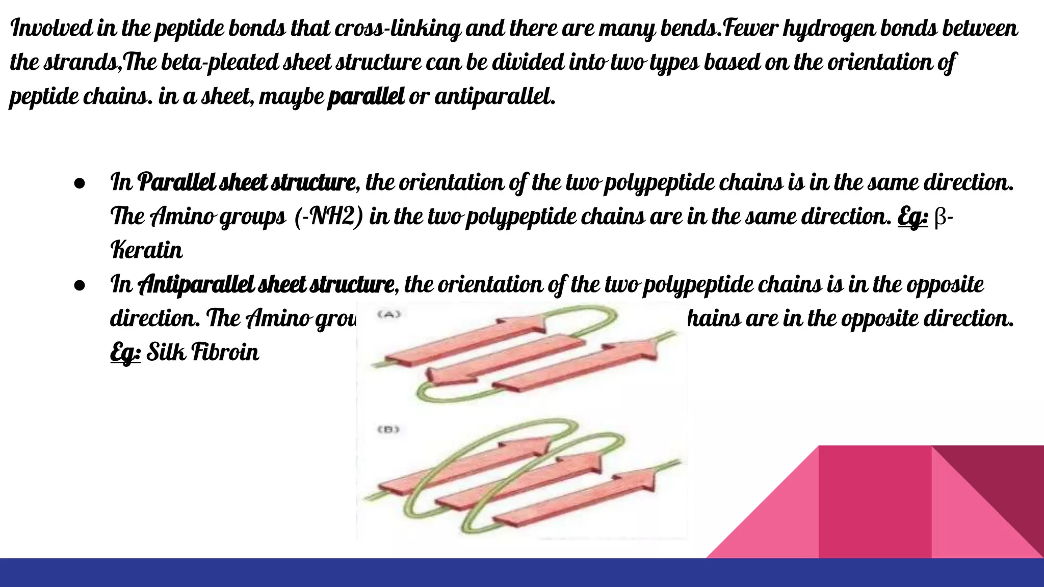
Involvedin the peptide bonds that cross-linking and there are many bends.Fewer hydrogen bonds between
the strands,The beta-pleated sheet structure can be divided into two types based on the orientation of
peptide chains. in a sheet, maybe parallel or antiparallel.
● In Parallel sheet structure, the orientation of the two polypeptide chains is in the same direction.
The Amino groups (-NH2) in the two polypeptide chains are in the same direction. Eg: β-
Keratin
● In Antiparallel sheet structure, the orientation of the two polypeptide chains is in the opposite
direction. The Amino groups (-NH2) in the two polypeptide chains are in the opposite direction.
Eg: Silk Fibroin
11.
RAMACHANDRAN PLOT
● Thisis made to visualize the backbone of amino
acid residues. The amino acids with larger side
chains will show less number of allowed region
within the ramachandran plot.
● A Ramachandran plot is a way to visualize
backbone dihedral angles ψ against φ of amino
acid residues in protein structure. It can be used to
show which values, or conformations, of the ψ and φ
angles are possible for an amino- acid residue in a
protein and to show the empirical distribution of
data points observed in a single structure.
● The darkest areas correspond to the "core" regions
representing the most favorable combinations of phi-
psi values.