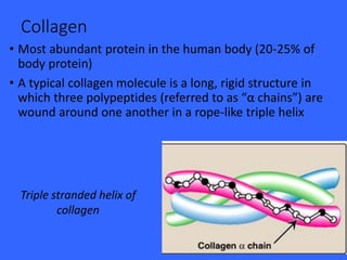
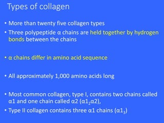
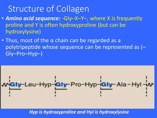
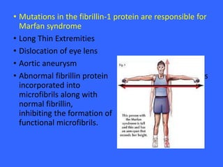
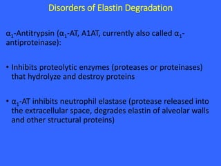
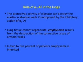
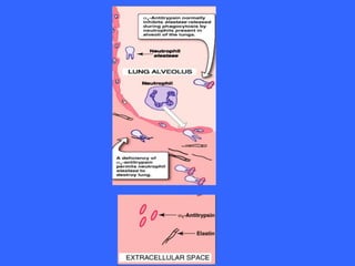
Structure and Function
of Fibrous Proteins
Source: Biochemistry by Lippincott
Maryam Fida (o-1827)
Collagen
• Most abundant protein in the human body (20-25% of
body protein)
• A typical collagen molecule is a long, rigid structure in
which three polypeptides (referred to as “α chains”) are
wound around one another in a rope-like triple helix
Triple stranded helix of
collagen
Types of collagen
• More than twenty five collagen types
• Three polypeptide α chains are held together by hydrogen
bonds between the chains
• α chains differ in amino acid sequence
• All approximately 1,000 amino acids long
• Most common collagen, type I, contains two chains called
α1 and one chain called α2 (α12α2),
• Type II collagen contains three α1 chains (α13)
Structure of Collagen
• Amino acid sequence: -Gly–X–Y–, where X is frequently
proline and Y is often hydroxyproline (but can be
hydroxylysine)
• Thus, most of the α chain can be regarded as a
polytripeptide whose sequence can be represented as (–
Gly–Pro–Hyp–)
Hyp is hydroxyproline and Hyl is hydroxylysine
Triple-helical structure:
• Elongated, triple-helical structure that places many of
its amino acid side chains on the surface
• Allows bond formation between the exposed R-
groups of neighboring collagen monomers, resulting
in their aggregation into long fibers
Hydroxyproline and Hydroxylysine:
• Result from the hydroxylation of some of the proline
and lysine residues after their incorporation into
polypeptide chains
• An example of posttranslational modification
• Hydroxyproline is important in stabilizing the triple-
helical structure of collagen because it maximizes
interchain hydrogen bond formation
These hydroxylation reactions require
molecular oxygen, Fe2+ and the
reducing agent vitamin C (ascorbic
acid) without which the
hydroxylating enzymes, prolyl
hydroxylase and lysyl hydroxylase, are
unable to function.
In the case of ascorbic acid deficiency
(and, therefore, a lack of prolyl and
lysyl hydroxylation), interchain H-
bond formation is impaired, collagen
fibers cannot be cross-linked, greatly
decreasing the tensile strength of the
assembled fiber. The resulting
deficiency disease is known as Scurvy.
Patients with ascorbic acid deficiency
also often show bruises on the limbs
as a result of subcutaneous
extravasation of blood
Collagen diseases
• 1,000 mutations have been identified in 22 genes
coding for twelve of the collagen types
• Defective Collagen Synthesis:
1. Ehlers-Danlos syndrome (EDS)
2. Osteogenesis imperfecta (OI)
Ehlers-Danlos syndrome (EDS)
• Can result from :
1. Deficiency of collagen-processing enzymes e.g. lysyl
hydroxylase deficiency or procollagen peptidase
deficiency
2. mutations in the amino acid sequences of collagen
types I, III, or V
• most clinically important mutations are found in the
gene for type III collagen (in blood vessels).
Osteogenesis imperfecta (OI) - Brittle bone
syndrome
• Bones that easily bend and fracture
• Retarded wound healing and a rotated and twisted
spine leading to a “humped-back” appearance are
common features of the disease
• Type I OI (osteogenesis imperfecta tarda):
Decreased production of α1 and α2 chains
Presents in early infancy with fractures secondary to
minor trauma
Prenatal ultrasound detects bowing or fractures of
long bones
• Type II OI is called osteogenesis imperfecta
congenita:
More severe. Patients die of pulmonary hypoplasia in
utero or during the neonatal period
• Most patients with severe OI have mutations in the
gene for either the pro-α1 or pro-α2 chains of type I
collagen
• The most common mutations cause the replacement
of glycine residues (in -Gly–X–Y–) by amino acids with
bulky side chains. The resultant structurally abnormal
pro-α chains prevent the formation of the required
triple-helical conformation
Lethal form (type II) of osteogenesis imperfecta in which the fractures
appear in utero, as revealed by this radiograph of a stillborn fetus.
Elastin
• Connective tissue protein with rubber-like properties
• Elastic fibers : Elastin and glycoprotein microfibrils
• Lungs, the walls of large arteries and elastic
ligaments.
Structure of Elastin
• Insoluble protein polymer synthesized from a precursor,
tropoelastin
• Tropoelastin: Linear polypeptide composed of about 700
amino acids that are primarily small and nonpolar (for
example, glycine, alanine, and valine)
• Also rich in proline and lysine
• Tropoelastin is secreted by the cell into the extracellular
space. There it interacts with specific glycoprotein
microfibrils, such as fibrillin, which function as a scaffold
onto which tropoelastin is deposited
• Mutations in the fibrillin-1 protein are responsible for
Marfan syndrome
• Long Thin Extremities
• Dislocation of eye lens
• Aortic aneurysm
• Abnormal fibrillin protein is
incorporated into
microfibrils along with
normal fibrillin,
inhibiting the formation of
functional microfibrils.
Disorders of Elastin Degradation
α1-Antitrypsin (α1-AT, A1AT, currently also called α1-
antiproteinase):
• Inhibits proteolytic enzymes (proteases or proteinases)
that hydrolyze and destroy proteins
• α1-AT inhibits neutrophil elastase (protease released into
the extracellular space, degrades elastin of alveolar walls
and other structural proteins)
Role of α1-AT in the lungs
• The proteolytic activity of elastase can destroy the
elastin in alveolar walls if unopposed by the inhibitory
action of α1-AT
• Lung tissue cannot regenerate; emphysema results
from the destruction of the connective tissue of
alveolar walls
• In two to five percent of patients emphysema is
inherited
Smokers
• A specific α1-AT methionine is required for the binding
of the inhibitor to its target proteases. Smoking
causes the oxidation and subsequent inactivation of
that methionine residue, thereby rendering the
inhibitor powerless to neutralize elastase
• Smokers with α1-AT deficiency, therefore, have a
considerably elevated rate of lung destruction and a
poorer survival rate than nonsmokers with the
deficiency
• The deficiency of elastase inhibitor can be reversed
by weekly intravenous administration of α1-AT
Fibrous proteins (biochem)

Fibrous proteins (biochem)

  • 1.
    Structure and Function ofFibrous Proteins Source: Biochemistry by Lippincott Maryam Fida (o-1827)
  • 2.
    Collagen • Most abundantprotein in the human body (20-25% of body protein) • A typical collagen molecule is a long, rigid structure in which three polypeptides (referred to as “α chains”) are wound around one another in a rope-like triple helix Triple stranded helix of collagen
  • 3.
    Types of collagen •More than twenty five collagen types • Three polypeptide α chains are held together by hydrogen bonds between the chains • α chains differ in amino acid sequence • All approximately 1,000 amino acids long • Most common collagen, type I, contains two chains called α1 and one chain called α2 (α12α2), • Type II collagen contains three α1 chains (α13)
  • 4.
    Structure of Collagen •Amino acid sequence: -Gly–X–Y–, where X is frequently proline and Y is often hydroxyproline (but can be hydroxylysine) • Thus, most of the α chain can be regarded as a polytripeptide whose sequence can be represented as (– Gly–Pro–Hyp–) Hyp is hydroxyproline and Hyl is hydroxylysine
  • 5.
    Triple-helical structure: • Elongated,triple-helical structure that places many of its amino acid side chains on the surface • Allows bond formation between the exposed R- groups of neighboring collagen monomers, resulting in their aggregation into long fibers
  • 6.
    Hydroxyproline and Hydroxylysine: •Result from the hydroxylation of some of the proline and lysine residues after their incorporation into polypeptide chains • An example of posttranslational modification • Hydroxyproline is important in stabilizing the triple- helical structure of collagen because it maximizes interchain hydrogen bond formation
  • 7.
    These hydroxylation reactionsrequire molecular oxygen, Fe2+ and the reducing agent vitamin C (ascorbic acid) without which the hydroxylating enzymes, prolyl hydroxylase and lysyl hydroxylase, are unable to function. In the case of ascorbic acid deficiency (and, therefore, a lack of prolyl and lysyl hydroxylation), interchain H- bond formation is impaired, collagen fibers cannot be cross-linked, greatly decreasing the tensile strength of the assembled fiber. The resulting deficiency disease is known as Scurvy. Patients with ascorbic acid deficiency also often show bruises on the limbs as a result of subcutaneous extravasation of blood
  • 8.
    Collagen diseases • 1,000mutations have been identified in 22 genes coding for twelve of the collagen types • Defective Collagen Synthesis: 1. Ehlers-Danlos syndrome (EDS) 2. Osteogenesis imperfecta (OI)
  • 9.
    Ehlers-Danlos syndrome (EDS) •Can result from : 1. Deficiency of collagen-processing enzymes e.g. lysyl hydroxylase deficiency or procollagen peptidase deficiency 2. mutations in the amino acid sequences of collagen types I, III, or V • most clinically important mutations are found in the gene for type III collagen (in blood vessels).
  • 11.
    Osteogenesis imperfecta (OI)- Brittle bone syndrome • Bones that easily bend and fracture • Retarded wound healing and a rotated and twisted spine leading to a “humped-back” appearance are common features of the disease
  • 12.
    • Type IOI (osteogenesis imperfecta tarda): Decreased production of α1 and α2 chains Presents in early infancy with fractures secondary to minor trauma Prenatal ultrasound detects bowing or fractures of long bones
  • 13.
    • Type IIOI is called osteogenesis imperfecta congenita: More severe. Patients die of pulmonary hypoplasia in utero or during the neonatal period • Most patients with severe OI have mutations in the gene for either the pro-α1 or pro-α2 chains of type I collagen • The most common mutations cause the replacement of glycine residues (in -Gly–X–Y–) by amino acids with bulky side chains. The resultant structurally abnormal pro-α chains prevent the formation of the required triple-helical conformation
  • 14.
    Lethal form (typeII) of osteogenesis imperfecta in which the fractures appear in utero, as revealed by this radiograph of a stillborn fetus.
  • 15.
    Elastin • Connective tissueprotein with rubber-like properties • Elastic fibers : Elastin and glycoprotein microfibrils • Lungs, the walls of large arteries and elastic ligaments.
  • 16.
    Structure of Elastin •Insoluble protein polymer synthesized from a precursor, tropoelastin • Tropoelastin: Linear polypeptide composed of about 700 amino acids that are primarily small and nonpolar (for example, glycine, alanine, and valine) • Also rich in proline and lysine • Tropoelastin is secreted by the cell into the extracellular space. There it interacts with specific glycoprotein microfibrils, such as fibrillin, which function as a scaffold onto which tropoelastin is deposited
  • 17.
    • Mutations inthe fibrillin-1 protein are responsible for Marfan syndrome • Long Thin Extremities • Dislocation of eye lens • Aortic aneurysm • Abnormal fibrillin protein is incorporated into microfibrils along with normal fibrillin, inhibiting the formation of functional microfibrils.
  • 19.
    Disorders of ElastinDegradation α1-Antitrypsin (α1-AT, A1AT, currently also called α1- antiproteinase): • Inhibits proteolytic enzymes (proteases or proteinases) that hydrolyze and destroy proteins • α1-AT inhibits neutrophil elastase (protease released into the extracellular space, degrades elastin of alveolar walls and other structural proteins)
  • 20.
    Role of α1-ATin the lungs • The proteolytic activity of elastase can destroy the elastin in alveolar walls if unopposed by the inhibitory action of α1-AT • Lung tissue cannot regenerate; emphysema results from the destruction of the connective tissue of alveolar walls • In two to five percent of patients emphysema is inherited
  • 21.
    Smokers • A specificα1-AT methionine is required for the binding of the inhibitor to its target proteases. Smoking causes the oxidation and subsequent inactivation of that methionine residue, thereby rendering the inhibitor powerless to neutralize elastase
  • 22.
    • Smokers withα1-AT deficiency, therefore, have a considerably elevated rate of lung destruction and a poorer survival rate than nonsmokers with the deficiency • The deficiency of elastase inhibitor can be reversed by weekly intravenous administration of α1-AT