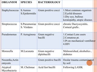
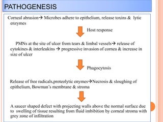
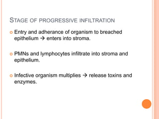
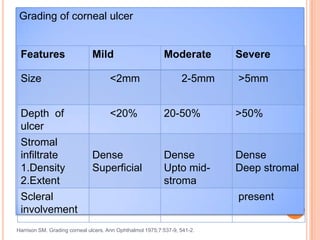
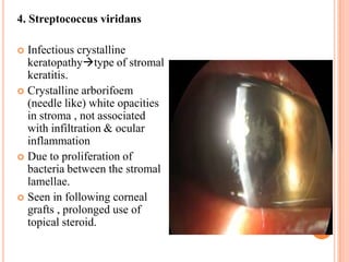
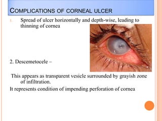
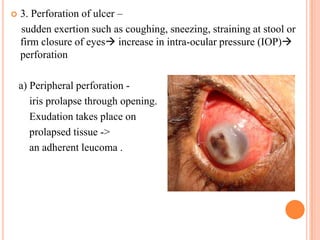
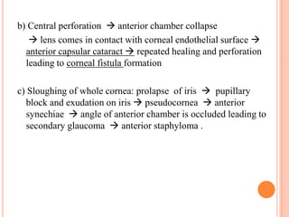
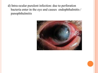
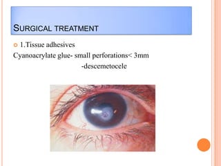
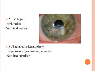
Dr. Bhushan Patil.
DEFINATION


A loss of epithelium with inflammation in the
sorrounding cornea is called as corneal ulcer.



Host cellular and immunologic responses to offending
agent which may be bacterial,viral,fungal or protozoal
organisms leads to formation of ulcer.



Sight threatening condition and should be considered as
ocular emergency.
NORMAL DEFENCE MECHANISM


Corneal epithelium- mechanical barrier



Conjunctiva- cellular & chemical components



Tear film- biological protective system

Major components of ocular defence system
BARRIERS OF MICROBIAL INFECTION

Anatomical

• Bony orbital rim,eyelids,
• Intact corneal & conjunctival
epithelium

Mechanical

• Tear film-mucus layer
• Lacrimal system

Antimicrobial

• Tear film constitutes-IgA,
complement components, and
enzymes lysozyme, lactoferrin,
betalysins have antibacterial effect
• CALT
PREDISPOSING FACTORS
OCULAR


1. Trauma-breach in corneal epithelium
-inoculation of organism



2.Eyelid & adnexal diseases- blepharitis, ectropion, entropion, trichiasis, lagophthalmos,
chronic dacryocystitis

Disturbed Tear film

Recurrent epithelial erosions
3. Ocular surface disorder- Dry eye, Steven-Johnson syndrome, ocular burn,
bullous keratopathy.





4. Contact lens use-Increased risk of bacterial keratitis with use of Extended
soft contact lens
corneal hypoxia & decompensation.
- Contamination of CL solution.



5. Local immune suppression due to topical corticosteroids.



6.Ocular surgery- cataract , LASIK.
SYSTEMIC FACTORS
1.Malnutrition
2.Diabetes
3.Immunosupression-Systemic steroids, AIDS
4.Chronic alcoholism
AETIOLOGY OF BACTERIAL ULCER


Caused by organisms which produce toxins causing
tissue death i.e. necrosis characterized by pus formation.



Such purulent keratitis is usually exogenous due to
infection by pyogenic bacteria such as pseudomonas,
staphylococcus,streptococcus, N. gonorrhoeae and C.
diphtheriae
AETIOLOGY OF BACTERIAL ULCER


Most of the bacteria are capable of producing corneal
ulcer only when the epithelium is damaged



N Gonorrhoeae, C Diphtheriae, Hemophilus , Shigella
and Listeria Monocytogenes – can penetrate intact
corneal epithelium.
ORGANISM

SPECIES

BACTERIOLOGY

Staphylococcus S.Aureus
Gram positive cocci
S.Epidermidis

1.Most common organism
2.Eyelid diseases
3.Dry eye, bullous
keratopathy, atopic disease.

Streptococcus

S.Pneumoniae Gram positive cocci
S. Viridans

chronic Dacryocystitis.
Corneal grafts .

Pseudomonas

P. Aeruginosa Gram negative
bacilli

1.Contact Lens users
2.Comatose pt.
3.Pt on mechanical ventillator
4.HIV

Moraxella

M.Lacunata

Malnourished, alcoholics ,
diabetes

Nocardia,Actin
omycets
Atypical
Mycobacteria

Gram negative
diplobacilli

Gram positive bacilli Ocular trauma contaminated
by soil
M. Chelonae

Acid fast bacilli

Following LASIK
PATHOGENESIS
Corneal abrasion Microbes adhere to epithelium, release toxins & lytic
enzymes
Host response
PMNs at the site of ulcer from tears & limbal vessels release of
cytokines & interleukins  progressive invasion of cornea & increase in
size of ulcer

Phagocytosis
Release of free radicals,proteolytic enymesNecrosis & sloughing of
epithelium, Bowman’s membrane & stroma

A saucer shaped defect with projecting walls above the normal surface due
to swelling of tissue resulting from fluid imbibition by corneal stroma with
grey zone of infiltration
STAGE OF PROGRESSIVE INFILTRATION


Entry and adherance of organism to breached
epithelium  enters into stroma.



PMNs and lymphocytes infiltrate into stroma and
epithelium.



Infective organism multiplies  release toxins and
enzymes.
STAGE OF ACTIVE ULCERATION


Necrosis occurs due to toxins and enzymes released by
infective organism.



Sloughing of epithelium and stroma  ulcer.



Ulcer Borders thickening due to infiltrates and edema.



It is associated with iritis due to diffusion of toxins of
infecting bacteria into AC.


Sometimes iridocyclitis is so severe that it is
accompanied by outpouring of leucocytes from uveal
blood vessels and these cells gravitate to bottom of the
AC to form hypopyon (sterile).
STAGE OF REGRESSION
Natural host defence & antimicrobial treatment
 Line of demarcation forms around ulcer which contains
leucocytes which phagocytose the organism & necrotic debris
 Necrotic material fall off- ulcer becomes larger -> infiltration and
swelling reduce and disappears -> margin & floor becomes
smooth.
 Vascularization develops from limbus to corneal ulcer to restore
lost tissue and to supply antibodies.

STAGE OF HEALING


Vascularization is followed by cicatrization due to
regeneration of collagen and formation of fibrous tissue



Newly formed fibers are laid down irregularly, not
conforming to normal pattern of stromal fibers.
Therefore this fibrous tissue refracts light irregularly and
forms opacity.
CLINICAL FEATURES
Clinical signs and symptoms are variable depends on the
 virulence of the organism
 duration of infection,
 pre-existing corneal conditions
 immune status of host
 previous use of local steroids
PRESENTATION
1. Diminution of vision, depending on location of
corneal ulcer
2. Watering due to reflex lacrimation
3. Photophobia
4. Pain due to exposed nerve endings
5. Mucopurulent / purulent discharge
WORK-UP
Evaluation of predisposing and aggravating Factors
1. A detailed history.
2.

Prior ocular history

3.

Review of related medical problems, current ocular
medications and history of systemic steroids.
OCULAR EXAMINATION
1.Visual acuity-reduced
2.Slit lamp Biomicroscope
Lids - edema

Conjunctiva – Ciliary congestion
4.

Cornea
-Location of the ulcer- central, paracentral , peripheral,total.

-Size , shape, depth, margins & floor- depends on stage of
ulcer.
-Density and extent of stromal infiltration.

5. Anterior chamber
- Cells/flare, mobile Hypopyon.
Iris- muddy
Toxin induced iritis

Pupil – miotic
Other:
-Sac syringing
-corneal sensation
-Fluorescein staining
Grading of corneal ulcer

Features

Mild

Size

<2mm

Depth of
ulcer

<20%

Stromal
infiltrate
1.Density
2.Extent
Scleral
involvement

Dense
Superficial

Moderate
2-5mm

Severe
>5mm

20-50%

>50%

Dense
Upto midstroma

Dense
Deep stromal

Harrison SM. Grading corneal ulcers. Ann Ophthalmol 1975;7:537-9, 541-2.

present
SPECIAL FEATURES
1.Staphylococcal
 Central,oval, opaque
 Distinct margins.
 Mild oedema of
remaining cornea.
 Stromal abscess in
longstanding cases.
 Mild to moderate AC
reaction.
 Affects compromised
corneas e.g. Bullous
keratopathy , dry eyes ,
atopic diseases.
2.Pneumococcal
 Ulcer serpens is greyish
white or yellowish disc
shaped ulcer occuring near
center of cornea.
 starts at periphery &
spreads towards centre
 Tendency to creep over the
cornea in serpiginous
fashion- Ulcus Serpen.
 Violent iridocyclitis is often
associated with it.
 Hypopyon – always present
 It has great tendency for
PERFORATION.
BACTERIAL ULCER WITH HYPOPYON

HYPOPYON.
3. Pseudomonas
Rapidly spreading.
 Extends periphery & deep
within 24 hrs.
 Stromal necrosis with shaggy
surface
 Spreads concentrically and
symmetrically to involve
whole depth of cornea-Ring
ulcer.
 Greenish-yellow discharge.
 Hypopyon is present.
 Untreated  corneal melting.

4. Streptococcus viridans
Infectious crystalline
keratopathytype of stromal
keratitis.
 Crystalline arborifoem
(needle like) white opacities
in stroma , not associated
with infiltration & ocular
inflammation
 Due to proliferation of
bacteria between the stromal
lamellae.
 Seen in following corneal
grafts , prolonged use of
topical steroid.

COMPLICATIONS OF CORNEAL ULCER
1.

Spread of ulcer horizontally and depth-wise, leading to
thinning of cornea

2. Descemetocele –

This appears as transparent vesicle surrounded by grayish zone
of infiltration.
It represents condition of impending perforation of cornea


3. Perforation of ulcer –
sudden exertion such as coughing, sneezing, straining at stool or
firm closure of eyes increase in intra-ocular pressure (IOP)
perforation
a) Peripheral perforation iris prolapse through opening.
Exudation takes place on
prolapsed tissue ->
an adherent leucoma .
b) Central perforation  anterior chamber collapse
 lens comes in contact with corneal endothelial surface 
anterior capsular cataract  repeated healing and perforation
leading to corneal fistula formation
c) Sloughing of whole cornea: prolapse of iris  pupillary
block and exudation on iris  pseudocornea  anterior
synechiae  angle of anterior chamber is occluded leading to
secondary glaucoma  anterior staphyloma .
d) Intra-ocular purulent infection: due to perforation
bacteria enter in the eye and causes endophthalmitis /
panophthalmitis
INVESTIGATIONS


Routine – Hemogram
BSL
HIV

Specific – Corneal scraping
Gram stain, Culture &
Antibiotic sensitivity
Culture of contact lens & solution
TREATMENT OF UNCOMPLICATED ULCER
Hospitalization
Treat the underlying cause/predisposing factor
LOCAL TREATMENT
Control of infection with appropriate antibiotic(s)
a. based on clinical judgment
b. based on finding of smear examination
c. based on culture and sensitivity report
Combination therapy with fortified broad spectrum antibiotics
1.Cephalosporin – gram positive cocci & some gram negative
rods
 Cefazolin 50mg/ml OR Ceftazidime 50mg/ml


2.Aminoglycoside - gram negative bacilli
 Tobramycin 14mg/ml
OR
Fluoroquinolone – broad spectrum-gram negative + gram
positive
 Moxiflox 5mg/ml
Topically every 30-60 min initially
 In severe cases- every 5 min for 30 min as a loading dose.

Vancomycin- reserved for very severe or recalcitrant infections
(50mg/ml)
 Amikacin (10-20mg/ml) for AF-bacilli


Fluoroquinolone monotherapy – 4th generation
< 3mm in diameter, peripheral & not associated with thinning
SYSTEMIC ANTIBIOTICS-FLUOROQUINOLONE
Indications
 Severe keratitis
 Scleral involvement
 hypopyon
 Impending perforation
 Frank perforation with risk of intraocular spread
 Infection in children
 P.aeruginosa infection
ADJUVANT THERAPY
1.Cycloplegic : Atropine 1% or cyclopentolate 1% or
Homatropine 2%- prevents ciliary spasm, relieves pain, breaks
adhesions and prevent synechia formation.
2.Analgesic anti-inflammatory
3. Oral vitamin C
4. Acetazolamide Tab - impending perforation or perforated
corneal ulcer and in cases where there is raised intra-ocular
tension .
TREATMENT OF IMPENDING
PERFORATION
1.
2.
3.
4.
5.

6.
7.

Straining should be avoided.
Pressure bandage
Lowering of IOP
Tissue adhesive glue (cynoacrylate)
Conjunctival flap
Soft contact lens Bandage
Penetrating keratoplasty
TREATMENT OF NON HEALING ULCER
Removal of any known cause.
->LOCAL
->SYSTEMIC
 Mechanical debridement of ulcer.
 Cauterisation of ulcer.
 Bandage soft contact lens.

TREATMENT OF PERFORATED CORNEAL ULCER
Tissue adhesives
 Conjunctival flap
 Soft bandage
 Keratoplasty

Modification of initial antimicrobial therapy:
-Should be based on clinical response not on culture sensitivity


If pt is responding  no change in initial treatment
If pt is not responding/ worsening drugs are changed according
to antimicrobial sensitivity
Signs of healing :
-resolution of lid edema, congestion
-decreased density of stromal infiltrate
-reduction of corneal oedema
-reduction in AC reaction/hypopyon
-re-epithelization
-corneal vascularization




Antibiotic frequency-tapered to 4hrly after 72 hrs
Signs of non-response
- Increase in infiltration, epithelial defect, height of hypopyon,
Corneal thinning, perforation
Treatment
 Re-evaluate for
Drug toxicity
Non-infectious causes or
Unusual organisms




Modification of anti-microbial therapy according to antimicrobial
sensitivity



Scraping of ulcer floor followed by cauterization with pure
(100%) carbolic acid or 10-20% trichloracetic acid.



Therapeutic keratoplasty
TOPICAL CORTICOSTEROIDS
Controversial in bacterial keratitis
 The rationale for using steroids - to decrease tissue destruction.
 Criteria for topical steroids in ulcer -1.Must not be used in presence of active infected corneal ulcer
2.If bacteria shows in-vitro sensitivity to the antibiotic being used
3.Patients compliance for follow-up
4. No other virulent organism is found




Monitor pt at 24 & 48 hrs after initiation
SURGICAL TREATMENT
1.Tissue adhesives
Cyanoacrylate glue- small perforations< 3mm
-descemetocele

2. Patch graft
-perforation –
5mm in diameter


3 . Therapeutic keratoplasty
-large areas of perforation, necrosis
-Non-healing ulcer

Bacterial corneal ulcer DrBP

Bacterial corneal ulcer DrBP

  • 1.
  • 2.
    DEFINATION  A loss ofepithelium with inflammation in the sorrounding cornea is called as corneal ulcer.  Host cellular and immunologic responses to offending agent which may be bacterial,viral,fungal or protozoal organisms leads to formation of ulcer.  Sight threatening condition and should be considered as ocular emergency.
  • 3.
    NORMAL DEFENCE MECHANISM  Cornealepithelium- mechanical barrier  Conjunctiva- cellular & chemical components  Tear film- biological protective system Major components of ocular defence system
  • 4.
    BARRIERS OF MICROBIALINFECTION Anatomical • Bony orbital rim,eyelids, • Intact corneal & conjunctival epithelium Mechanical • Tear film-mucus layer • Lacrimal system Antimicrobial • Tear film constitutes-IgA, complement components, and enzymes lysozyme, lactoferrin, betalysins have antibacterial effect • CALT
  • 5.
    PREDISPOSING FACTORS OCULAR  1. Trauma-breachin corneal epithelium -inoculation of organism  2.Eyelid & adnexal diseases- blepharitis, ectropion, entropion, trichiasis, lagophthalmos, chronic dacryocystitis Disturbed Tear film Recurrent epithelial erosions
  • 6.
    3. Ocular surfacedisorder- Dry eye, Steven-Johnson syndrome, ocular burn, bullous keratopathy.   4. Contact lens use-Increased risk of bacterial keratitis with use of Extended soft contact lens corneal hypoxia & decompensation. - Contamination of CL solution.  5. Local immune suppression due to topical corticosteroids.  6.Ocular surgery- cataract , LASIK.
  • 7.
  • 8.
    AETIOLOGY OF BACTERIALULCER  Caused by organisms which produce toxins causing tissue death i.e. necrosis characterized by pus formation.  Such purulent keratitis is usually exogenous due to infection by pyogenic bacteria such as pseudomonas, staphylococcus,streptococcus, N. gonorrhoeae and C. diphtheriae
  • 9.
    AETIOLOGY OF BACTERIALULCER  Most of the bacteria are capable of producing corneal ulcer only when the epithelium is damaged  N Gonorrhoeae, C Diphtheriae, Hemophilus , Shigella and Listeria Monocytogenes – can penetrate intact corneal epithelium.
  • 10.
    ORGANISM SPECIES BACTERIOLOGY Staphylococcus S.Aureus Gram positivecocci S.Epidermidis 1.Most common organism 2.Eyelid diseases 3.Dry eye, bullous keratopathy, atopic disease. Streptococcus S.Pneumoniae Gram positive cocci S. Viridans chronic Dacryocystitis. Corneal grafts . Pseudomonas P. Aeruginosa Gram negative bacilli 1.Contact Lens users 2.Comatose pt. 3.Pt on mechanical ventillator 4.HIV Moraxella M.Lacunata Malnourished, alcoholics , diabetes Nocardia,Actin omycets Atypical Mycobacteria Gram negative diplobacilli Gram positive bacilli Ocular trauma contaminated by soil M. Chelonae Acid fast bacilli Following LASIK
  • 11.
    PATHOGENESIS Corneal abrasion Microbesadhere to epithelium, release toxins & lytic enzymes Host response PMNs at the site of ulcer from tears & limbal vessels release of cytokines & interleukins  progressive invasion of cornea & increase in size of ulcer Phagocytosis Release of free radicals,proteolytic enymesNecrosis & sloughing of epithelium, Bowman’s membrane & stroma A saucer shaped defect with projecting walls above the normal surface due to swelling of tissue resulting from fluid imbibition by corneal stroma with grey zone of infiltration
  • 12.
    STAGE OF PROGRESSIVEINFILTRATION  Entry and adherance of organism to breached epithelium  enters into stroma.  PMNs and lymphocytes infiltrate into stroma and epithelium.  Infective organism multiplies  release toxins and enzymes.
  • 13.
    STAGE OF ACTIVEULCERATION  Necrosis occurs due to toxins and enzymes released by infective organism.  Sloughing of epithelium and stroma  ulcer.  Ulcer Borders thickening due to infiltrates and edema.  It is associated with iritis due to diffusion of toxins of infecting bacteria into AC.
  • 14.
     Sometimes iridocyclitis isso severe that it is accompanied by outpouring of leucocytes from uveal blood vessels and these cells gravitate to bottom of the AC to form hypopyon (sterile).
  • 15.
    STAGE OF REGRESSION Naturalhost defence & antimicrobial treatment  Line of demarcation forms around ulcer which contains leucocytes which phagocytose the organism & necrotic debris  Necrotic material fall off- ulcer becomes larger -> infiltration and swelling reduce and disappears -> margin & floor becomes smooth.  Vascularization develops from limbus to corneal ulcer to restore lost tissue and to supply antibodies. 
  • 16.
    STAGE OF HEALING  Vascularizationis followed by cicatrization due to regeneration of collagen and formation of fibrous tissue  Newly formed fibers are laid down irregularly, not conforming to normal pattern of stromal fibers. Therefore this fibrous tissue refracts light irregularly and forms opacity.
  • 17.
    CLINICAL FEATURES Clinical signsand symptoms are variable depends on the  virulence of the organism  duration of infection,  pre-existing corneal conditions  immune status of host  previous use of local steroids
  • 18.
    PRESENTATION 1. Diminution ofvision, depending on location of corneal ulcer 2. Watering due to reflex lacrimation 3. Photophobia 4. Pain due to exposed nerve endings 5. Mucopurulent / purulent discharge
  • 19.
    WORK-UP Evaluation of predisposingand aggravating Factors 1. A detailed history. 2. Prior ocular history 3. Review of related medical problems, current ocular medications and history of systemic steroids.
  • 20.
    OCULAR EXAMINATION 1.Visual acuity-reduced 2.Slitlamp Biomicroscope Lids - edema Conjunctiva – Ciliary congestion
  • 21.
    4. Cornea -Location of theulcer- central, paracentral , peripheral,total. -Size , shape, depth, margins & floor- depends on stage of ulcer. -Density and extent of stromal infiltration. 5. Anterior chamber - Cells/flare, mobile Hypopyon.
  • 22.
    Iris- muddy Toxin inducediritis Pupil – miotic Other: -Sac syringing -corneal sensation -Fluorescein staining
  • 23.
    Grading of cornealulcer Features Mild Size <2mm Depth of ulcer <20% Stromal infiltrate 1.Density 2.Extent Scleral involvement Dense Superficial Moderate 2-5mm Severe >5mm 20-50% >50% Dense Upto midstroma Dense Deep stromal Harrison SM. Grading corneal ulcers. Ann Ophthalmol 1975;7:537-9, 541-2. present
  • 24.
    SPECIAL FEATURES 1.Staphylococcal  Central,oval,opaque  Distinct margins.  Mild oedema of remaining cornea.  Stromal abscess in longstanding cases.  Mild to moderate AC reaction.  Affects compromised corneas e.g. Bullous keratopathy , dry eyes , atopic diseases.
  • 25.
    2.Pneumococcal  Ulcer serpensis greyish white or yellowish disc shaped ulcer occuring near center of cornea.  starts at periphery & spreads towards centre  Tendency to creep over the cornea in serpiginous fashion- Ulcus Serpen.  Violent iridocyclitis is often associated with it.  Hypopyon – always present  It has great tendency for PERFORATION.
  • 26.
    BACTERIAL ULCER WITHHYPOPYON HYPOPYON.
  • 27.
    3. Pseudomonas Rapidly spreading. Extends periphery & deep within 24 hrs.  Stromal necrosis with shaggy surface  Spreads concentrically and symmetrically to involve whole depth of cornea-Ring ulcer.  Greenish-yellow discharge.  Hypopyon is present.  Untreated  corneal melting. 
  • 28.
    4. Streptococcus viridans Infectiouscrystalline keratopathytype of stromal keratitis.  Crystalline arborifoem (needle like) white opacities in stroma , not associated with infiltration & ocular inflammation  Due to proliferation of bacteria between the stromal lamellae.  Seen in following corneal grafts , prolonged use of topical steroid. 
  • 29.
    COMPLICATIONS OF CORNEALULCER 1. Spread of ulcer horizontally and depth-wise, leading to thinning of cornea 2. Descemetocele – This appears as transparent vesicle surrounded by grayish zone of infiltration. It represents condition of impending perforation of cornea
  • 30.
     3. Perforation ofulcer – sudden exertion such as coughing, sneezing, straining at stool or firm closure of eyes increase in intra-ocular pressure (IOP) perforation a) Peripheral perforation iris prolapse through opening. Exudation takes place on prolapsed tissue -> an adherent leucoma .
  • 31.
    b) Central perforation anterior chamber collapse  lens comes in contact with corneal endothelial surface  anterior capsular cataract  repeated healing and perforation leading to corneal fistula formation c) Sloughing of whole cornea: prolapse of iris  pupillary block and exudation on iris  pseudocornea  anterior synechiae  angle of anterior chamber is occluded leading to secondary glaucoma  anterior staphyloma .
  • 32.
    d) Intra-ocular purulentinfection: due to perforation bacteria enter in the eye and causes endophthalmitis / panophthalmitis
  • 33.
    INVESTIGATIONS  Routine – Hemogram BSL HIV Specific– Corneal scraping Gram stain, Culture & Antibiotic sensitivity Culture of contact lens & solution
  • 34.
    TREATMENT OF UNCOMPLICATEDULCER Hospitalization Treat the underlying cause/predisposing factor LOCAL TREATMENT Control of infection with appropriate antibiotic(s) a. based on clinical judgment b. based on finding of smear examination c. based on culture and sensitivity report
  • 35.
    Combination therapy withfortified broad spectrum antibiotics 1.Cephalosporin – gram positive cocci & some gram negative rods  Cefazolin 50mg/ml OR Ceftazidime 50mg/ml  2.Aminoglycoside - gram negative bacilli  Tobramycin 14mg/ml OR Fluoroquinolone – broad spectrum-gram negative + gram positive  Moxiflox 5mg/ml Topically every 30-60 min initially  In severe cases- every 5 min for 30 min as a loading dose. 
  • 36.
    Vancomycin- reserved forvery severe or recalcitrant infections (50mg/ml)  Amikacin (10-20mg/ml) for AF-bacilli  Fluoroquinolone monotherapy – 4th generation < 3mm in diameter, peripheral & not associated with thinning
  • 37.
    SYSTEMIC ANTIBIOTICS-FLUOROQUINOLONE Indications  Severekeratitis  Scleral involvement  hypopyon  Impending perforation  Frank perforation with risk of intraocular spread  Infection in children  P.aeruginosa infection
  • 38.
    ADJUVANT THERAPY 1.Cycloplegic :Atropine 1% or cyclopentolate 1% or Homatropine 2%- prevents ciliary spasm, relieves pain, breaks adhesions and prevent synechia formation. 2.Analgesic anti-inflammatory 3. Oral vitamin C 4. Acetazolamide Tab - impending perforation or perforated corneal ulcer and in cases where there is raised intra-ocular tension .
  • 39.
    TREATMENT OF IMPENDING PERFORATION 1. 2. 3. 4. 5. 6. 7. Strainingshould be avoided. Pressure bandage Lowering of IOP Tissue adhesive glue (cynoacrylate) Conjunctival flap Soft contact lens Bandage Penetrating keratoplasty
  • 40.
    TREATMENT OF NONHEALING ULCER Removal of any known cause. ->LOCAL ->SYSTEMIC  Mechanical debridement of ulcer.  Cauterisation of ulcer.  Bandage soft contact lens. 
  • 41.
    TREATMENT OF PERFORATEDCORNEAL ULCER Tissue adhesives  Conjunctival flap  Soft bandage  Keratoplasty 
  • 42.
    Modification of initialantimicrobial therapy: -Should be based on clinical response not on culture sensitivity  If pt is responding  no change in initial treatment If pt is not responding/ worsening drugs are changed according to antimicrobial sensitivity
  • 43.
    Signs of healing: -resolution of lid edema, congestion -decreased density of stromal infiltrate -reduction of corneal oedema -reduction in AC reaction/hypopyon -re-epithelization -corneal vascularization   Antibiotic frequency-tapered to 4hrly after 72 hrs
  • 44.
    Signs of non-response -Increase in infiltration, epithelial defect, height of hypopyon, Corneal thinning, perforation Treatment  Re-evaluate for Drug toxicity Non-infectious causes or Unusual organisms   Modification of anti-microbial therapy according to antimicrobial sensitivity  Scraping of ulcer floor followed by cauterization with pure (100%) carbolic acid or 10-20% trichloracetic acid.  Therapeutic keratoplasty
  • 45.
    TOPICAL CORTICOSTEROIDS Controversial inbacterial keratitis  The rationale for using steroids - to decrease tissue destruction.  Criteria for topical steroids in ulcer -1.Must not be used in presence of active infected corneal ulcer 2.If bacteria shows in-vitro sensitivity to the antibiotic being used 3.Patients compliance for follow-up 4. No other virulent organism is found   Monitor pt at 24 & 48 hrs after initiation
  • 46.
    SURGICAL TREATMENT 1.Tissue adhesives Cyanoacrylateglue- small perforations< 3mm -descemetocele 
  • 47.
    2. Patch graft -perforation– 5mm in diameter  3 . Therapeutic keratoplasty -large areas of perforation, necrosis -Non-healing ulcer 

Editor's Notes

  • #5 CALT- conjunctiva associated lymphoid tissue.